Nucleic Acids Part 1
 Call Kai
 Learn
Learn Practice Test
 Spaced Repetition
 Match
Match Flashcards
 Knowt Play
1/23

There's no tags or description
Looks like no tags are added yet.
Name | Mastery | Learn | Test | Matching | Spaced | Call with Kai | Chat |
|---|
No analytics yet
Send a link to your students to track their progress
24 Terms
Info card: what this lecture is about
This lecture introduces the chemical and structural foundations of nucleic acids and sets the stage for understanding how DNA and RNA store, transmit, and express genetic information. Key topics include:
DNA and RNA Composition:
Chemical differences between DNA and RNA, including bases (purines and pyrimidines) and sugars (ribose vs. deoxyribose).
The concept of nucleosides and nucleotides and their role as monomers of nucleic acids.
Structural Organization:
The DNA double helix, antiparallel strands, hydrogen bonding, and base stacking.
Helical dimensions, grooves, and chemical interactions that stabilize the structure.
RNA and DNA Roles:
Overview of DNA as the genetic repository and RNA as a versatile functional molecule, including mRNA, tRNA, rRNA, and regulatory RNAs.
Information Flow:
The central dogma of molecular biology (replication → transcription → translation) and important exceptions found in viruses.
Gene Expression Basics:
Steps from DNA to RNA to protein in prokaryotes and eukaryotes, highlighting transcription, RNA processing, and translation.
Differences in mRNA organization (polycistronic in prokaryotes vs. monocistronic in eukaryotes).
Mutations:
Definitions and causes of mutations, including point mutations (transition and transversion).
Mechanisms such as tautomeric shifts that can lead to base mispairing and changes in the genetic code.
By the end of this lecture, students will have a strong conceptual understanding of nucleic acid chemistry, structure, and the flow of genetic information. This foundation will support later discussions on replication, gene regulation, and molecular techniques.
DNA vs RNA and what are some key notes in the sequence?
This slide introduces the basic chemical composition and structure of the two main types of nucleic acids: DNA (deoxyribonucleic acid) and RNA (ribonucleic acid).
DNA (left panel):
Contains the bases adenine (A), guanine (G), cytosine (C), and thymine (T).
Shown as nucleotides with a deoxyribose sugar (lacking an OH group at the 2′ position) and a phosphate backbone.
Bases are attached to the sugar and project inward; the phosphate groups link sugars via phosphodiester bonds.
RNA (right panel):
Contains the bases adenine (A), guanine (G), cytosine (C), and uracil (U) (instead of thymine).
Uses a ribose sugar, which has an additional OH group at the 2′ position.
Similarly, bases attach to sugars and are linked by phosphodiester bonds.
Key notes on sequence:
Nucleic acids are polymers (polynucleotides) linked 3′ to 5′ by phosphodiester bonds.
Sequences are always written and read 5′ to 3′ (indicated by the arrow on the slide).
Students should be comfortable with shorthand notations like TGCA (DNA) and ACGU (RNA).
Key message:
Understanding the basic chemistry of DNA and RNA is fundamental to all subsequent topics, including structure, replication, transcription, and function.

Which nitrogenous bases are purines and pyrimidines?
This slide introduces the nitrogen-containing bases that are fundamental to nucleic acids, emphasizing their chemical structures and classification.
Two categories of bases:
Pyrimidines (Py):
Single-ring structures.
Cytosine (C) – found in both DNA and RNA.
Uracil (U) – found only in RNA.
Thymine (T) – found only in DNA.
Purines (Pu):
Double-ring structures.
Adenine (A) – found in both DNA and RNA.
Guanine (G) – found in both DNA and RNA.
Chemical structures:
Panel (a) shows the pyrimidine ring, a single six-membered ring.
Panel (b) shows the purine ring system, composed of fused six- and five-membered rings.
Individual base structures are displayed with their functional groups and ring atoms numbered for clarity.
Key points:
Purines are larger, with two rings, while pyrimidines are smaller, with one ring.
The variety of functional groups (e.g., amino, keto, methyl) allows for specific base pairing in nucleic acids.
Key message:
Understanding purines and pyrimidines is essential for recognizing base-pairing rules (A–T/U, G–C) and for appreciating the chemical diversity that enables nucleic acids to store genetic information.

What difference between RNA and DNA effect stability?
Although DNA and RNA seem to have two distinguishing features — different pentoses and the presence of uracil in RNA and thymine in DNA — it is the pentoses that uniquely define the identity of a nucleic acid. If the nucleic acid contains
2′-deoxy-d-ribose, it is DNA by definition, even if it contains uracil. Similarly, if the nucleic acid contains d-ribose, it is RNA, regardless of its base composition. The presence of uracil or thymine is not a defining characteristic
This slide highlights the critical difference between the sugars that form the backbone of RNA and DNA, emphasizing its impact on nucleic acid properties.
Structures shown:
Left: Ribose, the sugar in RNA.
Right: Deoxyribose, the sugar in DNA.
Key distinction:
At the 2′ carbon position:
Ribose has a hydroxyl group (–OH).
Deoxyribose has a hydrogen atom (–H) (hence “deoxy,” meaning lacking oxygen).
Functional implications:
The extra hydroxyl group in ribose makes RNA more chemically reactive and less stable, especially under alkaline conditions or in the presence of nucleases.
DNA’s relative stability is due in part to the absence of the 2′-OH group, making it better suited for long-term information storage.
Key message:
This small structural change (2′-OH vs 2′-H) has major consequences for the function and stability of nucleic acids, explaining why DNA is the genetic repository while RNA serves more transient roles.

What is the difference between .
Nucleosides:
Composed of a nitrogenous base linked to a sugar (ribose or deoxyribose) through a glycosidic bond at the 1′ carbon.
Examples shown:
Cytidine (Cytosine + ribose)
Uridine (Uracil + ribose)
Adenosine (Adenine + ribose)
Guanosine (Guanine + ribose)
These are the basic building blocks before phosphorylation.
Nucleotides:
Nucleosides with one or more phosphate groups attached to the sugar.
They are the monomer units of DNA and RNA, carrying both energy and information.
Examples shown:
Adenosine 5′-monophosphate (AMP)
Cytidine 5′-monophosphate (CMP)
A nucleoside 3′-monophosphate (3′-AMP)
Highlighted concept: phosphoester bonds link the phosphate to the sugar.
Student tasks:
The slide encourages students to:
Draw deoxyribonucleosides and number each atom.
Draw adenosine 5′-triphosphate (ATP) and 3′,5′-cyclic AMP (cAMP) to practice understanding molecular forms and modifications.
Key message:
Nucleosides and nucleotides differ by the presence of phosphate groups. Understanding their structure is critical for learning how nucleic acids store energy (e.g., ATP) and information (DNA/RNA polymers).

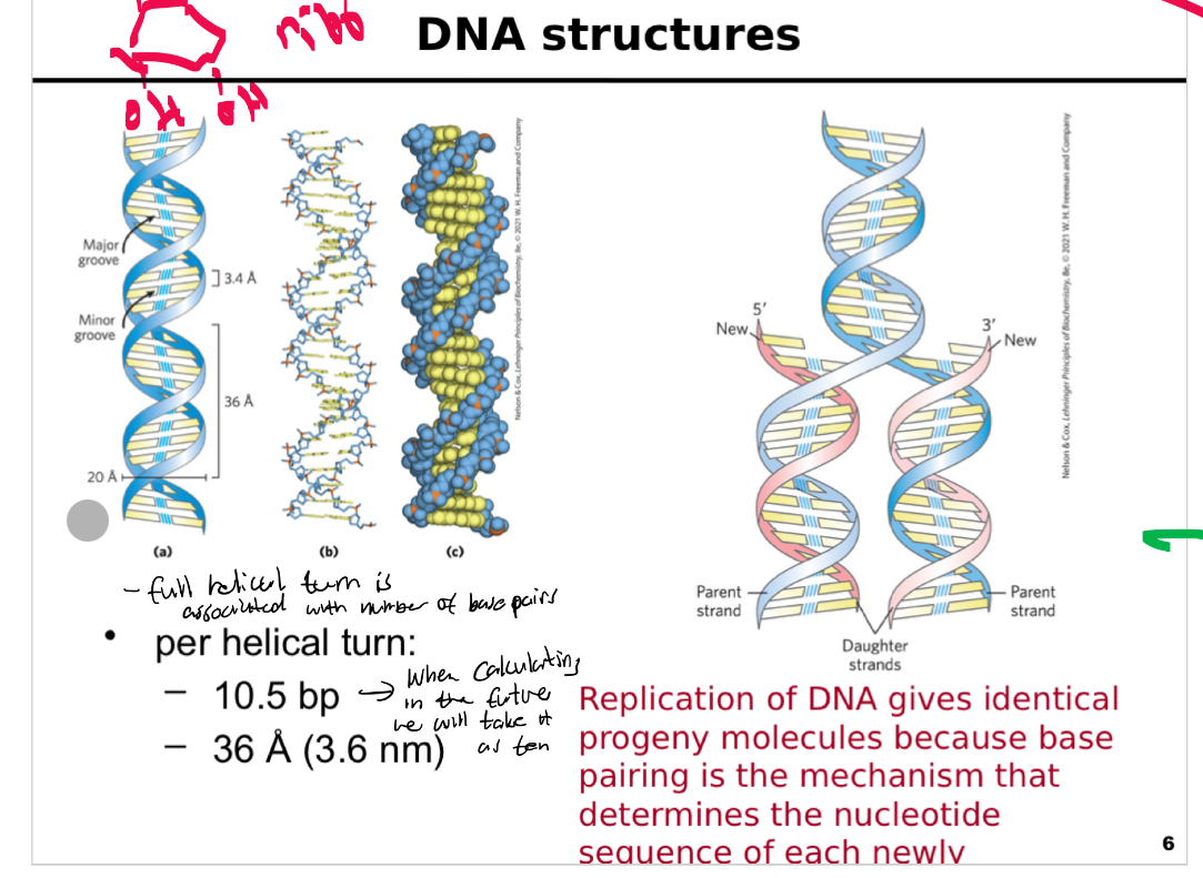
overview of the structure and replication of DNA,
This slide provides an overview of the structure and replication of DNA, illustrating its iconic double helix and how base pairing ensures accurate copying of genetic information.
Historical context:
A famous photograph of James Watson and Francis Crick is shown with their early DNA model, acknowledging their landmark contribution to discovering DNA’s structure.
DNA double helix:
Depicted in multiple formats to highlight key features:
Twisted ladder model showing base pairs (A–T, G–C) inside and sugar-phosphate backbones outside.
Molecular view showing major and minor grooves.
Space-filling model illustrating the arrangement of atoms: phosphate groups, sugars, and bases.
Key dimensions:
Diameter: approximately 2.0 nm (20 Å).
Helical pitch (one turn): 3.6 nm (36 Å) with about 10.5 base pairs per turn.
Distance between adjacent bases: 0.34 nm (3.4 Å).
Replication illustration (right):
Shows parental DNA strands separating and serving as templates for new complementary strands.
Highlights semiconservative replication: each daughter molecule contains one old and one newly synthesized strand.
Emphasizes the role of base pairing in ensuring accurate sequence copying.
Key message:
DNA’s elegant double-helical structure not only stores genetic information but also makes replication possible through complementary base pairing. Understanding its physical characteristics is critical for topics like replication, transcription, and genetic engineering.
What is the organization of the DNA double helix, focusing on base pairing, strand orientation, and the structural principles that enable accurate replication?
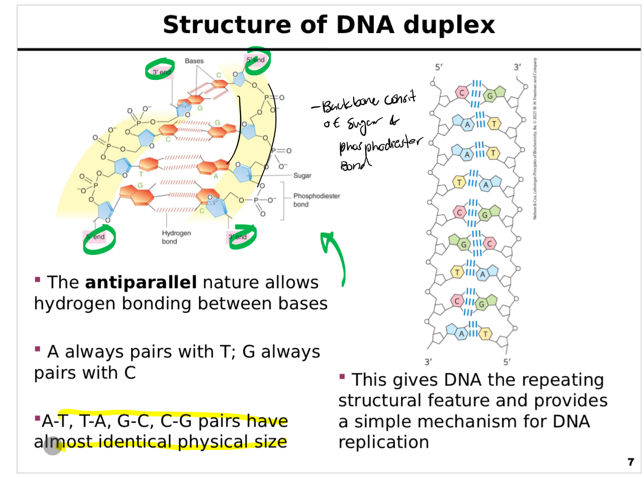
This slide explains the organization of the DNA double helix, focusing on base pairing, strand orientation, and the structural principles that enable accurate replication.
Antiparallel strands:
DNA is composed of two complementary strands running in opposite directions: one 5′ to 3′ and the other 3′ to 5′.
This antiparallel orientation is critical for hydrogen bonding between bases and enzymatic processes like replication and transcription.
Base-pairing rules:
Adenine (A) always pairs with Thymine (T) using two hydrogen bonds.
Guanine (G) always pairs with Cytosine (C) using three hydrogen bonds.
Pairings are complementary and consistent, maintaining a uniform helix diameter.
Size and uniformity:
Despite different chemical compositions, A-T and G-C pairs have nearly identical dimensions, allowing DNA to coil uniformly.
Structural features:
The diagram shows:
A 3D helical view of the DNA duplex.
An unwound segment illustrating the antiparallel orientation and phosphate backbone on the outside, bases on the inside.
Hydrogen bonds holding complementary bases together.
Functional significance:
This regular, repeating structure underlies DNA’s stability and explains how base pairing enables accurate copying during replication and repair.
Key message:
DNA’s antiparallel strands and complementary base-pairing are the foundation for its structure and function, providing a reliable mechanism for information storage and transfer.
What are the physical dimensions and structural properties of base pairs in the DNA double helix, focusing on the importance of geometry and hydrogen bonding?
This slide details the physical dimensions and structural properties of base pairs in the DNA double helix, focusing on the importance of geometry and hydrogen bonding.
Molecular structures:
Diagrams show the pairing between Guanine (G) and Cytosine (C) (top) and Adenine (A) and Thymine (T) (bottom).
Key atoms are color-coded (hydrogen, oxygen, nitrogen, carbon) for clarity.
Distances between hydrogen bonds are indicated (~0.28–0.30 nm).
Measured dimensions of B-DNA:
Helical turn:
One complete turn of the double helix spans approximately 3.32 nm and contains about 10.3 base pairs.
Helix diameter:
The diameter of B-DNA is about 2.37 nm, which allows optimal pairing of one purine with one pyrimidine. Pairing two purines would be too wide; two pyrimidines too narrow.
Base spacing:
The distance between stacked bases is 0.29–0.30 nm, measured as the length of hydrogen bonds, contributing to DNA’s stability and uniformity.
Key implications:
The consistent dimensions of base pairs allow the DNA double helix to maintain a regular shape, crucial for packaging, recognition by proteins, and accurate replication.
Proper base pairing (purine–pyrimidine) ensures a uniform helix diameter and structural integrity.
Key message:
The precise dimensions of DNA and base-pairing geometry are critical for molecular interactions, enzyme recognition, and overall genomic stability.

What are the interactions that help stabilize duplex DNA? And how does it work?
This slide summarizes the various noncovalent forces that maintain the stability and integrity of the DNA double helix.
Key stabilizing interactions:
Hydrophobic interactions:
Bases cluster internally, away from water, minimizing exposure of hydrophobic surfaces.
This helps shield the aromatic rings and contributes to helix formation.
Hydrogen bonds:
Specific pairing involves hydrogen bonds:
Three bonds between Guanine–Cytosine (G–C).
Two bonds between Adenine–Thymine (A–T).
While individually weak, their large numbers add stability.
Base stacking:
Planar bases are stacked vertically, stabilizing the helix through van der Waals forces and pi–pi interactions.
Base stacking also excludes water, further stabilizing the hydrophobic core.
Hydration:
DNA interacts with surrounding water molecules.
Water binds to available oxygen and nitrogen atoms on bases and the sugar-phosphate backbone, influencing DNA’s overall conformation and dynamics.
Electrostatic interactions:
DNA’s sugar-phosphate backbone carries negative charges due to phosphate groups.
Metal ions (e.g., Mg²⁺, Na⁺) can bind and help neutralize this repulsion, stabilizing the double helix.
Diagram:
Illustrates DNA strands, hydrogen bonds, and backbone arrangement, highlighting interactions that maintain structural integrity.
Key message:
Duplex DNA stability is not due to covalent bonds but to the sum of many weak interactions that, together, give DNA its unique balance of flexibility and strength.

What properties of purine and pyrimidine bases help determine the structure and stability of DNA and RNA?
Back:
The bases in DNA and RNA are purines and pyrimidines.
They are called bases because free purines and pyrimidines are weakly basic compounds.
1. The bases are aromatic
Purines and pyrimidines are aromatic molecules.
This means their ring electrons are delocalized, or spread out over the ring instead of belonging to just one bond.
Because of this:
many bonds have partial double-bond character
the rings become very rigid
pyrimidines are planar
purines are almost planar, with only a slight pucker
So the bases are basically flat molecules.
That flat shape becomes important because the bases can stack on top of each other.
Think:
Aromatic → flat rings → easy base stacking
2. Bases can exist as different tautomers
A tautomer is a slightly different arrangement of the same molecule.
The atoms are the same, but the positions of certain:
hydrogens
double bonds
can change.
For example, uracil can exist as:
lactam
lactim
double lactim
These forms can convert into one another.
Which tautomer is most common depends partly on pH.
At about pH 7, the normal tautomeric forms shown for A, G, C, T, and U are the forms that predominate.
This matters because the tautomer determines where hydrogen bonds can form.
So:
Tautomeric form → hydrogen-bonding pattern → which bases can pair
3. Bases absorb UV light
All nucleotide bases absorb ultraviolet light.
DNA and RNA absorb particularly strongly around:
260 nm
This happens because of their aromatic ring systems and delocalized electrons.
This is why scientists often measure absorbance at 260 nm to detect or estimate the amount of DNA or RNA in a sample.
Memory:
Nucleic acids → A₂₆₀
4. The bases are hydrophobic
At the near-neutral pH found inside cells, purines and pyrimidines are:
relatively hydrophobic
relatively insoluble in water
In contrast, at very acidic or alkaline pH, the bases can become charged.
Once charged, they interact better with water, so their solubility increases.
So:
Near-neutral pH → mostly uncharged → more hydrophobic
Acidic/basic pH → more charged → more water-soluble
5. Base stacking stabilizes nucleic acids
Because the bases are:
flat
aromatic
relatively hydrophobic
they tend to stack on top of each other inside nucleic acid structures.
Imagine a:
stack of coins
The flat rings lie roughly parallel to one another.
This is called base stacking.
Base stacking is stabilized by several interactions:
hydrophobic interactions
van der Waals interactions
dipole-dipole interactions
One major benefit is that stacking keeps the hydrophobic bases away from water.
So base stacking is very important for stabilizing the 3D structure of DNA and RNA.
6. What functional groups do the bases have?
The important functional groups on purines and pyrimidines include:
ring nitrogens
carbonyl groups
exocyclic amino groups
These groups determine how bases can form hydrogen bonds.
“Exocyclic” simply means:
outside the ring
7. Hydrogen bonding allows complementary base pairing
The amino and carbonyl groups are especially important for forming hydrogen bonds between bases.
The most common base-pairing pattern is Watson-Crick base pairing:
A pairs with T in DNA
A pairs with U in RNA
G pairs with C
So:
DNA: A–T and G–C
RNA: A–U and G–C
These are called complementary base pairs.
8. Why does a particular base pair with another base?
The normal tautomeric forms at about pH 7 place:
hydrogen-bond donors
hydrogen-bond acceptors
in the correct positions.
That is what allows:
A to fit with T/U
and
G to fit with C
So complementary pairing depends on both:
shape + hydrogen-bonding groups
9. Two major interactions stabilize nucleic acid structure
You should distinguish these:
Base stacking
occurs mainly between bases next to each other along the strand
bases lie roughly parallel
involves hydrophobic, van der Waals, and dipole interactions
strongly helps stabilize the overall structure
Hydrogen bonding
occurs between complementary bases
A–T or A–U
G–C
helps bases recognize their correct partner
A useful way to think about it:
Stacking helps hold the structure together.
Hydrogen bonding helps determine who pairs with whom.
10. Why is complementary base pairing biologically important?
Because A always pairs with T/U and G always pairs with C, one strand contains information that can be used to determine the sequence of another strand.
For example:
5′-A G C T-3′
pairs with
3′-T C G A-5′
That is what allows genetic information to be accurately copied and duplicated.
High-yield summary
Aromatic bases
→ delocalized electrons
→ flat structure
→ UV absorption near 260 nm
Flat + hydrophobic bases
→ base stacking
→ stabilizes DNA/RNA structure
Amino + carbonyl groups
→ hydrogen bonding
Watson-Crick pairing
→ A–T/U and G–C
Specific base pairing
→ allows accurate copying of genetic information
Easy memory line
“Flat bases stack, functional groups pair.”
Flat aromatic rings → stacking
Amino/carbonyl groups → hydrogen bonding
Together they help create and stabilize the 3D structure of nucleic acids.
What are the difference in the roles of DNA and RNA?
This slide contrasts the roles and types of DNA and RNA in cells, emphasizing their differences in structure and function.
DNA:
Described as one type with one primary purpose: to store and transmit genetic information.
DNA serves as the stable blueprint for all cellular activities and inheritance.
RNA:
Highlighted as having many types with many purposes, performing a variety of roles.
Key classes include:
Messenger RNA (mRNA):
Carries the genetic code from DNA to ribosomes, where it directs protein synthesis.
Ribosomal RNA (rRNA):
Structural and catalytic component of ribosomes, providing the scaffold and enzymatic activity for protein assembly.
Transfer RNA (tRNA):
Transports specific amino acids to the ribosome during protein synthesis, ensuring correct sequence assembly.
Other RNAs:
Small nuclear RNAs (snRNAs): involved in RNA processing (e.g., splicing).
Small non-coding RNAs: regulate gene expression and other cellular processes (e.g., microRNAs, siRNAs).
Key message:
While DNA is uniform and primarily a storage molecule, RNA exists in multiple forms, each specialized to fulfill critical roles in gene expression, catalysis, and regulation.

Give me an overview of transcription and translation
This slide illustrates the two main stages of gene expression: transcription and translation, showing how information in DNA is converted to proteins.
Transcription (left panel):
Occurs in the nucleus of eukaryotic cells (or cytoplasm in prokaryotes).
RNA polymerase unwinds the DNA double helix and uses one strand as a template.
Free ribonucleotides (A, U, G, C) are incorporated to form a complementary messenger RNA (mRNA) strand.
The mRNA carries the genetic message to the cytoplasm for protein synthesis.
Translation (right panel):
Takes place in the cytoplasm at ribosomes.
mRNA provides the sequence of codons, each coding for an amino acid.
Transfer RNAs (tRNAs) deliver specific amino acids, matching their anticodons to the mRNA codons.
Ribosomal RNA (rRNA) forms the core of the ribosome, catalyzing peptide bond formation to assemble the polypeptide chain.
Key points:
All RNA molecules are synthesized from DNA by transcription.
Several RNA types collaborate during translation:
mRNA – specifies the protein sequence.
tRNA – delivers amino acids.
rRNA – structural and catalytic component of ribosomes.
Key message:
Gene expression involves a two-step process: copying DNA into RNA and then decoding RNA to build proteins, with each RNA type playing a critical role.

How do prokaryotic mRNAs differentiate from eukaryotic mRNAs, focusing on transcription and translation in bacteria and archaea?
This slide explains how prokaryotic mRNAs differ from eukaryotic mRNAs, focusing on transcription and translation in bacteria and archaea.
Key features:
A single prokaryotic mRNA can encode multiple proteins – this is known as a polycistronic mRNA.
Genes are often organized in operons, where several genes are transcribed together into one mRNA.
Diagram overview:
At the top: a DNA segment containing Gene A, Gene B, and Gene C.
RNA polymerase transcribes the DNA, generating a continuous mRNA containing coding regions for all three genes.
At the bottom: the resulting mRNA molecule, with ribosomes binding to start translation even while transcription is ongoing (a prokaryote-specific feature).
Each ribosome reads the mRNA and synthesizes a different polypeptide (A, B, or C).
Important points:
Translation can begin before transcription finishes because prokaryotic cells lack a nuclear membrane.
Each coding region has its own ribosome binding site and start codon.
Multiple ribosomes can work on the same mRNA simultaneously, increasing efficiency.
Key message:
Prokaryotic mRNAs are versatile and efficient, allowing the coordinated expression of several genes in one transcript, supporting rapid growth and adaptation.

mRNA synthesis and processing in eukaryotic cells, highlighting how it differs from prokaryotes. Key stages of mRNA production…..
This slide outlines the process of mRNA synthesis and processing in eukaryotic cells, highlighting how it differs from prokaryotes.
Key stages of mRNA production:
Transcription:
RNA polymerase transcribes the DNA template into a primary transcript (pre-mRNA).
The transcript contains both exons (coding sequences) and introns (non-coding sequences).
Splicing and processing:
Introns are removed by splicing, and exons are joined to create a continuous coding sequence.
Additional modifications occur:
5′ cap (not shown here) for stability and translation initiation.
Poly(A) tail added at the 3′ end after transcription, enhancing stability and export.
Transport and translation:
The mature mRNA is exported to the cytoplasm, where ribosomes translate it into protein.
Each mature mRNA typically encodes only one protein (monocistronic).
Diagram details:
Shows the gene with Exon 1, Intron, and Exon 2.
Depicts transcription, splicing, and translation stages, ending with production of Protein A.
Key message:
Eukaryotic mRNAs are processed and refined before translation, ensuring correct protein coding. Unlike prokaryotic mRNAs, they are monocistronic and undergo splicing and chemical modifications.

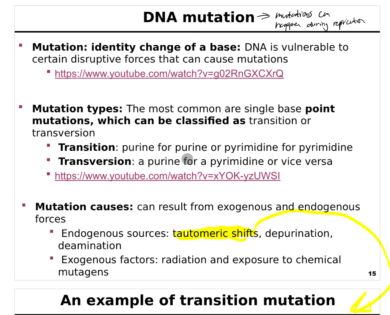
What is mutations, the types and what causes it?
This slide introduces the concept of mutations in DNA, their types, and possible causes, supported by video links for further explanation.
What is a mutation?
Defined as an identity change of a base in the DNA sequence.
DNA is inherently vulnerable to disruptive forces—both internal and external—that can alter its chemical structure and lead to mutations.
Types of mutations:
The most common are single-base (point) mutations.
Two key categories:
Transition:
Substitution of one purine for another (A ↔ G) or one pyrimidine for another (C ↔ T).
Transversion:
Substitution of a purine for a pyrimidine or vice versa (A/G ↔ C/T).
These changes can affect protein coding and regulation.
Mutation causes:
Endogenous sources (within the cell):
Tautomeric shifts: temporary rearrangements of base structure.
Depurination: loss of a purine base.
Deamination: conversion of cytosine to uracil, adenine to hypoxanthine, etc.
Exogenous sources (environmental factors):
Radiation: UV light, X-rays, gamma rays.
Chemical mutagens: reactive chemicals that modify DNA bases.
Additional resources:
Two YouTube links are provided for students to explore mutation mechanisms in more depth.
Key message:
Mutations occur naturally and under external influences. Understanding their types and causes is crucial for studying genetic variability, disease, and evolution.
What is tautomeric shift?
This slide illustrates how tautomeric shifts can lead to transition mutations, showing the chemical basis of base mispairing.
Tautomeric shifts:
Bases can exist in alternative chemical forms (tautomers) due to spontaneous proton rearrangements.
Examples include:
Amino ↔ Imino forms.
Keto ↔ Enol forms.
These shifts change the hydrogen-bonding properties of the bases.
Diagram explanation:
Panel (a): Normal pairing of adenine (amino form) with thymine or cytosine with guanine.
Panel (b): Adenine shifts to its imino form, changing its hydrogen bonding pattern.
Panel (c): The imino form of adenine can mispair with cytosine, leading to an incorrect base in the daughter strand after replication.
Key consequences:
If tautomeric forms arise during DNA replication, they can cause mispairing and result in a transition mutation (purine ↔ purine or pyrimidine ↔ pyrimidine).
These changes are rare but important sources of spontaneous mutations.
Summary points on the slide:
Tautomeric shifts occur spontaneously.
They can alter base-pairing rules temporarily.
Misincorporation during replication can lead to heritable changes in DNA.
Key message:
Small, transient chemical changes in bases can cause replication errors, emphasizing the delicate balance of DNA chemistry and the origins of spontaneous mutation.
