Introduction to the Nervous System
 Call Kai
 Learn
Learn Practice Test
 Spaced Repetition
 Match
Match Flashcards
 Knowt Play
1/121

There's no tags or description
Looks like no tags are added yet.
Name | Mastery | Learn | Test | Matching | Spaced | Call with Kai | Chat |
|---|
No analytics yet
Send a link to your students to track their progress
122 Terms
Nervous System Sections
Peripheral
Central
Peripheral Nervous System
comprises all neural tissue beyond the CNS (such as neurons that receive sensory information or that send information to muscles, and those that relay information to or from the spinal cord or the brain)
Autonomic
Somatic
Autonomic Peripheral Nervous System
controls self-regulated action of internal organs and glands
sympathetic (arousing)
parasympathetic (calming)
Contralateral
Opposite side of the brain
Ipsilateral
Same side of the brain
Unilateral
one side of the brain
Bilateral
both sides of the brain
Gray matter
neuronal cell bodies
White matter
axons, myelin, and glial cells
Axon tracts
occur within hemispheres, between hemispheres (commissure), and between cortical and sub-cortical regions (projection tracts)
Main commisure
corpus callosum —> massive white matter tract linking the two hemispheres
Major subdivisions of the brain

Brainstem
medulla
pons
midbrain
brainstem houses cell bodies of most cranial nerves
Cranial nerves
receive sensory information from the head and neck and control their movement
some are responsible for the neural control of internal organs
Medulla
connects brain to spinal cord
all the ascending (sensory) and descending (motor) nerve fibers connecting the brain and spinal cord pass through the medulla
controls many vital functions and reflexes such as respiration and heart rate
home to part of a set of neurons known as the reticular activating system (RAS)
directly superior to the spinal cord
the region of the brain that contains many of the cell bodies of the 12 cranial nerves
the region of the brain where most of the motor fibers cross from one side of the body to the other (—> right side of brain controls left side of body and vice versa)
Pons
connective bridge —> connects the rest of the brain to the cerebellum and cranial nerves
directly superior to the medulla and anterior to the cerebellum
important center for control of certain types of eye movements and vestibular functions (e.g., balance)
at the pons, information from both ears converges, allowing comparisons between the information received by each ear (thought to be important for localization of sounds)
cell bodies of cranial nerves V-VIII
head and face sensations
motor control of eyes, face, mouth
secretion of saliva and tears
sensory roles in hearing and taste
Midbrain
superior to the pons
contains the nuclei of the cells that form some of the cranial nerves
Orienting by sound and sight
two important structures on its dorsal side: the inferior colliculus and the superior colliculus
Inferior colliculus
a relay point for auditory information as it travels from the ear to the cortex
involved in sound localization
also contributes to reflexive movements of the head and eyes in response to sound
Superior Colliculus
allows us to perceive and orient toward large moving visual objects in the periphery
Cerebellum
located posterior to the medulla
important for regulation of muscle tone and guidance of motor activity
also important for fluidity and precision in mental processes and the brain’s internal clock (lateral cerebellum)
Damage to the Cerebellum
damage interferes with precision of movement and disrupts balance, motor control, and equilibrium
classic test used to detect damage: ask person to alternate between touching their nose, and then the doctor’s outstretched finger (path of hand will be imprecise and jagged)
punch-drunk syndrome - a common manifestation of temporary disruption to the cerebellum, in which a person temporarily loses balance and coordination after sustaining a blow to the head
The Diencephalon
consists of the thalamus and the hypothalamus
Thalamus
Main relay station of the brain (for almost all sensory information coming into the cortex and almost all motor information leaving it)
serves to reorganize information before it is sent elsewhere in the nervous system
the patterns of connections, both to and from the thalamus, are very specific —> one particular region of the thalamus receives information from just one sensory system and projects to only one particular region of the cortex
“gateway to the cortex”
all sensory pathways (except olfaction) make synaptic relays in the thalamus before continuing to the primary sensory receiving areas
lateral geniculate nucleus and medial geniculate nucleus
Lateral geniculate nucleus
receives information from retina, sends axons to primary visual cortex
Medial geniculate nucleus
receives information from inner ear, sends axons to primary auditory cortex
Hypothalamus
center for homeostasis (ex. provides signals telling the brain that it should drink/eat if thirsty/hungry)
main link between nervous and endocrine system
main site for control of hormone production
it synthesizes and secretes neurohormones (releasing hormones)
controls autonomic nervous system
connected to master gland: pituitary gland
Pituitary Gland
releases hormones in the bloodstream to influence other organs and tissues
involved in thermoregulation, appetite, thirst, fatigue, circadian cycles, stress response, reproduction
tightly linked with limbic system that plays major role in emotion regulation
Major Subcortical Systems
the basal ganglia and the limbic system
located in regions below the cerebral cortex
Limbic system
amygdala and hippocampus
Amygdala
essential for emotional, especially fear processing
implicated in the quick response to salient emotional information
Hippocampus
essential for conscious remembering
formation of new long-term memories
Basal Ganglia
groups of neurons that are central to motor functions
damage results in motor deficits (tremor, slowness, changes in posture, etc.) generally characterized by involuntary movements
Parkinson’s or Huntington’s Disease
The Cerebral Cortex
thin (1-3mm) layer, 2cm below scalp, that wraps surface of brain, that makes up 80% of the brain
divided into several lobes
surface: gray matter
the underlying tissue: white matter
all higher cognitive functions involve the cerebral cortex
convolution, or bump gyrus
valleys between bumps = sulcus or fissures
divided into two physically separated halves = cerebral hemispheres
The Four lobes of the cerebral cortex
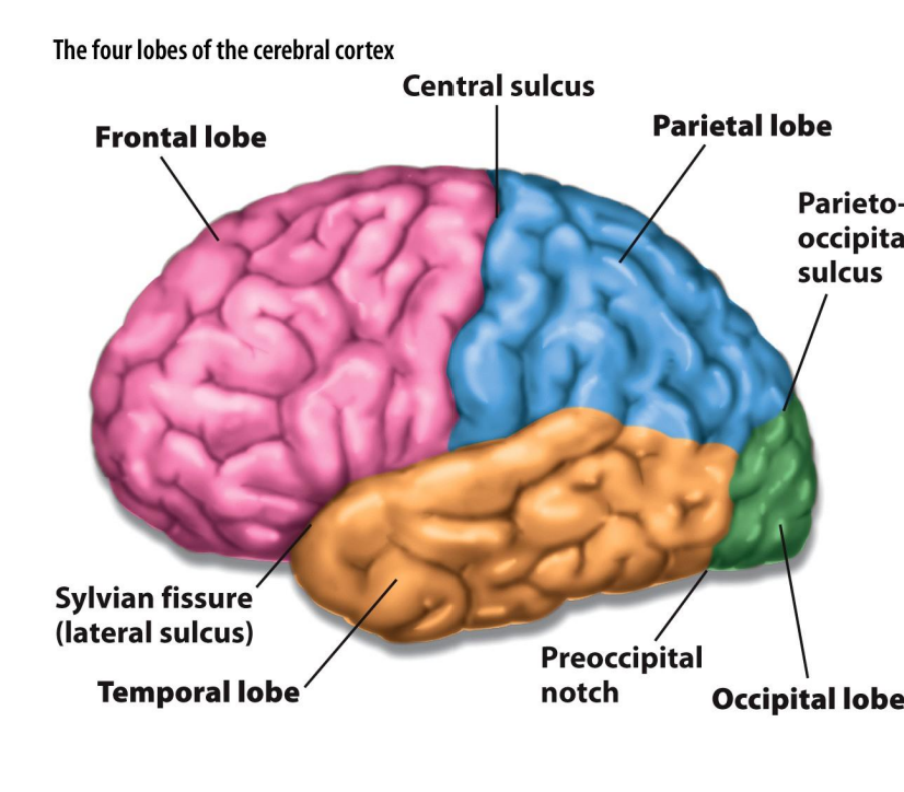
Longitudinal Fissure
separates the right cerebral hemisphere from the left
each hemisphere has a unique specialization in both cognitive and emotional functioning
Central Fissure
separates each hemisphere of the brain in an anterior-posterior dimension
in general, areas of the brain in front of the central fissure are more involved in motor processing
whereas, those behind are more involved in sensory processing
Sylvian (lateral) fissure
separates each hemisphere of the brain in the dorsal-ventral dimension
the division is important because the area of the brain below the Sylvian fissure is the temporal lobe, which plays a key role in memory, emotion, and auditory processing
Occipital Lobe
Visual processing
the remaining region of the brain (other than the other 3 lobes) behind the parieto-occipital sulcus
Temporal lobe
auditory processing, language, long-term memory, object recognition
the area below the Sylvian fissure
Parietal Lobe
Somatosensory processing (pressure, texture, warmth, proprioception, nociception); integrates information from various sensory modalities
the region directly behind the central fissure, but above the Sylvian fissure
Frontal lobe
all higher mental processes, executive functioning, cognitive control, motor planning, attention
the area in front of the central fissure
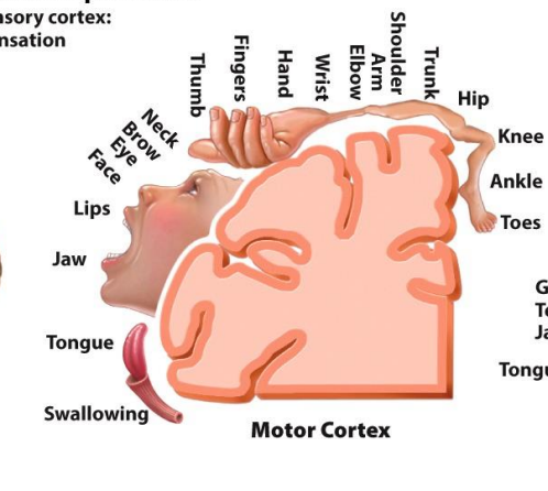
Motor Cortex
Movement
(topographic correspondence between cortical regions and body surface with respect to somatosensory and motor processes)
Somatosensory Cortex
somatic sensation
(topographic correspondence between cortical regions and body surface with respect to somatosensory and motor processes)

Anterior
front of the brain
Posterior
back of the brain
Rostral
regions toward the front of the brain and towards the head
Caudal
regions toward the rear of the brain and towards the tail
Superior
top of the brain
Inferior
bottom of the brain
Dorsal
top of the brain
toward an animal’s back
Ventral
bottom of the brain
toward an animal’s stomach
Medial
areas in the middle or center of the brain
Lateral
areas that are toward the outside of the brain
Coronal
when the brain is sliced ear-to-ear to separate the front from the back
Horizontal (or axial/tranverse)
if the brain is sliced so that the top of the brain is separated from the bottom
Sagittal
the brain is cut so that the left side of the brain is separated from the right side
Midsagittal
a sagittal slice down the middle of the brain
Lateral
a sagittal slice that is more toward one side
Contralateral
the opposite side of the brain
motor control occurs contralaterally
Ipsilateral
the same side of the brain
Unilateral
applies to only one side of the brain
Bilateral
applies to both sides of the brain
Proximal
Near the brain (or region)
Distal
far from the brain (or region)
ex: distal muscles are in your far extremities (like your hands)
Central Nervous System
brain and spinal cord
very fragile —> entire CNS is encased in bone
spinal cord is enclosed within the spinal column
brain is enclosed within the skull
Cerebrospinal Fluid (CSF)
between neurons and their bony encasement (CNS) —> brain essentially floats in CSF (makes it buoyant and cushions it)
similar in composition to blood plasma
also serves metabolic needs, allowing nutrients to reach neurons
Ventricles
the fluid-filled spaces that contain CSF
most prominent are in the lateral ventricles
What are the 7 main subdivisions of the CNS?
spinal cord
the medulla
the cerebellum
the pons
the midbrain
the hypothalamus and thalamus (diencephalon)
the cerebral cortex
Spinal Cord
the portion of the nervous system through which most sensory neurons relay information to the brain, and through which motor commands from the brain are sent to the muscles
Spinal Column
the bony structure housing the spinal cord
composed of vertebrae (sections)
at each vertebrae, sensory information enters the cord and motor information leaves it
if the spinal cord were cut in cross-sections, two clumps of nerve cells, one located ventrally and another dorsally, would be prominent
cells in the dorsal section receive sensory information
cells in the ventral section are responsible for conveying motor commands to the muscles and for receiving input from the brain and from other regions of the spinal cord
Damage to the Spinal Cord
leaves a person without sensation in or motor control for all body areas that are connected to the brain by spinal cord segments distal to the point of injury
12 cranial nerves
some are responsible for receipt of sensory information and motor control of the head
other are responsible for the neural control of internal organs
Damage to the Medulla
damage can be fatal
one common accompaniment of either diffuse or specific brain damage is swelling of the entire brain —> when this swelling puts enough pressure on the medulla to interfere with its functions, death can result
reticular activating system (RAS)
receives input from the environment as well as the internal milieu of the body, and then project diffusely to many other regions of the brain
allows RAS to contribute to overall arousal and attention, as well as regulation of sleep-wake cycles
Visual Periphery
plays a role in movement of the head and eyes in response to environmental stimuli —> with regards to large moving objects
Damage to the hypothalamus
Ventromedial region - eat more than is required to maintain a normal body weight —> obesity
Dorsal and Lateral regions - interfere with water intake
Gyrus
each convolution, or bump, of the brain is called a gyrus
basically a giant sheath of neurons wrapped around the other brain structures
serve to pack more brain tissue into a smaller space
every brain has the same basic gyral pattern; but subtle individual variations exist
Sulcus
each valley between the bumps (or gyrus) in the cerebral cortex
if its deep its called a fissure
Three major fissures in the brain
central fissure
sylvian (lateral) fissure
longitudinal fissure
these three major fissures also divide each hemisphere into four major regions, or lobes
The four major lobes
frontal lobe
temporal lobe
parietal lobe
occipital lobe
Primary Sensory Cortex
the first region in the cortex to receive information about a particular sensory modality
Primary Motor Cortex
the region of the cortex that is the final exit point for neurons responsible for fine motor control of the body’s muscles
in front of the central fissure in a long, narrow band called the motor strip
body parts for which we have a large degree of fine motor control have a disproportionately larger area of brain tissue devoted to their control
General characteristics of organization shared between the primary sensory and motor cortexes
all these brain areas are organized so that specific attributes of the physical world are “mapped” onto brain tissue (hair cells)
these maps are distorted relative to the physical world, reflecting the density of receptors (or effectors) within a system
ex: higher density of receptors at focal point in vision —> much more of the primary visual cortex is devoted to processing visual info from central part of vision (than periphery)
mapping of the world onto brain tissue occurs in an upside-down and backward manner for vision, touch, and motor control
Damage to the primary motor cortex
leads to muscle weakness on the contralateral side of the body
ex: damage to dorsal regions of the motor strip —> weakness of the bottom part of the body
massive destruction to the motor strip, along with damage to the basal ganglia (as often occurs after stroke) results in paralysis on the contralateral side of the body —> known as hemiplegia
Primary somatosensory cortex
portion of the cortex that receives information about tactile (touch) stimulation, proprioception, and pressure and pain sensations from internal organs and muscles
map of the body onto the primary somatosensory cortex is inverted left-to-right and top-to-bottom
distortion of body parts in the somatosensory map is proportional to the density of touch receptors (like motor homunculus)
Proprioception
the perception of the position of body parts and their movements
located directly posterior to the central fissure
Hair cells in the cochlea of the ear
are differentially sensitive to sounds of different frequencies, which we perceive as tones of different pitch
How does tactile information reach the somatosensory cortex?
skin contains various nerve endings, or receptors, that are sensitive to different aspects of tactile information
crude tactile information and pain and temperature information is sent to the cortex by neurons that synapse at dorsal regions of the spinal cord —> then carried to thalamus —> then to cortex
information about fine touch and proprioception enters to spinal column but doesn’t synapse until the medulla —> from which point it crosses over and is carried to the thalamus —> then to the cortex
Damage to the somatosensory cortex
impairs fine discriminations of touch on the side of the body contralateral to the damaged cortex
ex: can’t tell what material a cloth they are holding is
Primary Visual Cortex
the first region of the cortex that processes visual information, more specifically patterns of light across different regions of space
in the occipital lobe
How does information reach the primary visual cortex
when you look straight ahead information to the right of the fixation (right visual field) projects to the left half of the retinas in both your eyes (and vice versa)
except for information in the far periphery of the world, all visual information reaches both eyes (otherwise only detected by right if in right periphery and vice versa)
ultimately information from the right visual field is directed solely to the primary visual cortex of the left hemisphere (and vice versa)
Damage to the visual cortex
inability to perceive visual information
homonymous hemianopsia, quadranopsia, and scotomas
Homonymous Hemianopsia
if the entire occipital cortex of only one hemisphere is damaged, no visual information can be detected in the contralateral visual field
Quadranopsia
when just the dorsal or ventral portion of the occipital cortex is damaged, in which case just one quadrant of the visual world is lost
Scotomas
when only small portions of the visual cortex are damaged
particular regions of the visual field in which light-dark contrast cannot be detected
Auditory System
sensitive to sound (which is essentially pressure waves in the air)
physical energy in sound waves —> vibrations in the eardrum + bones in the ear
vibrations are transformed into pressure waves in a liquid in the cochlea, which contains hair cells that transduce pressure waves into a neural signal
organized so that there are both ipsilateral and contralateral projections from the ear to the brain (auditory information received at the right ear projects to both the left and right hemispheres
Heschl’s Gyrus
in superior portion of the posterior temporal lobe
where primary auditory cortex is
Primary Auditory Cortex
tonotopic —> lowest tones are processed rostrally and laterally and tones of increasing frequency are processed more caudally and medially
Tonotopic
organized according to the frequency of a tone, which we perceive as pitch
Damage to the primary auditory cortex
unilateral - does not preclude the ability to perceive sound because of the redundancy provided by both crossed and uncrossed connections in the auditory system
alters sound threshold (the softest intensity that can be perceived (it becomes higher contralateral to the damaged hemisphere)
ability to perceive location of a sound becomes poorer fir the contralateral side of space (partially because loudness of sound helps determine location)