7. Muscular Tissue FLASHCARDS
1/121
There's no tags or description
Looks like no tags are added yet.
Name | Mastery | Learn | Test | Matching | Spaced |
|---|
No study sessions yet.
122 Terms
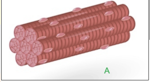
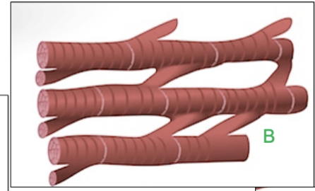
3 types of muscle tissues?
1.Skeletal Muscle
2.Cardiac Muscle
3.Smooth Muscle
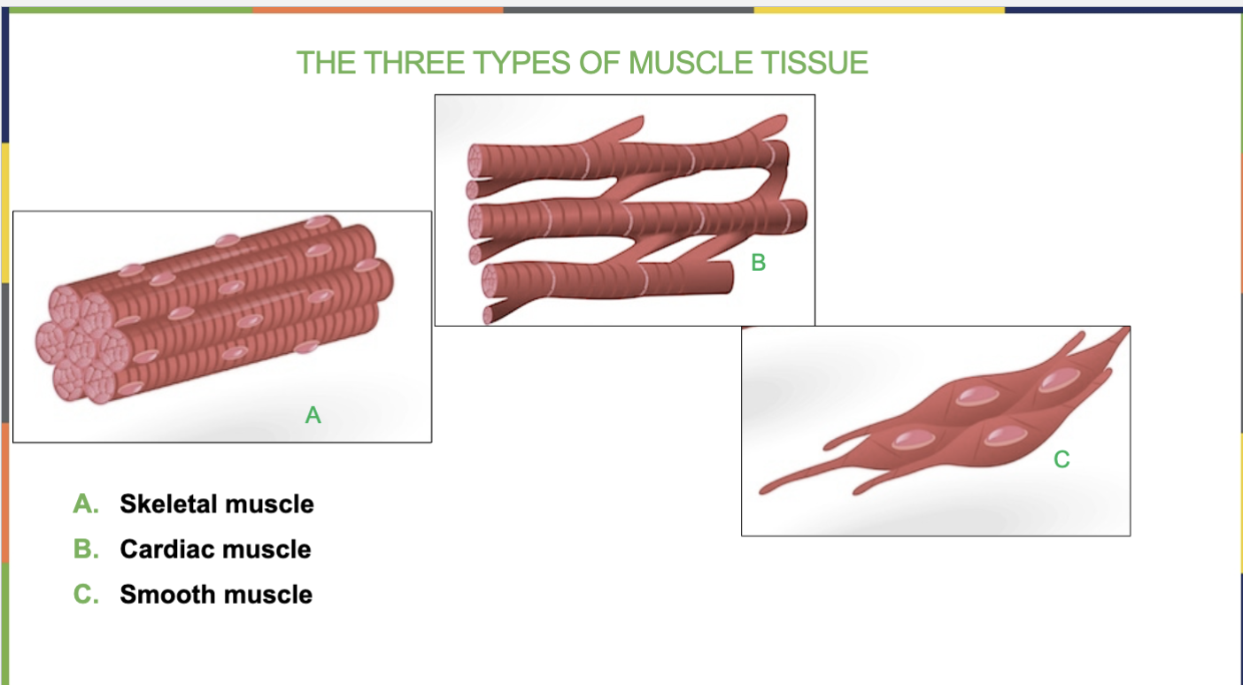
Location of Skeletal Muscle?
Attached to bones
Shape of Skeletal Muscle?
long, cylindrical

How many nuclei do Skeletal Muscle have?
Multiple (a lot), all located under the sarcolemma
Where are the nuclei of Skeletal Muscles located?
under the sarcolemma
Do Skeletal Muscles have striations(stripes)?
yes

4 Function of Skeletal Muscles?
Movement
Maintaining posture
Stabilizing joints
Generating body heat
Can you control the movement of Skeletal Muscles?
yes, you can control it
Location of Cardiac Muscle?
heart
Shape of Cardiac Muscle?
Branched
How many nuclei does the Cardiac Muscle have?
1, single, centrally located
Function of Cardiac Muscle?
Pumping blood
Does the Cardiac Muscle have striations(stripes)?
yes, its striated (has stripes)

Can you control the movement of Cardiac Muscles?
no, its involuntary (automatic)
happens automatically
Location of Smooth Muscle?
Lines some internal organs & walls of blood vessels
Shape of Smooth Muscle?
spindle shaped (pointed at both sides)
How many nuclei does Smooth Muscles have?
1, single, centrally located
Do Smooth Muscles have striations (stripes)?
No
Function of Smooth Muscles? (2 functions)
-contracts/pushes contents through organs (ex- intestines, urinary bladder, uterus)
-controls blood vessel diameter
Can you control the movement of Smooth Muscles?
No, it’s in involuntary
happens automatically
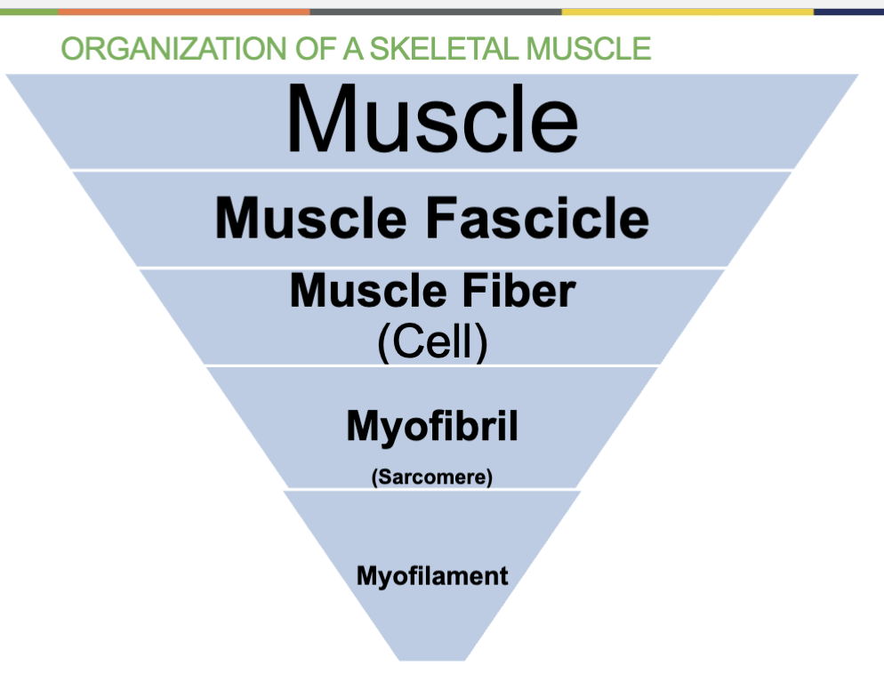
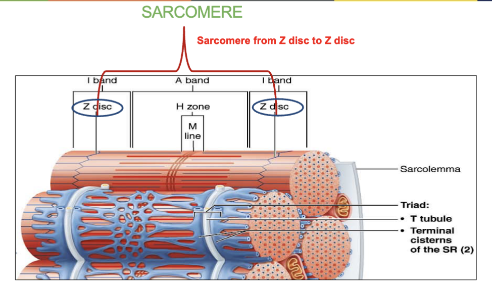
Organization of a Skeletal Muscle
(from biggest to smallest)
Muscle
Muscle Fascicle
Muscle Fiber (Cell)
Myofibril (made up of sarcomeres)
Myofilament (thick & thin)
Skeletal muscles is made up of smaller units called _____.
Fascicles

Fascicles contain _________.
(what are fascicles made up of?)
muscle fibers(cells)

What are the 3 Connective Tissue Coverings of Muscle?
Epimysium - covers entire muscle
Perimysium- Covers fascicles (bundle of fibers/cells)
Endomysium- Covers the individual muscle fibers (cells)
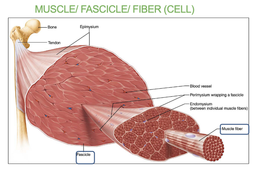
What is the Epimysium and what does it cover?
its connective tissue that covers the entire muscle.

What is the Perimysium and what does it cover?
its connective tissue that covers the fascicles.

What is the Endomysium and what does it cover?
its connective tissue that covers the individual muscle fibers that are in the fascicles.

4 Structures of the Muscle Fibers (cell)
(what it contains/is made up of)
Sarcolemma - plasma membrane that surrounds it
Sarcoplasm (cytoplasm) - the cytoplasm(fluid) of the muscle fiber/cell, enclosed by the sarcolemma, filled with mitochondria.
Nuclei - Multiple nuclei located under the sarcolemma.
Myofibrils- units that make up the muscle fibers.
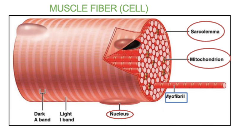
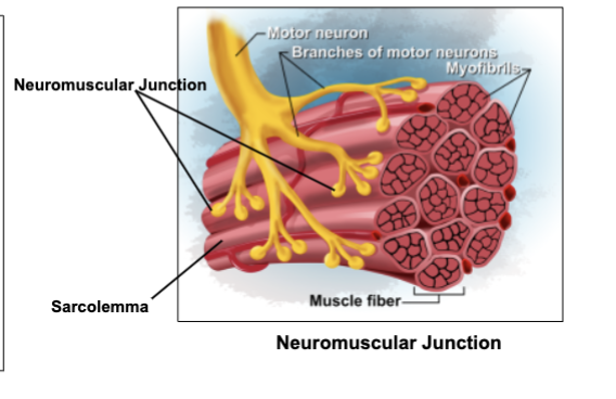
What’s the Sarcolemma and where can we find it?
it’s the plasma membrane of a muscle fiber (cell).
Found surrounding the muscle fibers and its beneath the endomysium.

The plasma membrane of the muscle cell is known as ______. This plasma membrane will become permeable to _______ ions.
Sarcolemma, sodium (Na⁺)
To which ions does the Sarcolemma become permeable to?
( These ions rush in and create an electrical signal (action potential) on the surface of the muscle cell for muscle contraction )
Sodium (Na⁺) ions
What’s the Sarcoplasm and where can we find it and what does it also contain a lot of?
it’s the cytoplasm of a muscle fiber (cell).
Enclosed by the Sarcolemma. it’s the "cell fluid" that fills the muscle cell.
Contains lots of mitochondria.

What are Myofibrils and where can we find them?
they are long, thread-like structures found inside muscle fibers (cells).
They make up the muscle fibers (cells)

What are Myofibrils made up of and what do they contain?
Myofibrils are Sarcomeres that are arranged side by side and these sarcomeres are made up of 2 Myofilaments (thick & thin)
3 structures of the Myofibrils?
Sarcoplasmic Reticulum (SR) - (the “rough er” of the muscle cells) Network of tubes all around/surrounding each myofibril.
Terminal Cisterns - when the SR enlarges into a pouch. It stores calcium.
Transverse Tubule (T-tubules) - Fold/tube coming from the sarcolemma that transmits electrical signals deep into muscle fiber (action potential)
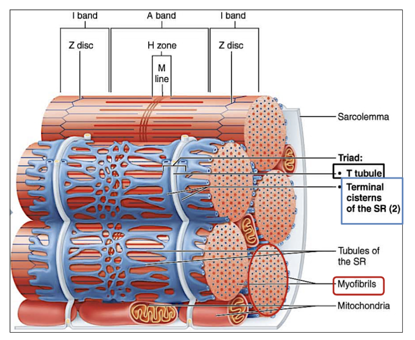
How is a Triad formed?
Formed by 2 Terminal Cisterns on either side of 1 T-tubule.
(triad = 2 terminal cisterns + 1 T-tubule)
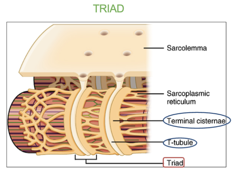
Job of Terminal Cisterns?
Stres calcium

Job of t-tubule?
A tube that comes from the sarcolemma that carries electrical signals to inside of muscle (action potential)

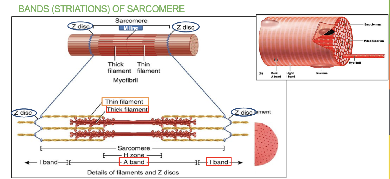
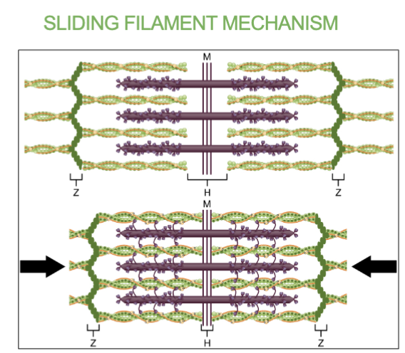
What are Sarcomeres and where are the found??
they are the basic functional units of muscle fiber
found in the Myofibrils (it’s what makes up the myofibrils & they’re arranged side by side)

2 Structures of Sarcomere?
Myofilaments- contain two overlapping myofilaments which are thick(myosin) & thin(actin).
(sarcomeres are the basic functional units of a myofibrils)
Z discs- Boundaries of the sarcomere, anchor the thin filaments (only in thin filaments)
______ are the basic functional unit of a muscle fiber.
Sarcomeres
What happens to Sarcomeres when muscles contract?
When a muscle contracts, sarcomeres shorten as actin and myosin slide past each other, pulling the Z discs closer together.
3 Arrangements/regions of Sarcomeres?
M line - middle of the sarcomere
A band - Contains thick filaments (myosin), appears dark (in the middle portion)
(red thick lines)
I Band - Contains thin filament (actin) - appears light , (at the ends of the centromeres)
(blue thin lines)
What does A band mean in Sarcomeres?
the region of the sarcomere that contains thick filaments (myosin)
(It appears dark→ dark band)
Red thick lines
→ does not move/change in length during contractions

What does I band mean in Sarcomeres?
the region of the sarcomere that contains only thin filaments (actin).
(appears light → Light band)
blue thin lines
→ shortens during contractions

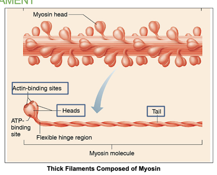
What does (myosin) mean in thick filaments and what does it contain and form during muscle contractions?
its the protein that makes up thick filaments in muscle fibers.
has myosin heads that can bind to actin (on thin filaments) to form cross-bridges during muscle contraction.

What does (actin) mean in thin filaments and what does it contain?
it’s the main protein that makes up thin filaments in muscle fibers.
contain the myosin-binding sites on actin, and it’s where myosin heads attach during contraction.
_______ (thick filaments) pull on _______ (thin filaments) to shorten the muscle and produce contraction.
(protein names)
Myosin , Actin
What are Z discs and where are they found?
they are the boundaries of a sarcomere, marking where one sarcomere ends and the next begins.
They anchor the thin filaments (actin) and help maintain the structure of the sarcomere.
Found at both ends of a sarcomere.
There are ___ thin filaments around 1 thick filament.
6

Do thick filaments move or not during contractions?
No. Thin filaments move towrdas midline.

Which type of filament is attached to Z discs?
ONLY thin filaments.
____ filaments will slide towards the midline when the muscle contracts.
Thin
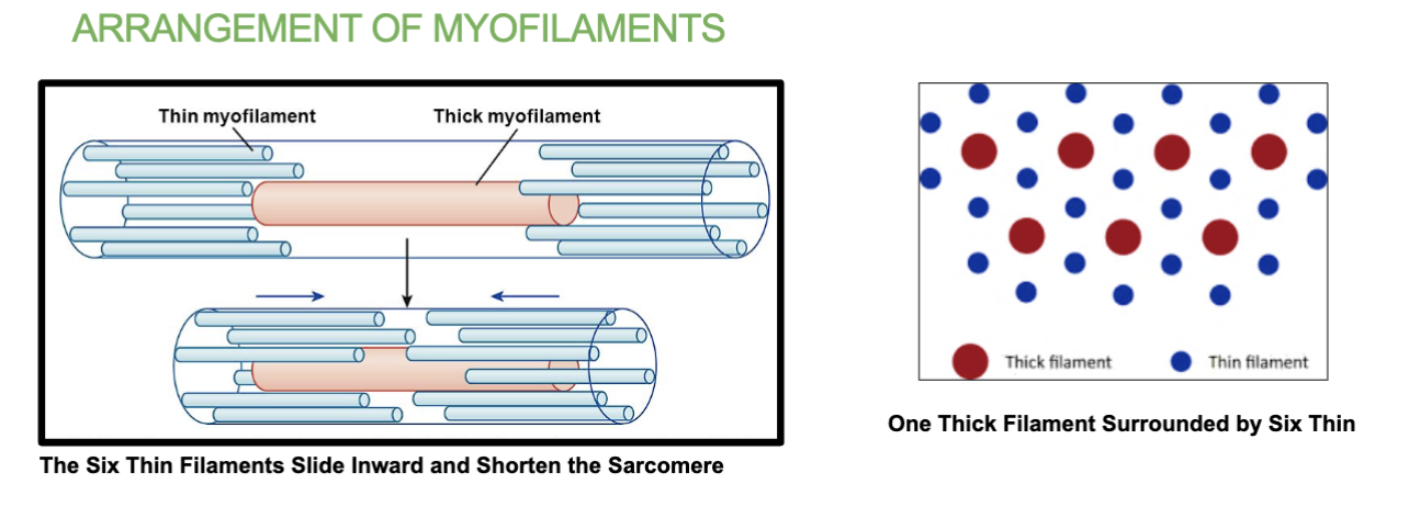
Thick filaments are in the _____ band region of a sarcomere and are composed of a number of protein molecules called _____.
A , Myosin

Structure of myosin molecule?
(in thick filament)
Each myosin molecule has 1 tail and 2 heads.
(Myosin is the protein molecule that makes up thick filaments)

Each myosin molecule has ____ tail and ___ heads.
1 , 2
What happens when muscle contraction begins in thick filaments and what does it form?
Actin-binding sites on myosin heads will attach to
actin(on thin filaments) forming a cross-bridge.

Where are the Actin-binding sites on a myosin molecule on thick filaments?
On the 2 heads of 1 myosin molecule

When muscle contraction begins, ___ binding sites on ____ ____ of ____ filaments will attach to ____ on ____ filaments forming a ________.
Acting binding sites, myosin heads, actin , thin filaments, crossbridge ,
When muscle contraction begins, Actin binding sites on Myosin heads of Thick filaments will attach to Actin on Thin filaments forming a Crossbridge.
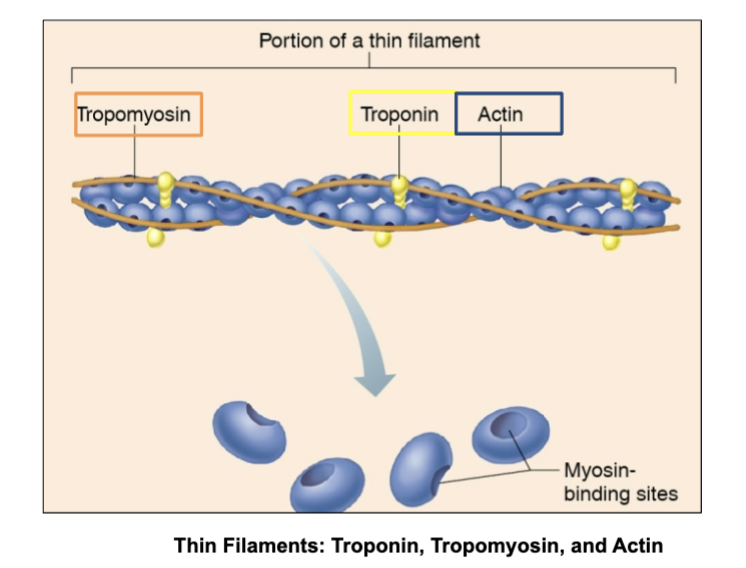
How many types of proteins are in thin filament and what are they?
3 types → Actin, Tropomyosin & troponin
Actin- Contains myosin-binding sites. Its where the myosin heads from the thick filament binds to.
Tropomyosin - long strand of protein that Covers binding sites on actin when muscle is relaxed.
Troponin- Anchors tropomyosin in place, binds to calcium and moves/pulls the tropomyosin exposing the binding sites for contraction.

Name of 3 proteins in thin filaments?
Actin, Tropomyosin, & Troponin

Job of Actin in thin filaments?
the main protein in thin filaments and serves as the binding site for myosin(on thick filaments) during muscle contraction.

Job of Tropomyosin in thin filaments?
long strand of protein that Covers binding sites on actin when muscle is relaxed.

Job of Troponin in thin filaments?
Anchors tropomyosin in place, but when ready to contract it binds to calcium and moves/pulls the tropomyosin exposing the binding sites .
Actin binds to ____.
Myosin heads in thick filaments.
Troponin binds to ____.
Calcium, when ready to contract.
Calcium binds to _____.
troponin on thin filaments.

Motor area in the brain sends electrical
signals called ______ through neurons (nerve cells).
Action potential
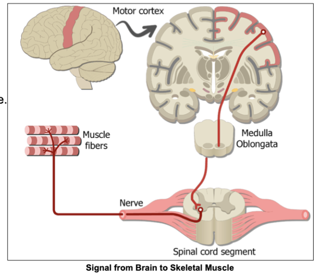
What are neurons?
nerve cells
Neurons transmit the action potential to other ________ and finally to the ________.
nuerons , skeletal muscle
The _______ junction is the connection between a motor neuron and a skeletal muscle fiber, where nerve signals trigger muscle contraction.
neuromuscular

What is the junction/area where a motor neuron and a muscle fiber meet called?
the neuromuscular junction.
It is the area where the nerve signal (action potential) is transmitted from the neuron to the muscle, triggering contraction.
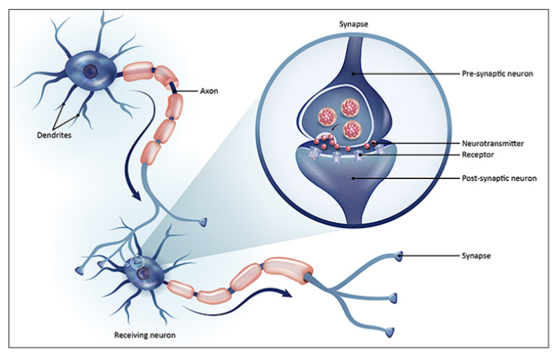
What is the junction between 2 neurons or neuron with muscle called?
the synapse.
It allows nerve signals to pass from one neuron to the next using neurotransmitters.

What are neurotransmitters and what’s their function?
Chemical messengers that transmit signals across a synapse from one neuron to another or from a neuron to a muscle cell→ ex : ACH
Function:
Released from the presynaptic neuron into the synaptic cleft.
Bind to receptors on the postsynaptic neuron or muscle.
Can be excitatory (stimulate action potential) or inhibitory (prevent action potential)
Role of Motor Area in the brain?
Sends action potential through neurons to the muscles.

What does the Motor Unit consist of?
one somatic motor neuron (nerve cell) and all the muscle fibers it
activates.
3 Structures at neuromuscular junction.
Synaptic Knob (tip of Axon terminal/end of motor neuron)
Motor End-Plate (part of the sarcolemma on the muscle fiber/cell that receives signals from the neuron.)
Synaptic Cleft ( space between the synaptic knob and the motor end plate)

Location of the Synaptic Knob (axon terminal)?
(neuromuscular junction)
at the end of motor neuron resting on the junctional folds in sarcolemma of the skeletal muscle
What is the Synaptic Knob(axon terminal) and what does it contain?
This is the end of the motor neuron where communication with the muscle fiber happens.
It contains synaptic vesicles, which are small sacs filled with acetylcholine (ACh)—the neurotransmitter responsible for stimulating muscle contraction.
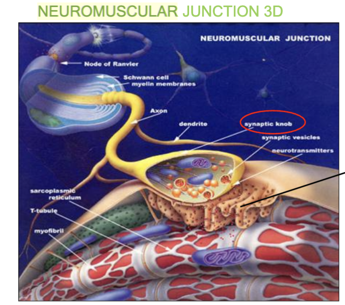
What is the Motor end plate and what does it contain?
This is the part of the sarcolemma on the muscle fiber/cell membrane that receives signals from the neuron.
→ It contains the junctional folds, which increases surface area and contain ACh receptors/ligand-gated NA++ channels to bind ACH (acetylcholine)
What is the Synaptic Cleft and what’s it’s purpose?
This is the space between the synaptic knob and the motor end plate.
ACh molecules travel across this gap to reach the muscle cell.
(also contains the ACHe that removes left over ACH to relax muscle)
5 steps of Excitation at the Neuromuscular Junction.
(Sending the signal from the neuron to the muscle, how the nerve tells the muscle to contract, or wake up)
“NAASA”
Nerve Signal Arrives
Action potential (electrical signal) reaches the axon terminal of the motor neuron.
This opens voltage-gated Ca²⁺ channels, allowing calcium ions (Ca²⁺) to enter the neuron.
ACh is Released
Calcium ions trigger synaptic vesicles to release acetylcholine (ACh) into the synaptic cleft by exocytosis (vesicle bursting).
ACh Binds to Receptors
ACh diffuses across the synaptic cleft and binds to ACh receptors(on the ligand gated sodium channels) in the junctional folds of the muscle sarcolemma.
Sodium Channels Open
ACh binding opens ligand-gated Na⁺ channels.
Sodium (Na⁺) rushes into the muscle fiber, creating a new action potential in the sarcolemma.
Action Potential Spreads
The action potential travels down the sarcolemma and into the T-tubules, spreading the signal deep into the muscle fiber.

1st step of - Excitation at the Neuromuscular Junction.
NAASA
Nerve Signal Arrives - An action potential (electrical signal) reaches the axon terminal of the motor neuron. This opens voltage-gated Ca²⁺ channels, allowing calcium ions (Ca²⁺) to enter the neuron.

2nd step of - Excitation at the Neuromuscular Junction.
NAASA
ACh is Released - Calcium ions trigger synaptic vesicles to release acetylcholine (ACh) into the synaptic cleft by exocytosis (vesicle bursting).

3rd step of- Excitation at the Neuromuscular Junction.
NAASA
ACh Binds to Receptors - ACh diffuses across the synaptic cleft and binds to ACh receptors that are on the ligand gated Sodium (Na++) Channels. These channels are in the junctional folds of the muscle sarcolemma (motor end plate).
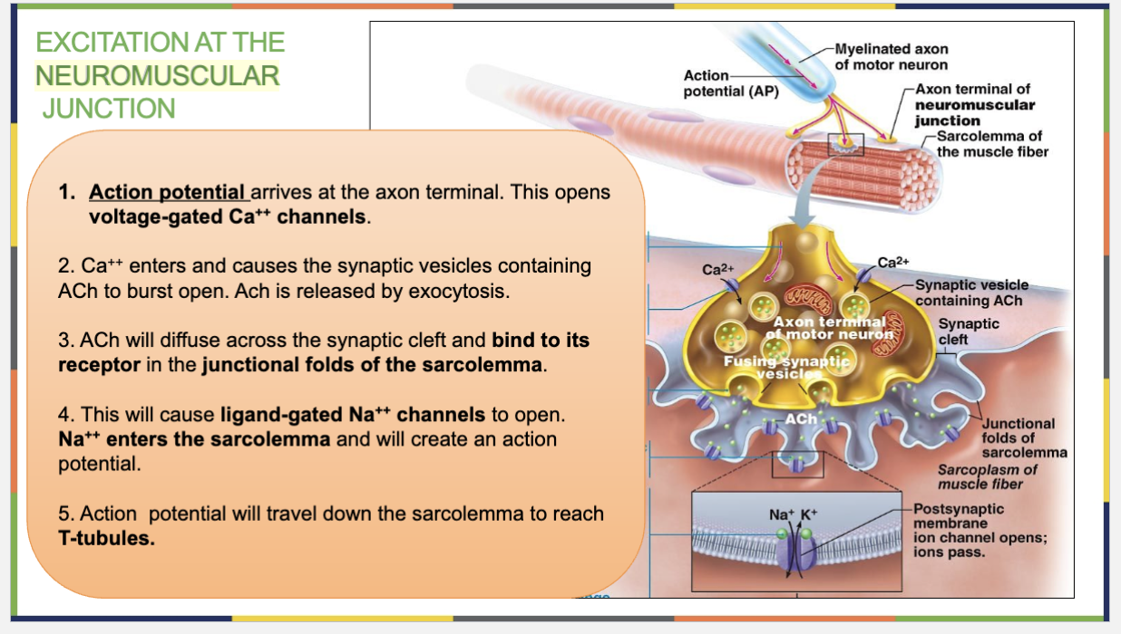
4th step of - Excitation at the Neuromuscular Junction.
NAASA
The ACh binding opens ligand-gated Sodium (Na⁺) channels.
Then, Sodium (Na⁺) rushes into the muscle fiber, creating a new action potential in the sarcolemma.

5th (last) step of - Excitation at the Neuromuscular Junction.
(Sending the signal from the neuron to the muscle, how the nerve tells the muscle to contract)
NAASA
Action Potential Spreads
The action potential travels down the sarcolemma and into the T-tubules, spreading the signal deep into the muscle fiber.

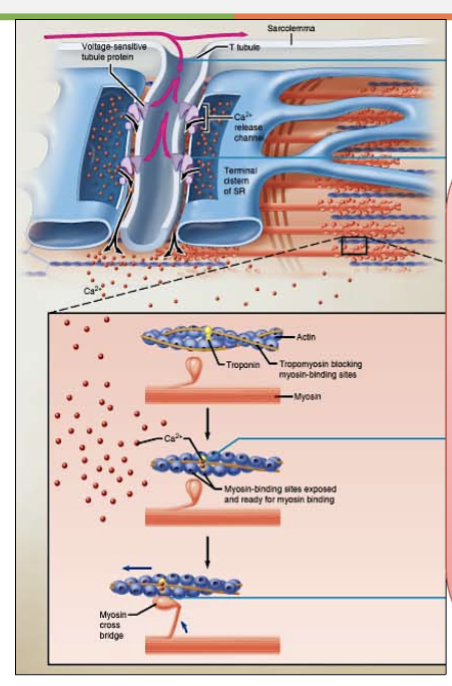
what does “Excitation-Contraction Coupling” refer to?
the process that links the electrical signal (excitation) to muscle contraction.
It describes how an action potential in the muscle fiber triggers the release of calcium ions (Ca²⁺), leading to muscle contraction

4 steps of Excitation-Contraction Coupling.
ACTA - process that describes how an action potential in the muscle fiber triggers the release of calcium ions (Ca²⁺), leading to muscle contraction
Action potential travels down T-tubule, releasing Ca++ from terminal cisterns
Ca++ binds to troponin
Troponin pulls tropomyosin, exposing myosin-binding sites
A cross-bridge forms between actin and the myosin heads. The heads will do a “power-stroke” causing the thin filaments to slide inwards. (thick filaments don’t move)
1st step of Excitation-Contraction Coupling
(how a nerve signal turns into a muscle contraction, turning the signal into movement)
ACTA
Action potential travels down T-tubule, releasing Ca++(calcium) from terminal cisterns
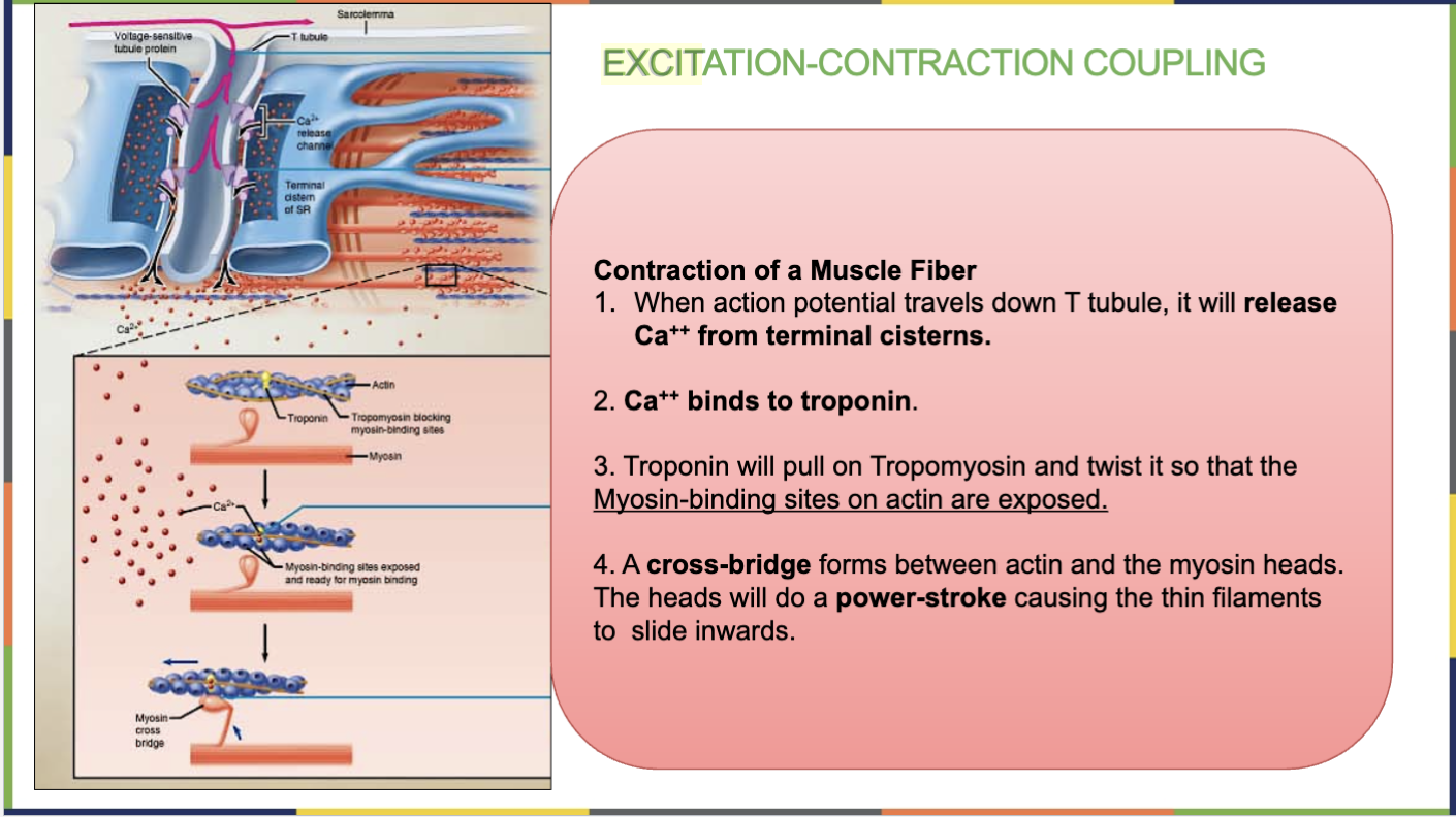
2nd step of Excitation-Contraction Coupling
ACTA
Ca++ binds to troponin

3rd step of Excitation-Contraction Coupling
(how a nerve signal turns into a muscle contraction, turning the signal into movement)
ACTA
Troponin pulls tropomyosin, exposing myosin-binding sites

4th (last) Step of- Excitation-Contraction Coupling
ACTA
A cross-bridge forms between actin and the myosin heads. The heads will do a “power-stroke” causing the thin filaments to slide inwards. (thick filaments don’t move)

4 steps in the role of Calcium for Sliding thin FIlament
Calcium binds to troponin : Ca²⁺ is released from the sarcoplasmic reticulum (SR) and binds to troponin on the thin filament.
Exposure of Myosin-Binding Sites : Troponin pulls tropomyosin away so that the myosin binding sites on actin are exposed.
Cross-Bridge Formation & Power Stroke: Myosin heads attach to actin, forming cross-bridges. Myosin pulls actin inward using a power stroke, shortening the sarcomere.
Sarcomere shortens creating muscle contraction: Thin filaments slide inward toward the M line.
Z discs move closer together, I band shrinks, while A band stays the same width.
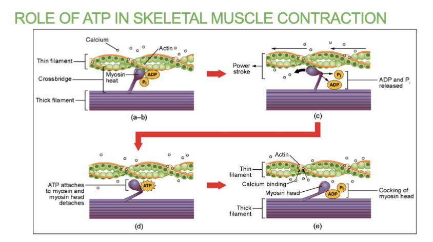
2 Roles of ATP in Skeletal Muscle Contraction
1 ATP will help the myosin head to attach and form the cross bridge. it will enable the myosin head to bend and pull on actin.
A new ATP causes the cross bridge to detach, allowing process to repeat.
What’s Acetylcholine (ACh) & where is it found?
a neurotransmitter that transmits signals from a motor neuron to a muscle fiber, triggering muscle contraction.
→ Location : Stored in synaptic vesicles inside the synaptic knob of the motor neuron.
Released into the synaptic cleft at the neuromuscular junction (NMJ).
Binds to ACh receptors on the motor end plate of the muscle sarcolemma.
→ Function : Starts muscle contraction by opening ligand-gated Na⁺ channels, leading to an action potential in the muscle fiber.
What Opens the ligand-gated Na⁺ channels, leading to an action potential in the muscle fiber?
the neurotransmitter Acetylcholine (ACh).
Whats Acetylcholinesterase (AChe) , its location and function ?
an enzyme that breaks down acetylcholine (ACh) in the synaptic cleft after it has transmitted a nerve signal to the muscle.
→ Location : Inside the Synaptic Cleft → The space between the axon terminal and the muscle fiber's motor end plate.
→ Function : stops muscle contraction by breaking down ACh into acetate and choline. Prevents continuous stimulation of the muscle, allowing it to relax.