PTRS 828 - Medical Imaging Final Review
 Call Kai
 Learn
Learn Practice Test
 Spaced Repetition
 Match
Match Flashcards
 Knowt Play
1/107

There's no tags or description
Looks like no tags are added yet.
Name | Mastery | Learn | Test | Matching | Spaced | Call with Kai | Chat |
|---|
No analytics yet
Send a link to your students to track their progress
108 Terms
Which of the following statements accurately describes MRI (Magnetic Resonance Imaging) technology?
- MRI scanners utilize ultrasound waves to create images of internal body structures.
- Images in MRI are primarily generated from the reflection of gamma rays within the body.
- MRI machines create images by using strong magnetic fields and radio waves to capture signals from protons in fat and water molecules.
- MRI scans typically last between 5-15 minutes, regardless of the body part being imaged.
MRI machines create images by using strong magnetic fields and radio waves to capture signals from protons in fat and water molecules
Which of the following best explains radiologic density in relation to the absorption of x-rays?
- Radiologic density refers to the brightness of the image produced by the x-ray machine.
- It is the ability of an object to reflect x-rays back to the source.
- Radiologic density is determined by the thickness of the body part being imaged.
- Increased density of an object leads to greater absorption of x-rays during penetration.
Increased density of an object leads to greater absorption of x-rays during penetration.
Which of the following best describes the process involved in medical radiography?
- Emitting x-rays from the body to create an image.
- Using sound waves to visualize internal organs.
- Passing an x-ray beam through the body and recording the transmitted pattern.
- Directly visualizing internal structures without the need for x-rays.
Passing an x-ray beam through the body and recording the transmitted pattern.
Which of the following scenarios warrants diagnostic imaging?
- When the patient history, systems review, and objective measures indicate a need for further diagnostics.
- Imaging solely based on patient preference.
- When the initial intervention yields appropriate response regardless of the clinical scenario.
- To add to an established diagnosis already available.
When the patient history, systems review, and objective measures indicate a need for further diagnostics.
Which of the following scenarios is medical imaging decision making involves aiding in making a final diagnosis or drafting a treatment plan?
- Contributing to determining the prognosis in oncology.
- Monitoring the clinical course of disease and/or screening for at-risk health.
- Proactive imaging.
- Identifying the presence of a stroke.
Identifying the presence of a stroke.
A patient presents with chronic lower back pain and undergoes a radiographic examination to assess for potential vertebral abnormalities. Which radiographic feature would most likely represent the bones of the spine?
- Black shade
- White
- Gray shade
White
Which of the following clinical scenarios best illustrates the appropriate use and precautions associated with ionizing radiation in diagnostic imaging?
- A 25-year-old athlete with a recent ankle injury opts for multiple x-ray scans over a short period without considering the potential cumulative effects of ionizing radiation exposure.
- A 40-year-old patient with a history of heart disease undergoes routine chest x-rays every month for monitoring purposes, despite the absence of clinical symptoms or changes in health status.
- A 55-year-old patient presenting with chronic lower back pain undergoes repeated x-ray examinations without adequate shielding, resulting in prolonged exposure to radiation.
- A 30-year-old pregnant woman with suspected appendicitis undergoes a computed tomography (CT) scan with proper shielding and abdominal lead apron, minimizing fetal exposure to ionizing radiation.
A 30-year-old pregnant woman with suspected appendicitis undergoes a computed tomography (CT) scan with proper shielding and abdominal lead apron, minimizing fetal exposure to ionizing radiation.
True or False: Fluoroscopy, also known as Digital Motion X-ray, enables the real-time visualization of physiological functions or medical procedures and is commonly utilized for examinations such as barium x-rays, cardiac catheterization, and image-guided surgical techniques.
True
In pediatric patients, why is exposure to ionizing radiation a particular concern compared to adults?
- Pediatric patients are less likely to require medical imaging procedures involving ionizing radiation.
- Pediatric patients have a higher tolerance for ionizing radiation due to their developing physiology.
- Pediatric patients have a shorter lifetime for the effects of radiation exposure to manifest as cancer.
- The cancer risk per unit dose of ionizing radiation is higher for pediatric patients compared to adults.
The cancer risk per unit dose of ionizing radiation is higher for pediatric patients compared to adults.
When is a CT scan typically warranted in medical practice?
- As a routine screening tool for general health assessment in asymptomatic individuals.
- To assess the extent of musculoskeletal injuries such as sprains or strains.
- To evaluate the progression of chronic conditions like hypertension or diabetes.
- For diagnosing acute conditions such as traumatic injuries, suspected strokes, or abdominal pain of unknown origin.
For diagnosing acute conditions such as traumatic injuries, suspected strokes, or abdominal pain of unknown origin.
A 45-year-old male patient presents with chronic lower back pain that has persisted for over six months despite conservative treatment with physical therapy and non-steroidal anti-inflammatory drugs (NSAIDs). He reports occasional tingling and weakness in his left leg. Physical examination reveals decreased sensation in the left lower extremity and a positive straight leg raise test suggesting nerve root involvement. What is the most appropriate imaging to suggest to his healthcare provider?
- Request a CT scan to distinguish soft tissue structures
- Refer patient for an x-ray of the lumbar spine
- Suggest an MRI to isolate the level of involvement in the spine of the nerve root
- Obtain a PET scan to rule out autoimmune disease
Suggest an MRI to isolate the level of involvement in the spine of the nerve root
True or False: PET scans and SPECT scans both use radioactive tracers to visualize metabolic processes in the body, but PET scans provide higher resolution images and more precise quantification of biological functions compared to SPECT scans.
True
Which of the following statements about the use of PET scans in autoimmune diseases is correct?
- FDG PET can differentiate between normal thyroid parenchyma and malignant tumors in patients with autoimmune thyroid diseases.
- FDG PET/CT is a standard diagnostic tool in sarcoidosis workup.
- FDG uptake in rheumatoid arthritis reflects disease severity but does not correlate with clinical parameters or response to therapy.
- FDG PET/CT is efficient in assessing cardiac involvement and evaluating reversible granulomas in sarcoidosis.
FDG PET can differentiate between normal thyroid parenchyma and malignant tumors in patients with autoimmune thyroid diseases.
What is the primary function of the strong magnetic field in an MRI?
- To produce images directly by emitting radiation.
- To detect the energy released by protons as they realign with the magnetic field.
- To stimulate protons and cause them to spin out of equilibrium.
- To force protons in the body to align with the magnetic field.
To force protons in the body to align with the magnetic field.
True or False: Magnetic Resonance Angiography (MRA) is a non-invasive imaging technique primarily used to visualize blood vessels and diagnose conditions such as aneurysms, stenosis, and vascular malformations.
True
True or False: In a clinical ultrasound, the image is generated by the reflection of sound waves off body structures, with the strength (amplitude) of the sound signal and travel time of the wave through the body providing the necessary information for image formation.
True
All of the following are indications for use of ultrasound imaging EXCEPT:
- Visualize the appendix
- Assess blood flow and valve integrity of the heart
- Determine extent of achilles tendon rupture
- Assess a bleed in the brain
Assess a bleed in the brain
A 45-year-old male presents to the emergency department with severe abdominal pain following a car accident. The attending physician suspects internal bleeding. Which of the following diagnostic tools is most appropriate for quickly identifying the presence of internal bleeding in this patient?
- Ultrasound
- MRI
- X-ray
- CT
Ultrasound
A 65-year-old postmenopausal woman presents to her primary care physician for a routine check-up. She has a family history of osteoporosis and has recently experienced a minor fracture from a low-impact fall. The physician recommends a diagnostic test to assess her bone density. Which of the following is the most appropriate test to evaluate for osteoporosis in this patient?
- MRI
- Plain film x-ray
- CT scan
- DEXA scan
DEXA scan
Which of the following is a key benefit of using interventional radiology for medical procedures?
- Longer recovery times
- Higher risk of infection
- Minimally invasive techniques
- Limited imaging guidance
Minimally invasive techniques
True or false: A patient who presents to clinic with persistent cough and unexplained weight loss over the past three months with + CT scan with right lower lobe mass and biopsy would use histology to examine the sample and assist with diagnosis.
True
What is histology primarily concerned with?
- The study of the morphology of cells within their natural tissue environment.
- The examination of the electrical activity of neurons.
- The analysis of the biochemical reactions within cells.
- The study of the genetic makeup of cells.
The study of the morphology of cells within their natural tissue environment
True or false: Staphylococcus aureus, a gram-positive bacterium, is a common cause of skin and soft tissue infections.
True
Which type of tissue is commonly examined in histology to diagnose and understand diseases?
- Connective tissue
- All answers are correct
- Epithelial tissue
- Nervous tissue
All answers are correct
Which microbiologic technique is commonly used to identify the causative organisms in a wound infection?
- Polymerase Chain Reaction (PCR)
- Serology testing
- Wound culture
- Blood culture
Wound culture
How do health professionals use imaging?
- Clinical imaging examinations and diagnosis of findings play an important role in patient centered interdisciplinary care
- Clinical imaging tests (x-ray, CT, MRI, nuclear medicine scans) are performed to assist in initial diagnosis and sometimes follow up care
What is an example of how diagnostic tests can help determine outcomes of an imaging investigation to contribute to the intermediate goal of determine the prognosis?
Oncology usage - imaging to see progress with treatment
What is an example of how diagnostic tests help to monitor the clinical course of disease and/or the screening for at-risk health?
Proactive screening
What are indications for diagnostic imaging?
- If patient history, systems review, and objective measures make it evident that further diagnostics are needed
- If imaging results will change the course of treatment or alter the outcome of the problem
- In the "absence of appropriate response to intervention"
What are reasons that diagnostic tests may NOT be indicated?
- Injury response to treatment is consistent with expected prognosis
- If diagnosis suggests surgical intervention but the patient is unwilling or is a poor candidate for surgical intervention
What are the Ottawa rules?
A set of rules individualized for different parts of the body with respect to pain/tenderness/inabilities to weight bear, etc. that determines whether or not imaging is required in that area.
Can physical therapists order imaging in Kansas?
No :(
What is medical imaging?
Several different technologies that are used to view the human body in order to diagnose, monitor, or treat medical conditions
What is a radiograph?
A picture formed from x-ray beams passed through the body, where some rays are absorbed or scattered by the internal structures, so that the remaining pattern is transmitted to a detector to form an image.
What is a radiograph used for?
Static images
- EX: dental examination, verification of correct placement of surgical markers prior to invasive procedures, mammography, orthopedic evaluations, spot film or static recording during fluroscopy
ALSO:
- assess the presence or absence of disease, foreign objects, and structural damage or anomaly
How does an x-ray work?
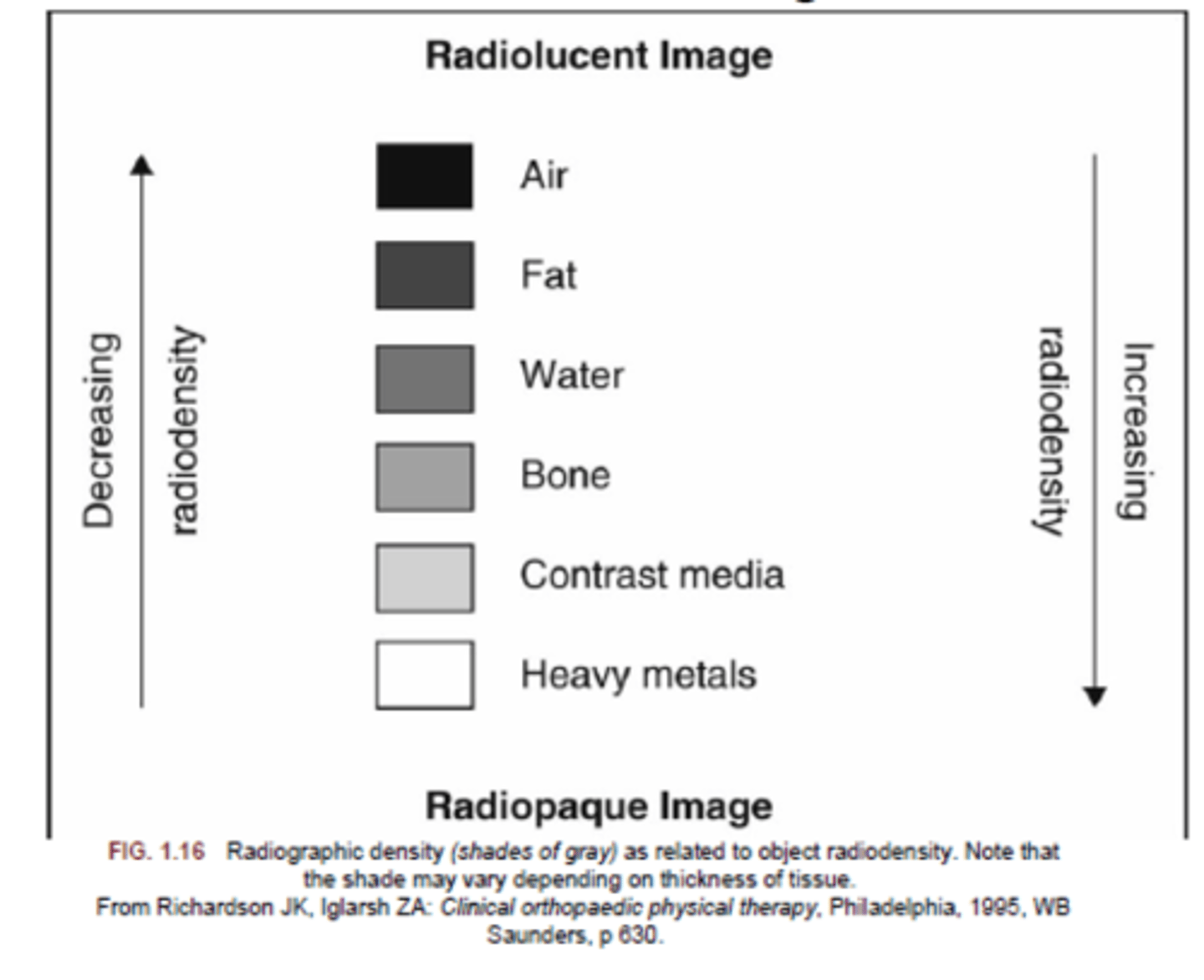
- The more radiation a structure absorbs, the lighter the structure would appear on the film.
- Attenuation: the absorption of the rays by objects (increased density = absorb more rays = lighter color)
What is computed tomography (CT)?
A noninvasive medical examination or procedure that uses specialized x-ray equipment to produce cross-sectional images of the body
What does a CT scan do?
Provide images of internal organs, bones, soft tissues, and blood vessels in greater clarity and detail than conventional x-ray images.
What is a CT scan good for?
- Diagnosing disease, trauma, or abnormalities
- Planning and guiding interventional or therapeutic procedures
- Monitoring the effectiveness of therapy (ex. cancer treatment)
- Show bony misalignments (ex. scoliosis)
- Degenerative conditions
- Post-operative conditions
- Neoplastic conditions
- Infectious conditions
- Image guidance during injections, biopsies, and aspirations
- Imaging with contraindications (MRI can't be used)
What is magnetic resonance imaging (MRI)?
A non-invasive medical imaging procedure for making images of the internal structures of the body, using strong magnetic fields and radiowaves to make 3D images.
Why are MRIs so good for visualizing soft tissue?
The signal in an MRI comes mainly from the protons in fat and the water molecules in the body.
What can be used for contrast in an MRI?
Gadolinium-based contrast agents (GBCAs) or intravenous (IV) drugs
What non-intentional factors can affect images?
- physical as well as geometric factors
- results in thickness, motion, scatter, magnification, and distortion
What intentional factors can affect images?
Those that are planned to highlight area of evaluation with contrast enhancement
What's another form of a radiograph and how is it used?
Fluoroscopy
- allows viewing of physiological function or medical procedure in real time
- used for examinations and procedures such as barium x-rays, cardiac catheterization, placement of IV catheters, image guided injections, and image guided surgical techniques
What are risks of radiographic imaging?
- developing a radiation-induced cancer or cataracts sometime later in life
- causing a disturbance in the growth or development of an embryo or fetus when performed on a pregnant patient
True or false: The risk of cancer is never exceeded by the benefit of radiography, and should be considered before imaging, as it can't be prevented.
False: when needed, the benefit of radiography far exceeds the small cancer risk associated with the procedure, and risks can usually be reduced or eliminated with proper shielding.
What is radiodensity?
The density of the object and how it correlates to the amount of radiation absorbed from an x-ray beam.
What does radiopaque mean?
X-rays are absorbed by tissue/substance and object appears white on radiograph, such as bones, heavy metals, and surgical implants
What does radiolucent mean?
X-rays pass through tissue/substance and object appears black on radiograph, such as air filled organs and soft tissues
What is heel effect?
A source of visual error related to x-ray production, because x-rays released by the machine are not uniform.
What is artefact?
An error in the perception of the visual image of the radiograph, usually seen as an abnormal finding or foreign body. Can happen with metal or implants in the body.
What is exposure?
A measure of the amount of ionizing radiation determined by 3 factors: time, x-ray energy, and the quantity of the x-ray photons. Can be manipulated to highlight structures of interest.
-over penetration enhances bone visibility, under penetration enhances soft tissue visibility
What is movement (processing error)?
A blurring in the image as a result of movement by the patient the moment the x-ray exposure is made
What is a film processing error?
An error that occurs during the processing of the film can result in disturbances in the contrast, detail, or density of the image displayed.
What are the 6 body positions for an x-ray?
1. Erect - standing or sitting
2. Supine
3. Trendelenburg position - supine on an inclined radiographic table with head lower than feet
4. Prone
5. Lateral - side touches the camera
6. Lateral decubitus - lying on one side, with camera view anterior/posterior
What are the 5 image projections for an x-ray?
1. Antero-posterior (AP)
2. Postero-anterior (PA)
3. Lateral - left to right or right to left
4. Oblique - at an angle
5. Axial - superior to inferior or vice versa
How does an MRI work?
- Powerful magnets produce a strong magnetic field that forces protons in the body to align with the field.
- A radiofrequency current is pulsed through the patient, stimulating the protons to spin out of equilibrium and strain against the pull of the magnetic field.
- Once the radiofrequency is turned off, the MRI sensors can detect the energy released as the protons realign with the magnetic field
- The time it takes for the protons to realign + the amount of energy released changes depending on the environment and chemical nature of the molecules, which tells the physicians the difference between various types of tissues based on their magnetic properties
What is the difference between a T1 and T2 MRI?
T1-weighted = enhances the signal of the fatty tissue and suppresses the signal of the water
T2-weighted = enhances the signal of the water
What is intensity in regards to an MRI?
The shade of grey of tissues or fluid:
- high signal intensity = white
- intermediate signal intensity = grey
- low signal intensity = black
The faster protons realign in an MRI, the _____ the image
brighter
darker
Brighter
How are MRIs different than CT scans?
They do not use the damaging ionizing radiation of x-rays
Why are MRIs the modality of choice for frequent imaging when used for diagnosis or therapy?
The magnetic aspect is much less damaging than ionizing radiation used in x-rays and CT scans
What is a downside of MRIs?
They're really freaking expensive!!!!
What are MRIs really good at visualizing?
The brain, spinal cord and nerves, muscles, ligaments, and tendons
How are MRIs used in regard to the brain?
They can differentiate between white and grey matter and can be used to diagnose aneurysms and tumors
What is a functional MRI (fMRI)?
A way to observe brain structures and determine which areas of the brain activate (use more oxygen) during various cognitive tasks - MOSTLY FOR RESEARCH
What is a magnetic resonance angiography (MRA)?
- A form of an MRI that is used to image arteries with good diagnostic accuracy and is less invasive.
- Can be used to image the thoracic and abdominal aorta and arteries of the brain, neck, abdominal organs, kidneys, and lower extremities
What gives the best images of venous abnormalities, such as thrombosis and anomalies?
Venous imaging - Magnetic Resonance Venography (MRV)
What are risks of MRIs?
- The magnetic field is very strong and can be harmful to patients with metal or implants in their body
- Very loud (up to 120 decibels)
- Can stimulate nerves from rapidly switching magnetic fields
- Contrast agents (rarely) can cause severe renal failure and require dialysis
- Pregnant patients should not get MRIs at all as a precaution
- Claustrophobia - open MRI machine available sometimes
What is a Positron Emission Tomography (PET) scan?
An imaging test that can help reveal the metabolic or biochemical function of tissues and organs using a radioactive drug called a tracer.
What are risks of PET scans?
- Patient should avoid eating or drinking anything with calories or sugar for several hours before scan
- While radioactive tracers are only in body for a short time, patient should avoid close contact with pregnant women, babies, and young children for six hours post-scan.
What are risks of SPECT scans?
- The radioactive tracer injection can cause bleeding, pain, or swelling at the injection site, and in rare cases, an allergic reaction.
- Small amounts of radiation are used
What are PET scans used for?
- Primarily in oncology
- See changes in metabolism, blood flow, and regional chemical composition
How do PET scans work?
- Radioactive tracer is injected, swallowed, or inhaled, and tracer gets trapped in various tissues of body based upon the affinity
- Areas of higher activity show higher uptake and brighter spots on images
Unstable nuclei of tracers emit positrons that produce gamma rays when combined with neighboring electrons, which are detected by a ring of detectors
- that info is used to make a 3D image of the tracer in the body
What is an FDG-PET?
A PET scan using fluorodeoxyglucose as the tracer, usually used for diagnosis staging and monitoring cancers
What does a positive FDG-PET scan mean?
A tissue biopsy is indicated in that location when cancer is suspected
What things can PET scans detect?
- Alzheimer disease
- Neuropsychology
- Psychiatry
- Stereotactic surgery and radiosurgery
- Cardiology
- myocardial perfusion and metabolism
- Infectious diseases
- Autoimmune diseases
- Musculoskeletal system diseases
What are Single Photon Emission Computed Tomography (SPECT) scans primarily used for?
To diagnose and track the progression of heart disease, as well as detect disorders in bone, gall bladder disease, and intestinal bleeding, and Parkinson's disease (recently)
What are the main differences between PET and SPECT scans?
- Type of radiotracers used
- SPECT scans measure with photons, while PET scans measure with positrons
What is a diagnostic ultrasound?
An imaging system that uses high-frequency sound waves to view inside the body in real time
What can ultrasounds show?
- abdominal tissues and organs
- bone fragility (bone sonometry)
- breast tissue
- doppler fetal heart rate monitors (listen to fetal heart beat)
- doppler ultrasound (visualize blood flow)
- echocardiogram
- fetal ultrasound
- ultrasound-guided biopsies (to collect a sample of tissue)
- ophthalmic ultrasound (visualize ocular structures)
- ultrasound-guided needle placement
How does an ultrasound work?
The image is produced based on the reflection of the waves off of the body structures, where the strength (amplitude) of the sound signal and the time it takes for the wave to travel through the body provide the information necessary to produce an image.
What is a Point of Care Ultrasound (POCUS)?
A portable ultrasound to perform limited imaging and direct care in a bedside setting
Yes or no: Do ultrasounds use ionizing radiation?
No
How does a CT scan work?
An x-ray source and detector rotate around a patient, producing snapshots of the patient. A computer puts together the snapshots to form cross-sectional images of the internal organs and tissues.
Why are CT scans utilized often?
- Show more detail
- Easier to see smaller fractures
- Very quick procedure, used a lot in trauma
When is an MRI better than a CT scan?
In less urgent situations, MRI takes longer but has clearer images and is better for creating pictures of organs and soft tissues, as compared to CT images.
What is histology?
The study of the morphology of cells within their natural tissue environment
What does cell shape predict?
Function - that's why alternations can identify and explain dysfunction and disease
What is medical histology used for?
Medical diagnosis, scientific study, autopsy, and forensic investigation
What steps do tissues go through before being able to be analyzed by pathologists?
Fixation, processing/dehydration, embedding, sectioning, and staining
Why is histology important?
An understanding is crucial to comprehend human biochemical and physiological processes, as well as to gain insights into how structural abnormalities lead to disorders resulting in disease
What are the protein stains, and what color do they stain proteins?
Hematoxylin - blue
Eosin - pink
What do microbiologists do?
Compare samples to microbiologic studies to determine pathogens and how to treat the infectious disease
What is most commonly encountered in microbiologic studies in health professions?
Gram stains and cultures
How do gram stains work?
Gram positive takes the stain, includes staphylococcus and streptococcus species
Gram negative doesn't take the stain, includes organisms that frequently require different antimicrobial drugs
What does acid-fast testing test for?
Tuberculosis infection - tested with sputum or through fluid from bronchoscopy
What is used to identify a fungal infection?
Potassium hydroxide
What are cultures used for?
To detect the presence of bacteria in blood, sputum, pleural fluid, throat, urine, skin, and wounds. They are performed when clinical presentation is suggestive of infection, and should be performed before antibiotic therapy is initiated.