Chapter 1 : Anatomy of the Nervous System
1/139
Earn XP
Name | Mastery | Learn | Test | Matching | Spaced | Call with Kai | Chat |
|---|
No analytics yet
Send a link to your students to track their progress
140 Terms
What is the Nervous System ?
It is known as the body’s command centre and communication network. It mainly helps regulate eveyhtign that you feel, think and do form conscious movements and thoughts to autonomic life functions.
The Brain
the primary location where movement, thoughts, dreams and consciousness originate
160mm long
90mm tall
1400 cm ³
adult Brian weighs up to 1.5 kg an dis less than 2% of the body’s weight
White Matter
also known as myelinated axons
myelin : a fatty lipid layer that covers axons to help nerve signals move faster
cause the tissue to look white
helps with the communication between different regions of the Brian
Grey Matter
they are composed of neuronal cells bodies + dendrites + synapses
they appear darker
this is where information processing occurs
Corpus Callosum
the largest white matter trac tin the brine
connects the left and right hemisphere
the information passes rapidly between both sides of the Brian
What is dessucation ?
When white matter pathways crosses form one hemisphere to the other, it mainly helps with communication
Rostral
forward
caudal
back
dorsal
the top of the head
medial
towards the centre of the head
ventral
bellow the top of the head
superior
the structure not he top
inferior
the structure bellow
Types of Brian Slices
Coronal
Horizontal
Parasagittal
Coronal Section
front ( anterior)
back ( posterior)
Horizontal
top ( superior)
bottom ( inferior)
Parasagittal
left
right
tuns parallel to the milling meaning it is not symmetrical
shows 1 hemisphere at a time
What are the stages of brine development
Germ Layer
Neural Tube
Three Vesicle Stage
Five Vesicle Stage
Germ Layer
the embryo begins with three layers '
ectoderm ( where the nervous system evolves form
mesoderm
endoderm
Neural Tube
forms during the 3-4 weeks of gestation
develops form the ectoderm
gives rise to the central nervous system
Three vesicle stage
the neural tube developes and forms three pars of the brine
prosencephalon
The prosencephalon manages higher-order cognition, sensory processing, and homeostasis. [1]
Cognition: Controls conscious thought, memory, and voluntary movement.
Sensory Integration: Relays and interprets visual, auditory, and tactile information.
Homeostasis: Regulates body temperature, hunger, thirst, and hormone release
mesencephalon : The mesencephalon functions as a reflex center and tracking hub for visual and auditory stimuli. [1, 2, 3, 4]
Visual Reflexes: Coordinates tracking movements of the eyes and head.
Auditory Reflexes: Directs your head toward sudden or unexpected sounds.
Motor Pathways: Contains major conduits for descending motor signals. [1, 2, 3, 4, 5]
rhombencephalon : The rhombencephalon regulates autonomic survival functions and motor coordination. [1]
Autonomic Control: Governs heart rate, blood pressure, and breathing.
Coordination: Refines voluntary muscle movements and maintains balance.
Conduction: Serves as the primary traffic bridge between the brain and spinal cord. [1, 2, 3, 4, 5]
The five vesicle stage
Diencephalon : Thalamus + Hypothalamus
Telencephalon : Basal Ganglia + Cerebral Cortex
Mesencephalon : periadquetal grey ( pain response), red nucleus, substansia nigra ( movement ), tectum ( visual reflex), reward + motivation
Myelencephalon : Medulla Oblongta
Mesencephalon : Pons + cerebellum
Thalamus
the Brian main relay station in which all sensory information goes through
Hypothalamus
controls the endocrine system
maintain hometstais
maintain body chemistry
Basal Ganglia
motor + habit learning
emotional processing
action selection
Cerebral Cortex
attention
memory
language
thinking
Pons
hearing
breathing
taste
Cerebellum
balance
coordination
posture
motor learning
Gyri
raised bumps
sulci
fissures or grooves
Longitude fissure
separate the left and right hemisphere
Central Sulcus
seperate the frontal and parietal lobe
Lateral Fissure
separate temporal lobe from the frontal/pareital lobe
occipital lobe
posterior of the Brian
vision
primary visual cortex
color + shape + motion + orientation
Temporal Lobe
located on the side of the brine
primary auditory cortex
hippocampus
Hearing + memory + language comprehension
Parietal Lobe
top of the brain
primary somatosensory cortex
touch + temperature + pain + vibration + proprioception
Frontal Lobe
front of the brine ]
primary motor cortex
movement + planning + personality + descirion making + working memory
Spinal Cord
carry information both upwards towards the brain, and downwards
towards the body’s other organs and muscles.
process sensations and form an
appropriate motor response in the absence of
brain input.
The spinal cord originates roughly at
the level of your neck and runs down to the small
of your back,
length around 44 cm (17.5 inches).
The diameter of the spinal cord is
not uniform all the way down, being ~13 mm (0.5
inches) at its widest and ~6.5 mm (0.25 inches)
Five sections of the spinal cord
Cervical
Thoriac
Lumbar
Sacral
Cocygeal
Cervical Section
Location: The neck region, right at the base of the skull down to the shoulders.
Function: Contains 8 pairs of nerves that control the diaphragm for breathing, neck and head movements, and the shoulders, biceps, wrists, and hands.
Thoriac Section
Location: The upper and middle back, extending through the chest area.
Function: Contains 12 pairs of nerves that control the trunk muscles, abdomen, and chest wall. These nerves are highly involved in core stability and posture
Lumbar Section
Location: The lower back area.
Function: Contains 5 pairs of nerves that control the hips, thighs, and some muscles in the front of the legs.
Sacral Section
Location: Just above the tailbone, nestled between the pelvic bones.
Function: Contains 5 pairs of nerves that handle the back of the legs, buttocks, and pelvic organs, including bowel and bladder control and sexual function.
Coccygeal Section
Location: The very bottom of the spine, known as the tailbone.
Function: Contains 1 single pair of nerves that carry sensory information from the skin overlying the tailbone
Parts of the Spinal Cross Section
Gray Matter
White Matter
Central Canal
Spinal Roots + Nerves
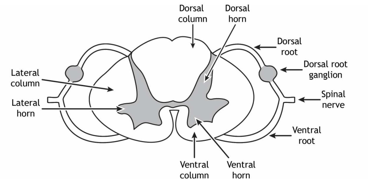
Gray Matter
Posterior (Dorsal) Horn: Processes sensory information.
Anterior (Ventral) Horn: Contains cell bodies of motor neurons that control voluntary and involuntary muscle movement.
Lateral Horn: Present primarily in the thoracic and lumbar regions, this area contains autonomic motor neurons that regulate involuntary body functions (like heart rate and digestion). [1, 2, 3, 4, 5]
White Matter
Posterior Funiculus: Carries sensory signals (such as touch and limb position) up to the brain.
Lateral Funiculus: Contains pathways for pain and temperature sensations, as well as pathways that control movement.
Anterior Funiculus: Contains both ascending and descending tracts for various sensory and motor signals.
Central Canal
A small, fluid-filled channel located in the center of the gray matter ("crossbar" of the butterfly) that runs the length of the spinal cord and contains cerebrospinal fluid (CS
Spinal Roots and Nerves
Dorsal (Posterior) Root: Carries sensory information into the spinal cord. It features a noticeable swelling called the dorsal root ganglion, which holds sensory cell bodies.
Ventral (Anterior) Root: Carries motor commands out of the spinal cord to the muscles and glands.
What is the function of the Peripheral Nervous System (PNS)?
t acts as the intermediary between the Central Nervous System (CNS) and the rest of the body, including the skin, internal organs, and muscles.
What does proximal mean?
close to the CNS
What does distal mean ?
far from the CNS
What does contralateral mean?
A connection that crosses to the opposite side of the body (e.g., the left brain processes information from the right side of the body).
What does ipsilateral mean?
Located on the same side of the body.
Is most of the nervous system organized contralaterally or ipsilaterally?
contralaterally
What are the three main branches of the PNS?
The somatic nervous system, autonomic nervous system, and enteric nervous system.
What does the somatic nervous system do?
It handles interactions with the external environment, including sensing and voluntary movement.
What is the afferent somatic nervous system responsible for?
Carrying sensory information from the body to the CNS.
What is the efferent somatic nervous system responsible for?
Sending motor commands from the CNS to skeletal muscles.
Why is the somatic nervous system called the "voluntary nervous system"?
Because it controls intentional skeletal muscle movements.
What does the autonomic nervous system control?
he body's internal environment and involuntary functions.
What types of structures does the autonomic nervous system control?
Smooth muscles, glands, and internal organs.
Give three examples of autonomic functions.
Sweating, pupil dilation, and blood pressure regulation.
What response does the sympathetic nervous system produce?
A: The fight-or-flight response.
Q: When is the sympathetic nervous system
A: During perceived or real threats.
What happens to heart rate during the sympathetic response?
A: It increases.
Where do sympathetic nerves originate?
In the thoracic and lumbar regions of the spinal cord (thoracolumbar region).
What are the clusters of sympathetic nerve cells called?
A: Sympathetic ganglia.
What response does the parasympathetic nervous system produce?
A: The rest-and-digest response.
When is the parasympathetic nervous system most active?
A: During relaxation and digestion.
Where do most parasympathetic signals originate?
Brain + sacral regions
Which cranial nerve provides much of the parasympathetic innervation?
A: The vagus nerve (CN X).
What is the enteric nervous system?
A: A network of neurons that controls the digestive tract.
Approximately how many neurons are in the enteric nervous system?
A: About 500 million neurons.
Which organs are controlled by the enteric nervous system?
A: The esophagus, stomach, and intestines.
What is CN I, and what is its function?
A: Olfactory nerve — Sensory — Responsible for the sense of smell.
What is CN II, and what is its function?
A: Optic nerve — Sensory — Responsible for vision.
What is CN III, and what is its function?
A: Oculomotor nerve — Motor — Controls most eye movements, pupil constriction, and lens shape.
What is CN IV, and what is its function?
A: Trochlear nerve — Motor — Controls the superior oblique muscle, moving the eye downward and laterally.
What is CN V, and what is its function?
A: Trigeminal nerve — Sensory + Motor — Provides touch and pain sensation from the face and controls chewing muscles.
What is CN VI, and what is its function?
A: Abducens nerve — Motor — Controls the lateral rectus muscle, moving the eye outward.
What is CN VII, and what is its function?
A: Facial nerve — Sensory + Motor — Controls facial expressions and taste from the anterior 2/3 of the tongue.
What is CN VIII, and what is its function?
A: Vestibulocochlear nerve — Sensory — Responsible for hearing and balance.
What is CN IX, and what is its function?
A: Glossopharyngeal nerve — Sensory + Motor — Controls swallowing and provides sensation and taste from the posterior 1/3 of the tongue.
What is CN X, and what is its function?
A: Vagus nerve — Sensory + Motor — Controls internal organs through parasympathetic activity.
What is CN XI, and what is its function?
A: Accessory nerve — Motor — Controls the neck and shoulder muscles.
What is CN XII, and what is its function?
A: Hypoglossal nerve — Motor — Controls tongue movements.
Why does the brain need a large blood supply?
A: Because it requires a lot of oxygen and nutrients to function.
What percentage of body weight is the brain?
A: About 2%.
What percentage of cardiac output does the brain receive?
A: About 15%.
Which two pairs of arteries supply blood to the brain?
A: The vertebral arteries and internal carotid arteries.
What do the vertebral arteries form?
A: The basilar artery.
What is the Circle of Willis?
A: A circular network of arteries that provides backup blood flow if one artery becomes blocked.
What areas do the anterior cerebral arteries supply?
A: The dorsomedial cortex and deep brain structures.
What areas do the middle cerebral arteries supply?
A: The lateral cerebral cortex.
What areas do the posterior cerebral arteries supply?
A: The occipital lobe.
What is a stroke?
A: A loss of blood flow to the brain.
What is the number one risk factor for stroke?
A: High blood pressure.
What are the two main types of stroke?
A: Ischemic stroke and hemorrhagic stroke.
What percentage of strokes are ischemic?
A: About 80%.