Edexcel GCSE 9-1 Biology - SB8: Exchange and Transport in Animals
1/40
Earn XP
Description and Tags
Paper 2 Specification: https://www.wardleacademy.co.uk/wp-content/uploads/2020/09/Biology-KS4-GCSE-Specification.pdf (for some miraculous reason the pearson spec wasn't working)
Name | Mastery | Learn | Test | Matching | Spaced | Call with Kai |
|---|
No analytics yet
Send a link to your students to track their progress
41 Terms
Recall 4 substances that need to be transported in and out of the body.
- CO2
- Oxygen
- Urea
- Water/mineral ions
Describe the functions of Oxygen, CO2 and water in the body.
- Oxygen = Aerobic respiration.
- Carbon dioxide = Released in aerobic respiration.
- Water = chemical reactions.
Describe the adaptations of the lungs for gas exchange.
- Moist lining = Dissolving gases.
- Good blood supply = maintain concentration gradient.
- Thin walls = Minimising distance that gases have to diffuse.
- Large surface area = Increase diffusion rate.
Do larger organisms have a larger SA:V ratio or a smaller one? Why is this a problem.
Smaller SA:V ratio - will not be able to exchange enough substance across surface area to supply the entire volume of the organism - won't get all things it needs.
Define concentration
the amount of substance present in a certain volume (mol dm-3).
Describe how surface area affects the rate of diffusion.
More surface for molecules to move across, therefore they can get from one side to the other faster. (proportional)
Describe how concentration gradient affects the rate of diffusion.
Bigger concentration gradient = faster rate of diffusion - lots more particles on one side - more can move across to the other side. (proportional)
Describe how distance affects the rate of diffusion.
Increased distance = slower rate of diffusion, as the particles have to move further. (inversely proportional).
Recall the components of the circulatory system.
- The heart.
- Blood vessels (arteries, veins, capillaries).
- Blood .
Recall the functions of the different types of blood vessels.
Arteries = Carry blood away from the heart.
Veins = Carry blood back to the heart.
Capillaries = Carry blood through tissues and organs.
Describe the functions of different types of blood cells.
- Lymphocytes = Produce antibodies and antitoxins (destroy harmful microorganisms and invading pathogens).
- Phagocytes = Engulf and digest invading pathogens (phagocytosis - change shape).
- Erythrocytes = Transport oxygen from lungs to body cells (haemoglobin in blood binds to oxygen at lungs to produce oxyhaemoglobin, oxyhaemoglobin breaks down into haemoglobin and oxygen at cells so cells can get oxygen).
Describe the functions of blood platelets and plasma.
Platelets = Small, irregularly shaped fragments of cells (no nucleus) - help blood clot at wound - prevent blood loss and microorganisms getting in.
Plasma = Straw coloured liquid that carries blood cells and dissolved substances like glucose, carbon dioxide and urea.
Describe how blood cells are adapted to their functions.
Arteries = Narrow tube (small lumen size), thick layer of elastic and muscle fibres, walls thick to withstand pressure and stretch and recoil to aid blood flow.
Veins = Thin walls, large lumen size, flexible wall, muscles in skeleton help to push the blood along.
Capillaries = Small, thin walls, narrow, sit in between gaps in cells and speeds up diffusion, narrow lumen.
Recall the parts of the heart and describe the pathway of blood flow.
- Deoxygenated blood enters right atrium from body via the vena cava.
- Flows to the right ventricle then into the lungs via the pulmonary artery.
- Oxygenated blood goes into left atrium via pulmonary vein, then to left ventricle and then to rest of the body via the aorta.
How is the heart adapted to its function?
Has valves to stop blood flowing the wrong way/prevent backflow.
Lef ventricle thicker muscle - has to pump blood to the rest of the body and not just to the lungs.
Recall the equation relating cardiac output, stroke volume and heart rate.
Cardiac output (cm 3 min -1). = heart rate (bpm) x stroke volume (cm 3)
Why do organisms need to respire?
To give them energy for life processes (MRS GREN).
State the word equation for aerobic respiration.
glucose + oxygen -> carbon dioxide + water
State the word equation for anaerobic respiration.
Glucose - lactic acid.
Why is respiration exothermic?
Because it transfers energy to the environment
Compare anaerobic and aerobic respiration.
Aerobic:
- With oxygen.
- More energy released, more efficient.
- Mitochondria.
Anaerobic:
- Without oxygen.
- Less energy released (less efficient).
- Glucose not fully broken down.
- Lactic acid build up as a product (in muscles as cramp).
why do multicellular organisms need transport systems and exchange surfaces?
- because of their smaller surface area: volume ratio, larger organisms need transport systems to move substances, such as oxygen, around the body to where they are needed
- they also need specialised exchange surfaces where substances can enter and leave the transport system by diffusion
give some examples of transportation of substances being needed in organisms
in plants:
- oxygen must diffuse out of palisade cells and then out of the leaf
- carbon dioxide must diffuse into the leaf and then into palisade cells for photosynthesis
- water and mineral ions must diffuse into the roots
in many animals:
- oxygen must diffuse into the blood from alveoli in the lungs to be carried throughout the body
- carbon dioxide must diffuse out of the blood into the alveoli to be dispelled
- urea must diffuse from liver cells into the blood to be removed in the kidneys
how can you increase the rate of diffusion?
- high surface area
- high concentration gradient
- short distance to diffuse across
rate of diffusion formula
rate of diffusion is proportional to (s.a x concentration difference) / thickness of membrane
how are alveoli adapted for gas exchange?
- very small size, giving it a large surface area to volume ratio
- high number, around 700 million alveoli
- high area of contact with capillaries
- short distance to diffuse across, one cell thick walls
- steep concentration gradient because of contant ventilation and blood flow
what is the blood composed of? what
plasma, red blood cells, white blood cells (phagocytes and lymphocytes), and platelets
what do red blood cells do and how are they adapted for this?
red blood cells (erythrocytes) transport oxygen for aerobic respiration
- contain the protein haemoglobin which can bond to oxygen as the blood passes through the lungs, and release the oxygen when it reaches the cells
- no nucleus so they can contain more haemoglobin
- biconcave (disc) shape maximises their surface area for oxygen
what do the white blood cells do?
phagocytes engulf and digest foreign cells/pathogens that enter the blood, by phagocytosis
lymphocytes produce antibodies which neutralise pathogens
these antibodies neutralise pathogens in a number of ways:
- they bind to pathogens and damage or destroy them
- they clumping them together so that they are easily ingested by phagocytes
- they bind to the pathogens and release chemical signals to attract more phagocytes
what do platelets do?
- they have proteins on their surface that enable them to stick to breaks in a blood vessel and clump together
- they secrete proteins that make blood clot
what does plasma do?
- makes the blood thinner so it can flow
- carries everything: nutrients, waste, hormones, etc
blood travels from the heart in ...
blood travels to the heart in ...
blood travels from the heart in arteries
blood travels to the heart in veins
what is the structure and function of the arteries?
function: carry blood away from the heart, at high pressures
structure:
- thick, muscular, elastic walls so it can handle high pressure
- connective tissue makes it stronger
- narrow lumen
what is the structure and function of the veins?
function: carry blood to the heart at low pressures
structure:
- thin elastic walls, as veins dont need to be that strong
- valves prevent backflow
- less connective tissue
- wide lumen
what is the structure and function of the capillaries?
function: connect artieries and veins to tissues, exchange nutrients, oxygen and carbon dioxide with cells
structure:
- one cell thick walls for fast diffusion
- permeable walls
- tiny lumen
- high cross sectional area
- lower blood pressure for diffusion
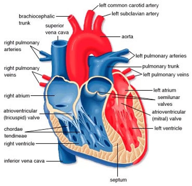
heart diagram
- deoxygenated blood coming from the body enters the right atrium through the vena cava
- then it enters the right ventricle
- it leaves through the pulmonary arteries to the lungs
- oxygenated blood from the lungs enters the left atrium through the the pulmonary veins
- then it enters the left ventricle
- it leaves through the aorta to the rest of the body
the heart valves (tricuspid and semilunar) stop blood moving backwards through the lungs
the walls of the left ventricle are thick and contain lots of muscle to create pressure to pump blood all through the body
the right ventricle doesn't need walls as thick, as it recieves low pressure blood from the body and only has to transport it to the lungs, which are close
cardiac output formula
cardiac output = heart rate (bpm) x stroke volume (cm3)
cellular respiration definition
an exothermic reaction which occurs continuosly in living cells to release energy for metabolic processes
compare the types of respiration
aerobic:
- glucose is oxidised in mitochondria to release energy
- glucose + oxygen → carbon dioxide + water (+ lots of energy)
- C6H12O6 + 6O2 → 6CO2 + 6H2O
this us used almost all of the time, except for when sprinting, as the blood can't transport oxygen fast enough, and anaerobic respiration begins to increase
anaerobic:
- glucose in muscle is converted to lactic acid
- glucose → lactic acid (+ little energy)
this is very inefficient and can only be used for short periods of time (sprints), as it builds up toxic lactic acid in muscles
how do our bodies respond to excercise?
- breathing becomes faster and deeper
- aerobic respiration increases (during vigourous exercise, anaerobic increases)
- heart rate + stroke volume increase, so cardiac input increases
- increasing temperature
- breathing rate increases
- glycogen reserves deplete
measuring respiration core prac
1. add soda lime, then cotton wool and gauze to a boiling tube
2. add a known number/weight of an organism to the boiling tube
3. insert a bung connected to a capillary tube, with a scale
4. set up a control tube without the organisms
5. place in a water bath (at an angle) for a few minutes at a set temp to adjust
6. hold a beaker of coloured liquid to the capillary tube so some enters
7. record the position of the liquid on the scale
8. record again after 5 minutes
9. repeat at different temps