inflammation
1/29
There's no tags or description
Looks like no tags are added yet.
Name | Mastery | Learn | Test | Matching | Spaced |
|---|
No study sessions yet.
30 Terms
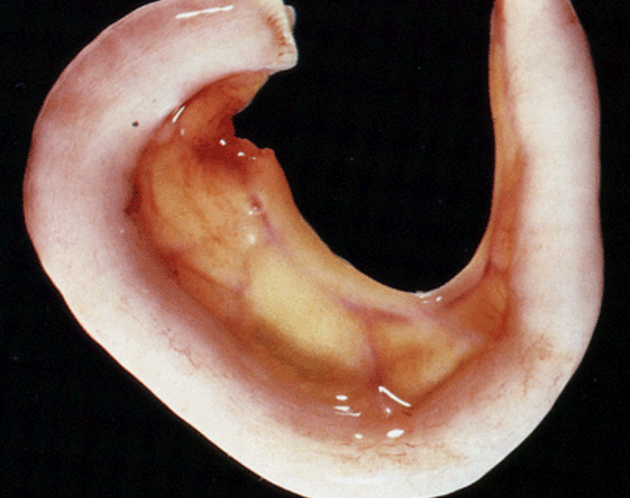
normal appendix
smoothy, shiny serosa
pink colour
no swelling
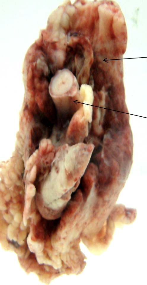
inflammed appendix inacute appendicitis
thickened wall
dull surface loss of shine
red brown colour
edema + pus
possiple fibrin
وصفيه وهين يظهر؟
وهيش فيه
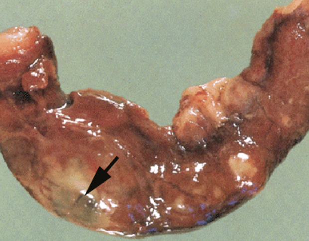
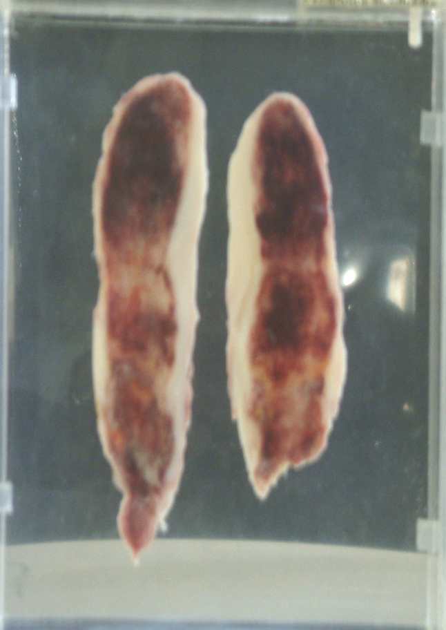
A.Inflamed appendix, fixed in
formalin.

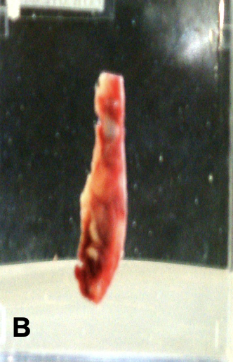
B.Inflamed appendix, special
fixative, therefore the difference in colour.
inflamed meso appendix
———————————-
inflamed appendix
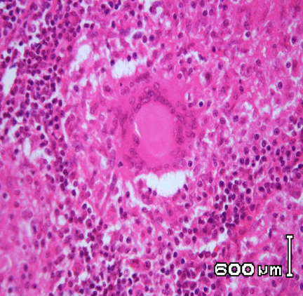
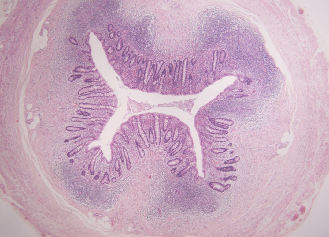
Transverse section of a normal appendix.
tubal origin has mucosa/submucosa/serosa/mascularnus
epithelial crypts lined by columer epithelial cells
what is this?
crypts lined by?
what have
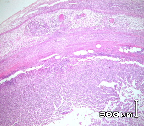
Acute appendicitis – note oedema, neutrophilic infiltration and congestion
cogulative necrosis
Acute appendicitis – note oedema, neutrophilic infiltration and congestion
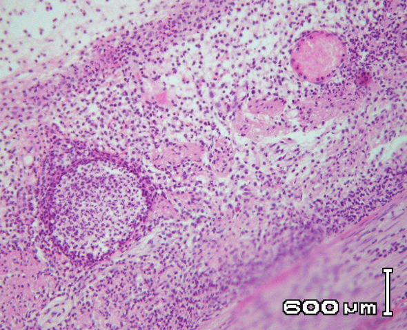
Acute appendicitis – note oedema, neutrophilic infiltration and congestion
periappendicular abscess
perforation-fatal
chronic appendicits»»»» appendicular mass
outcomes of inflamed appendix?
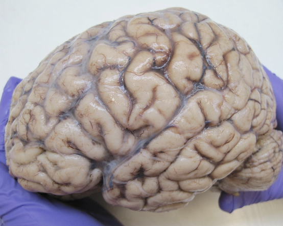
meningitis
SUPERIOR SURFACE OF THE EREBRAL HEMISPHERS TO
SHOW THICK HAZY MENINGES

meningitis
SUPERIOR SURFACE OF THE EREBRAL HEMISPHERS TO
SHOW THICK HAZY MENINGES
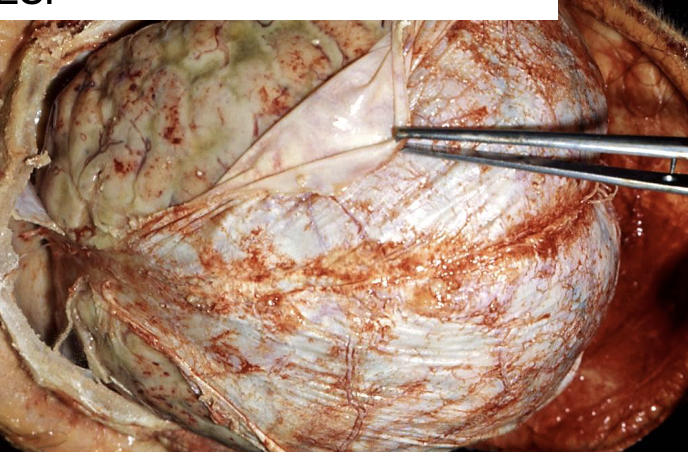
meningitis
SUPERIOR SURFACE OF THE EREBRAL HEMISPHERS TO
SHOW THICK HAZY MENINGES
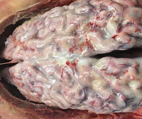
meningitis
SUPERIOR SURFACE OF THE EREBRAL HEMISPHERS TO
SHOW THICK HAZY MENINGES
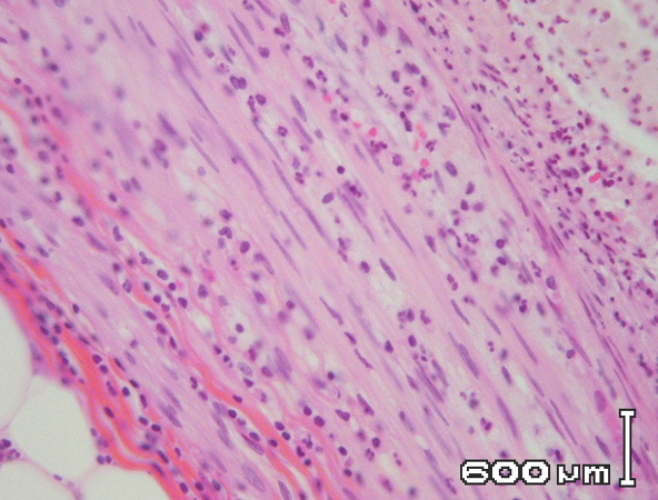
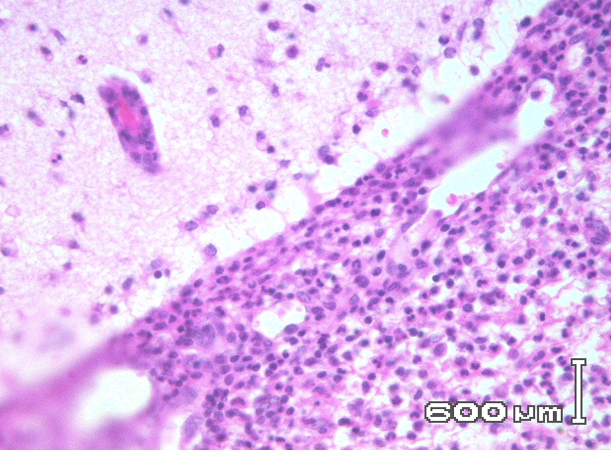
acute meningitis
CEREBRAL CORTEX WITH MENINGES. NOTE WIDE SUB-ARACHNOID
SPACE WITH البنفسجي الي تحت INFLAMMATORY CELLS neutrophils+exduate AND الدايرة CONGESTED VESSELS.
congestion = dilation = more blood flow.
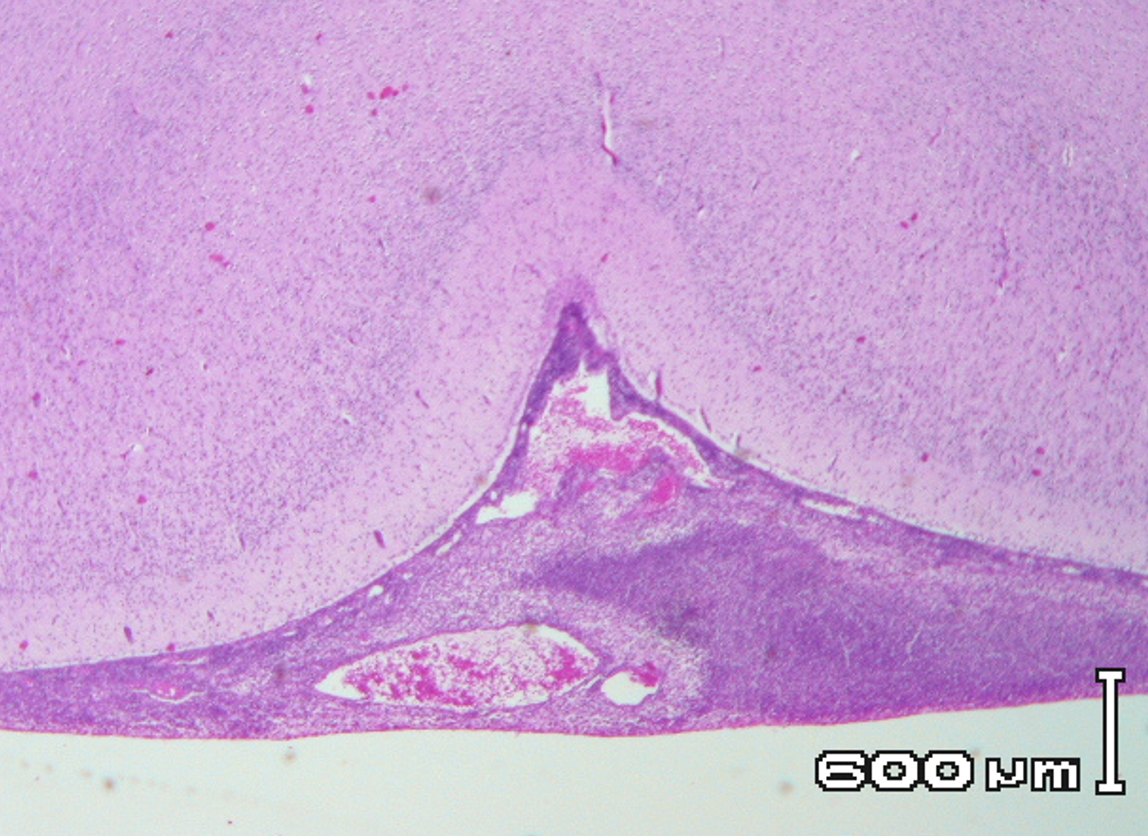
SUB-ARACHNOID EXUDATE WITH NEUROTPHILS.
acute meningitis
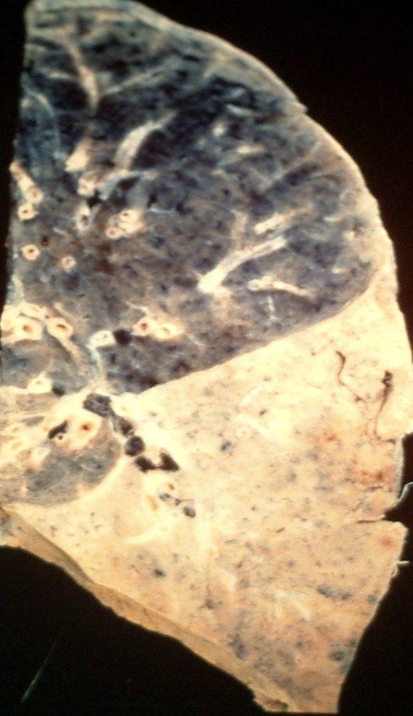
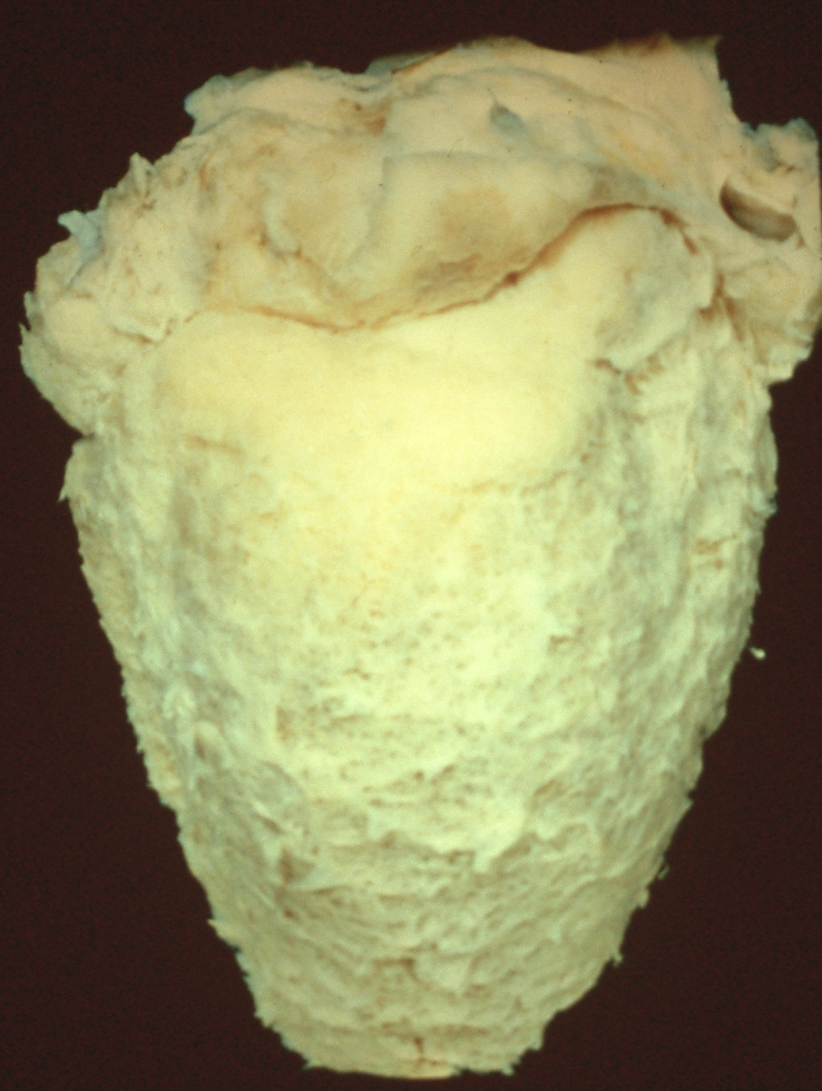
Lung showing lower lobe pale solid
Consolidation. Upper lobe is
Normal.
heals by resolution
lobar pneumonia
vascular congestion + netruphil
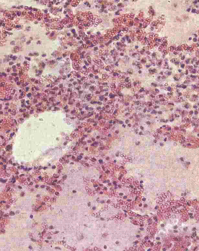
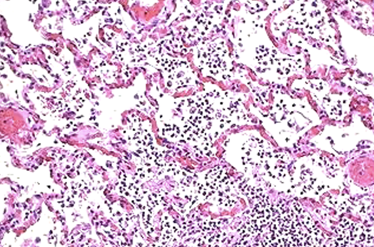
Higher power of consolidation. Note the inflammatory
acute pneumonia
cells (neutrophils) packing the alveolar spaces
Higher power of consolidation. Note the inflammatory
acute pneumonia
cells (neutrophils) packing the alveolar spaces

Higher power of consolidation. Note the inflammatory
acute pneumonia
cells (neutrophils) packing the alveolar spaces
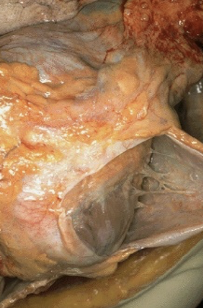
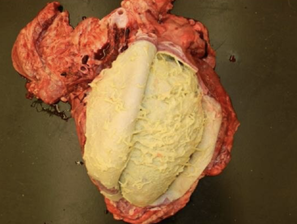
The pericardium is rough due
to fibrinous pericarditis.
exudate b/w visceral and parietal
serous ( excess water )
The pericardium is rough due
to fibrinous pericarditis.
The pericardium is rough due
to fibrinous pericarditis.
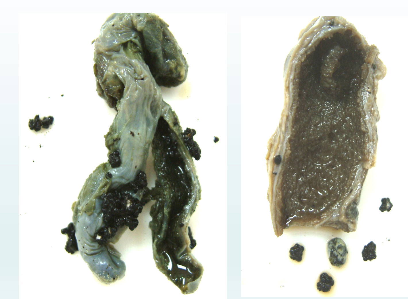
Gallbladder cut open to show
thickened wall and inflammed
mucosal surface
chronic inflammation abcess »» repeated acute inflammation repeat attacks of pain and contraction of bladder
gallbladder thick wall
stones removed
green colour is dye
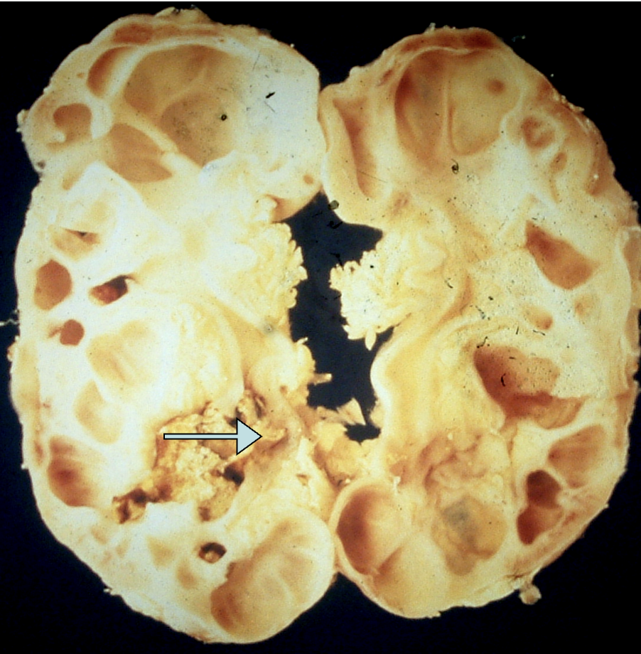
kidney showing many cystic spaces
and rough irregular structures in calyces (arrow)
الي تحت renal stones
وفوق dilated calyx
acute inflammation followed by repair
accumulation of urine
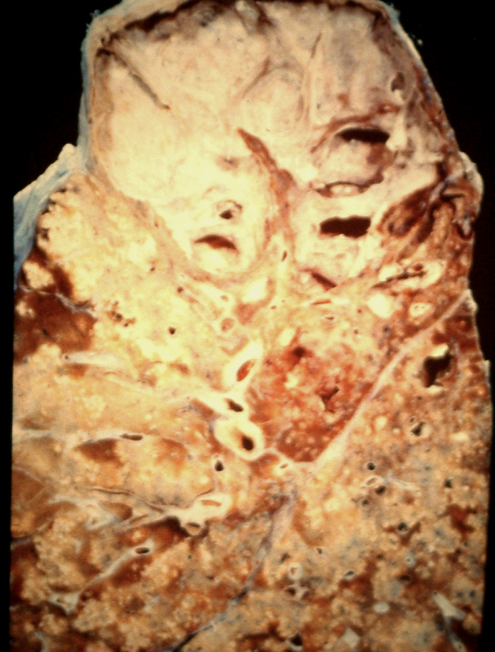
caceous necrosis white material
granulation»» repair by fibrosis
haemoptysis
الي يسار فوق هيش
epitheloid granuloma

caseation necrosis

langhan type giant cells