final exam - neuro content
 Call Kai
 Learn
Learn Practice Test
 Spaced Repetition
 Match
Match Flashcards
 Knowt Play
1/51

There's no tags or description
Looks like no tags are added yet.
Name | Mastery | Learn | Test | Matching | Spaced | Call with Kai | Chat |
|---|
No analytics yet
Send a link to your students to track their progress
52 Terms
Guillain-Barre Syndrome s/s
weakness in both legs, decreased reflexes, weakness that moves upward (ascending paralysis), N/T, pain, paresthesia's of hands and feet, neuromuscular respiratory failure, weakness continuous until it reaches a plateau (can take up to 2 years), cranial nerve involvement
Guillain-Barre Syndrome treatments
medical emergency and requires admission to ICU.
Pt requires respiratory support (RT, mechanical ventilation), plasmapheresis (filters antibodies out of the blood), IVIG (intravenous immunoglobin to lower harmful antibodies), continuous ECG monitoring, tx HTN and tachycardia, tx hypotension w/ fluids, IS, chest physiotherapy, monitor vital capacity and negative inspiratory force (measures inspiratory muscle strength – nml is greater than –60), monitor for respiratory failure.
Amyotrophic Lateral Sclerosis (ALS) s/s
sx depend on which motor neurons are affected first.
Early sx include a gradual onset of fatigue, muscle weakness, and weakness will be asymmetric.
If the arms are effected, the pt will have reduced hand dexterity, difficulty writing or buttoning clothes, weak grip.
If the legs are affected first, the pt will have tripping, falling, difficulty walking, gait changes.
Upper motor signs include spasticity, hyperreflexia. Lower motor sx include muscle weakness, muscle wasting (atrophy), muscle twitching.
Bowel and bladder remains nml.
Bulbar ALS includes cranial nerve involvement which effects about 25% of patients. These pts may have swallowing problems, tongue and palate problems, speech difficulty, aspiration risk, emotional changes, resp. Problems.
Amyotrophic Lateral Sclerosis (ALS) treatment
no cure, tx if focused on maintaining function, improving QOL, managing sx, preventing complications. Riluzole may be used to protect motor neurons early in the disease. Meds for spasticity may be baclofen, dantrolene, diazepem. Enteral feedings may be needed if swallowing worsens. A PEG tube must be placed before the forced vital capacity (FVC) falls below 50%), may need respiratory support (CPAP, BiPAP initially)
myasthenia gravis s/s
hallmark sx is fluctuating muscle weakness that gets worse with activity and better after rest. Drooping eyelids (ptosis), double vision, weak facial muscles
bulbar sx (weakness after talking, chewing, swallowing) which causes hoarse/weak voice, difficulty speaking and swallowing. Aspiration risk, generalized weakness
myasthenia gravis treatment
pyridostigmine is the first line tx (prevents breakdown of acetylcholine = muscle strength improves – taken 4 times daily). If that does not work, pts will be put on immunosuppressants. plasmapheresis is used for severe sx and exacerbations (removes antibody containing plasma), IVIG , thymectomy (surgical removal of the thymus)
MS s/s
exacerbations and remissions
fatigue, weakness, N/T, pain, visual problems, poor coordination, balance problems, spasticity, bladder and bowel problems, sexual dysfunction, cognitive changes, depression.
Visual sx may be the first sign (optic neuritis = sudden vision loss in one eye, pain behind or around the eye, pain that increases w/ eye movement)
Lhermitte sign (electric shock sensation that travels down the neck, back, arms, lower trunk)
MS treatments
no cure; Treating acute relapses, Reducing the number of relapses, Slowing, disease progression, managing symptoms, maintaining independence, preventing immobility complications, Supporting emotional health.
For tx of an acute relapse we would give the pt IV methylprednisolone.
parkinson’s s/s
TRAP: T = tremor, R = rigidity, A = akinesia (little or no movement), P = postural instability. Other motor sx are hypokinesia, freezing, decreased arm swing, mask-like face, speech problems, swallowing problems.
parkinson’s treatment
levodopa-carbidopa is the most effective medication, anticholinergics, amantadine, deep brain stimulation, entacapone and tolcapone (given with levodopa)
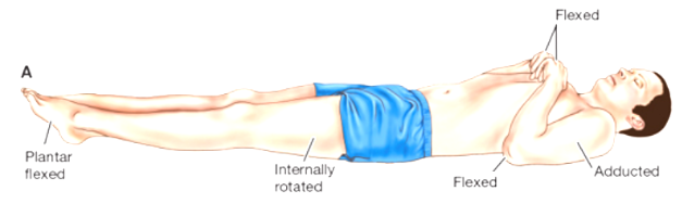
Decorticate posturing
involves abnormal flexion of the upper extremities and extension of the lower extremities and indicates damage to the upper midbrain
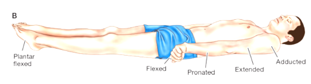
Decerebrate posturing
involves extreme extension of the upper and lower extremities and indicates severe damage to the brain at the lower midbrain and upper pons
bacterial meningitis
s/s: common early sx include HA, high fever, altered LOC. Classic triad is fever, stiff neck or nuchal rigidity, and altered LOC. Positive Kernig sign (flex hip and extend the knee = pain and resistance to knee flexion), positive Brudzinski sign (flexing one leg causes the opposite leg to flex), photophobia, meningococcal rash, neuro changes, increased ICP
Tx: 100% fatal if not treated. IV antibiotics, fluids, seizure tx, GI bleeding prevention
Precautions: respiratory isolation until 24 hours after treatment
viral meningitis
s/s: HA, low grade fever, stiff neck, photophobia, malaise, sx of recent upper resp. Viral infection. Not usually associated with alerted mental status or seizures
Tx: no specific treatment; management is based on sx relief and supportive care.
Precautions: direct contact through respiratory droplets.
tests for seizures - CT scan
CT scan to help with ID bleeding, tumors, structural abnormalities, and brain injury. Perfusion CT to evaluate blood flow through the brain. Looks for underlying reasons why pt has seizure
tests for seizures - MRI
detect tumors, scarring, lesions, and structural abnormalities.
Magnetic resonance spectroscopy
evaluate the chemical and metabolic changes in brain tissue.
EEG
record the brains electrical activity (helps support the sx of epilepsy, classify the type of seizure and locate abnml electrical activity).
depth electrodes
microelectrodes may be placed deep inside the brain to record the activity of individual brain cells. These are usually used when more detailed information is needed, especially when surgery is being considered.
video EEG monitoring
helps to determine what the seizure looks like, where it may begin, how long it lasts, how severe it is, where the physical movements match abnml electrical brain activity
PET scan
can help to identify areas of abnml brain function
SPECT scan
Single-photon emission computed tomography evaluates blood flow and activity in the brain. It may help locate the epileptogenic zone, which is the area where seizures begin. If medications do not control the seizures, this area may sometimes be surgically removed.
common stroke-like sx
sudden numbness or weakness that usually effects the face, arm, leg, etc., sudden trouble speaking, sudden vision changes difficulty walking, sudden severe HA
-Hemiparesis: partial paralysis on one side (ischemic)
-Exploding headache & decreases LOC (hemorrhagic)
-Aphasia: dysfunction in communication
-Dysphasia: impaired ability to communicate & think
-Dysarthria: impairment w/ muscular control of speech (pronunciation, articulation, phonetics)
-Penumbra: area around infarcted tissue that still is viable (stunned)
· Motor: Impaired mobility & self-care
-Dysphagia: swallowing, gag, respiratory
-Affect: Labile emotions, Difficulty controlling emotions, Exaggerated or unpredictable
-Intelligence: Memory—left side with language; Judgment: Right—impulsive, move quickly; Left—more cautious
-Visual: Field cuts, Visual/spatial relationships
-Spatial: Right side more likely, Incorrect perception of self and illness
-Sensory: Loss sensation, don't know where body parts are, may not know previously known objects
-Elimination: Temporary or permanent
stroke acronyms
FAST: F = face drooping, A = arm weakness, S = speech difficulty, T = time to call 911 immediately
BFAST: balance, eyes, face, arms, speech, time
diagnostic testing for stroke
CT scan of the head WITHOUT CONTRAST. First line to determine ischemic vs hemorrhagic stoke. MRI, Cerebral angiography, Carotid ultrasound.
Labs: CBC, electrolytes, glucose, PT/INR/APTT coag studies, lipids.
common causes of stroke and complications
causes: biggest one is Hypertension!!, atherosclerosis, a fib, DM, smoking, high cholesterol, obesity, CAD, blood clots, brain aneurysm, trauma.
complications: cerebral edema!!! increased ICP, seizures, aspiration pneumonia weakness, language problems, depression, cognitive changes, bowel/bladder habits, DVT/PE, contractures.
stroke sx to assess and tx
LOC changes!!! Increased ICP signs are a decreased LOC, N/V, HA, PERLA changes, increased BP with decreased HR(Cushing’s triad)!!!!
treatment of stroke: ABCs!!
ischemic stroke
happens when blood vessel supplying the brain becomes blocked and prevents oxygen rich blood from reaching the brain tissue
hemorrhagic intracerebral stroke
Bleeding into brain tissue (parenchyma) caused by rupture, HTN most common cause, coagulation disorders, anticoagulants and thrombolytics, brain tumors, aneurysms.
During periods of activity with sudden onset of symptoms, Poor outcome 40%-80% die within 30 days, After stroke from damaged brain tissue, AVM—arteriovenous malformations
hemorrhagic subarachnoid stroke
Bleeding in cerebrospinal fluid filled space (between pia mater and arachnoid Mater), Most often by cerebral aneurysm (40% die initially, 15% die from continual
bleeding), Illicit drugs (cocaine) and trauma, WORST HEADACHE OF MY LIFE, Incidence higher in women, Vasospasm after
causes for ischemic
Large-artery thrombosis, Small, penetrating artery thrombosis, Cardiogenic embolic, Cryptogenic (no known cause)
Low flow (hypotension) or no flow
-Thrombotic & Embolic
-Large artery or penetrating artery (small artery)
-Injury to vessel wall which causes clot formation
-Blood vessel is narrowed
-Develops where atherosclerotic deposits (plaque) exists
-50-60% of all stroke's majority in cases of HTN or DM 30%-50% history of TIA
causes for hemorrhagic
Intracerebral hemorrhage, Subarachnoid hemorrhage, Cerebral aneurysm, arteriovenous malformation
Bleed
-Intracerebral & Subarachnoid
-HTN most common cause
-Coagulation disorders, anticoagulants & thrombolytics, brain tumors, aneurysms
-During periods of activity w/ sudden onset of symptoms
-AVM—arteriovenous malformations
-Most often by cerebral aneurysm
tx for ischemic
goal is to restore blood flow as quickly as possible.
Thrombolytic therapy (tPA) aka alteplase (can only be used within 3 hours of sx onset)
Tenecteplase
thrombectomy (cath is inserted into an artery and is guided to the clot)
tx for hemorrhagic
decrease ICP to prevent brain herniation, secondary brain injury, death. Airway. Stop the bleeding, control BP, surgery, prevent seizures, fever control, keep NPO, early rehabilitation.
delirium s/s
Hyperactive delirium: confused, agitated, restless, tires to get out of bed, pulls out IVs, tubes or catheters, may become combative.
Hypoactive: sleepy, quite, difficult to wake, inattentive, less responsive
DELERIUM
Dementia, dehydration
Electrolyte imbalance/emotional stress
Lung, liver, heart, kidney, brain
Infection, ICU
Rx drugs
Injury/immobility
Untreated pain/unfamiliar environment
Metabolic disorders
PINCHME
Pain
Infection
Nutrition
Constipation
Hydration
Medication
Environment
delirium prevention and risk factors
SLEEP, reorient the pt, promote nml sleep, encourage early mobility, maintain hydration and nutrition, tx the underlying cause, control pain, ensure pt can see and hear prevent hypoxia, avoid restraints if possible
Dementia, stress, surgery, sleep deprivation, anesthesia, mechanical ventilation.
-Controlling HTN
-Lowering the amount of cholesterol & saturated fat in your diet
-Smoking cessation
-Managing DM
-Maintaining a healthy weight
-Eating a diet rich in fruits & vegetables
-Exercising regularly
-Limiting alcohol consumption
-Treating obstructive sleep apnea
-Avoiding illegal drugs
dementia
Dysfunction or loss of, memory, language, orientation, judgement, attention, reasoning (progressive organic mental disorder)
dementia causes
Alzheimer’s disease, vascular conditions (vascular dementia), dementia with lewy bodies, and frontotemporal lobar degeneration.
Reversible:
Hyper and hypothyroidism
Meningitis
Brain tumors
Vitamin deficiencies
Non-reversible:
Down Syndrome
ALS, Parkinson’s, and other chronic neurologic disorders
Alcoholism
dementia s/s
gradual months to years. Short term memory loss, trouble with daily tasks, communication issues, confusion about time and place, poor judgement, mood shifts/personality.
-Memory losses affect job skills (Forget people, Forget familiar objects/places)
-Difficulty w/ normal tasks (Forgot they ate, Forgot to shower)
-Problems w/ language (Wrong words)
-Disorientation to time & place
-Poor judgement (Sweater in summer, Impulsive behavior)
-Problems w/ abstract thinking (Simple calculations difficult)
-Misplaced items
-Changes mood/behavior (Rapid mood swings)
-Changes personality (Depression, paranoid, hostile, wandering)
-Loss of initiative
-Get lost in familiar environment
-Repeat same stories over and over
Alzheimer's s/s
early signs are forgetfulness, mild memory loss, difficulty at work, difficulty in social situations, depression. As the disease progresses, they may forget familiar faces, places, objects, get lost in familiar surroundings, have difficulty finding words, and have trouble carrying on conversations. Thinking problems, difficulty with daily activities, personality changes.
behavioral problems with Alzheimer's
Overreaction to excessive stimulation - keep environment calm and comfortable. Sundowning is when the patient gets confused in the late afternoon/evening.
nursing interventions for AD
safety, oral care, eating and swallowing difficulties, infection prevention, skin care, and elimination problems.
nml ICP
normal is 5-15 mm Hg
Greater than 20 needs to be treated
causes of an increased ICP and how to decrease it
fever, pain, stimulation, shivering, pain, seizures, blood, tumor, edema, increased CSF, agitation.
-Head injury
-Brain tumors
-Hydrocephalus
-Bleeding into the brain
-Swelling of the brain
-Aneurysm
-Infection such as encephalitis or meningitis
decrease it → Low stimulation environment, burr holes, craniotomy and or drains treatment all decrease ICP. Elevate Hob decreases ICP. Can use a bubble level string as part of the External Ventricular Drain or ICP monitoring system. Can give osmotic diuretic (manatol!!!). lower the volume of CSF. Ventriculostomy, fiberoptic catheter, subarachnoid bolt.
basilar skull fracture sx
base of the scull.
Bleeding nose pharynx, ears, conjunctiva.
Raccoon eyes-bruising around the eyes with swelling.
Rhinorrhea- drainage from nose
Otorrhea- drainage from the ear
Battle’s sign- bruising behind base of the ear
Halo sign – place draining onto white gauze. Blood will go to center with yellow ring around. Shows CSF leak.
basilar interventions and considerations
do not turn them, we need to watch out for infection. Tell them do not blow nose. No nasal suctioning. No NG tube. Elevate the HOB (decrease ICP), check for glucose.
Do not blow nose
-HOB elevates 30 degrees
-Do not suction nose
-If NG tube needs to be placed insert it orally
complications of a spinal cord injury
spinal shock-areflexia(no reflexes)- muscles paralyzed below injury.
Neurogenic shock- no perspiration, hypotension, decrease CO, venous pooling.
DVT prophylaxis and assessment- pneumatic devices (SCD), ROM, compression stockings.
Orthostatic hypotension.
Autonomic dysreflexia- excessive hypertension when damage t6 or above- bradycardia, sweating above lesion, nausea.
Nurses must prevent bladder fullness, inspect for areas of skin pressure or damage, remove tight clothing. Promote adequate breathing improve mobility, skin integrity, urinary elimination and bowel function.
-DVT/PE thrombophlebitis
-Orthostatic hypotension
-Autonomic dysreflexia
Cushing’s triad
suggests increased ICP
systolic hypertension and widening pulse pressure
bradycardia with bounding pulse
irregular respirations
*medical emergency-brainstem compression-impending death.
sx of brain tumors
altered LOC, neuro changes, confusion, sensory issues, headaches, seizures, N/V/Vision changes, numbness and weakness, balance problems, personality shifts.
permissive HTN
220/120
Permissive HTN is when clinicians intentionally do NOT lower the blood pressure in an ischemic stroke because the brain needs that higher pressure to push blood around the clot and maintain oxygenation