Wk3 L8 - Synaptic Integration and Summation
1/28
There's no tags or description
Looks like no tags are added yet.
Name | Mastery | Learn | Test | Matching | Spaced | Call with Kai |
|---|
No analytics yet
Send a link to your students to track their progress
29 Terms
Axon Hillock
The most excitable region of a nerve cell where action potentials are typically initiated.
Dendrites
Parts of neurons that have very few voltage-sensitive Na+ channels and conduct signals passively.
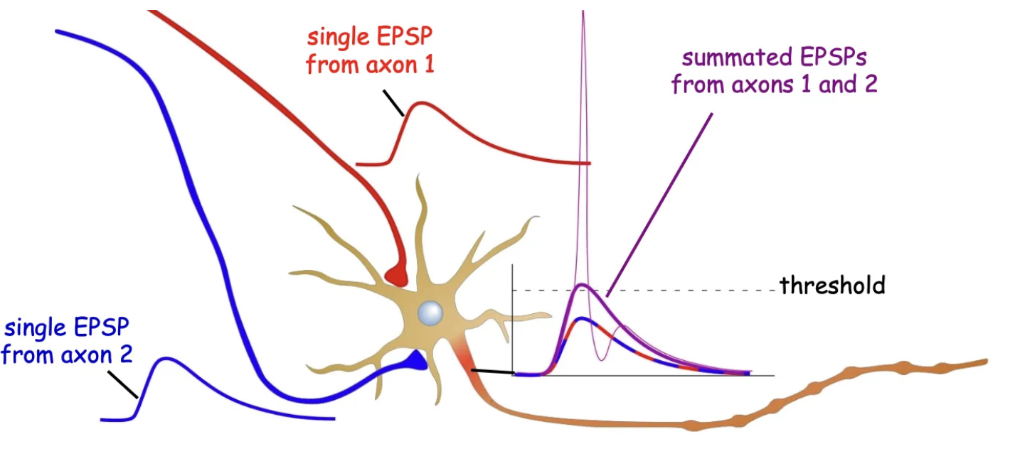
Excitatory Postsynaptic Potential (EPSP)
Occurs when excitatory neurotransmitters (e.g., glutamate) bind to postsynaptic receptors, causing depolarization.
Caused by the influx of positively charged ions (e.g., Na+).
Increases the likelihood of reaching threshold potential and triggering an action potential.
Short-lived and decays with distance from the synapse.
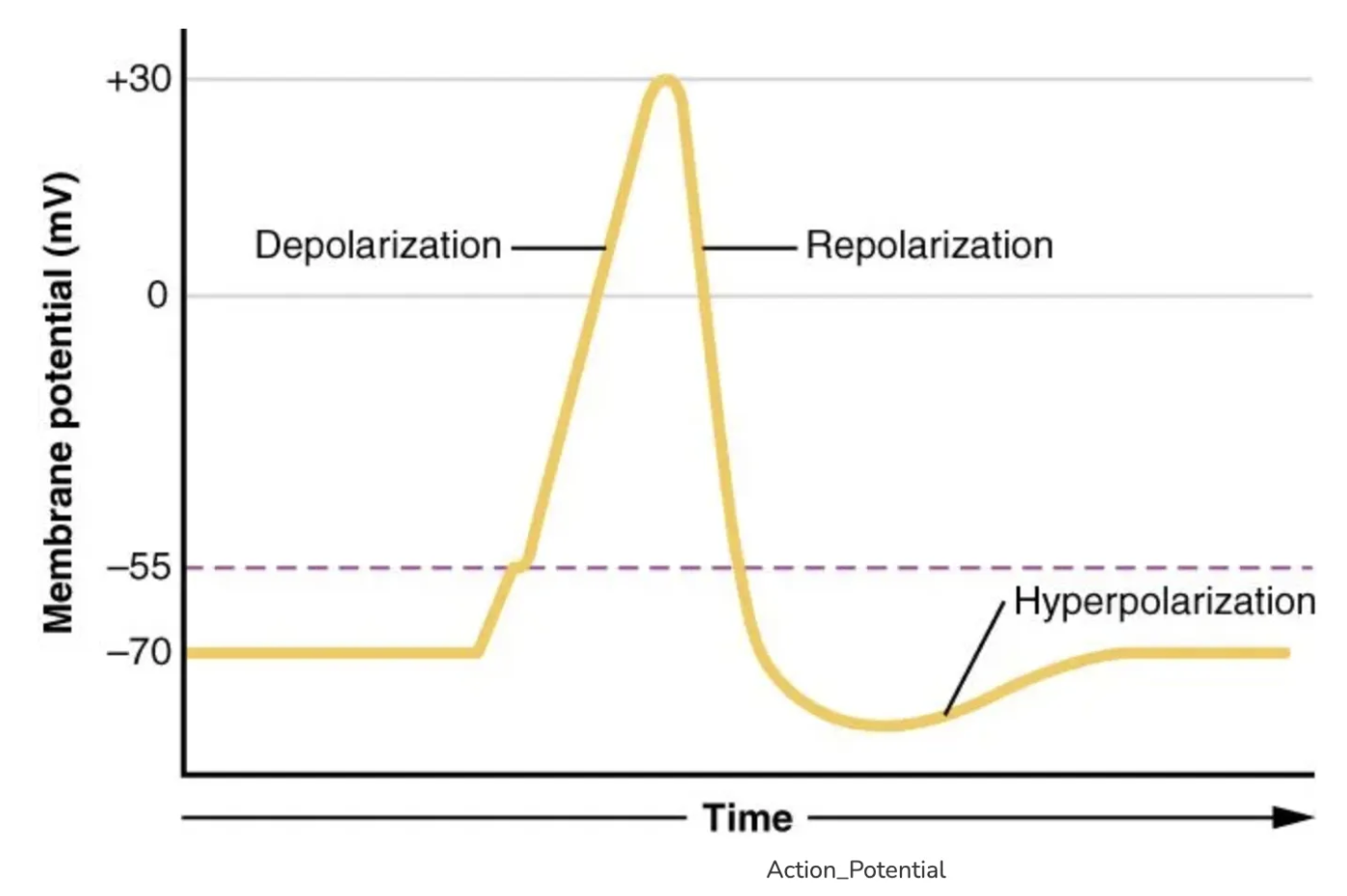
Depolarisation
A reduction in membrane potential, making the inside of a cell less negative.
Occurs when positive ions (e.g., Na+) enter the cell, making the inside less negative.
Increases the likelihood of reaching threshold potential and triggering an action potential.
Action Potential Threshold
The critical level to which the membrane potential must be depolarized to trigger an action potential - around -55 mV in neurons.
Reached when excitatory signals depolarize the membrane, activating voltage-gated Na+ channels.
Amplitude Decrement
The decrease in the strength of an electrical signal as it travels from the point of entry towards the axon hillock, due to charge leakage.
Reduced by myelination and larger axon diameter.
Inhibitory Postsynaptic Potential (IPSP)
Occurs when inhibitory neurotransmitters bind to postsynaptic receptors, causing hyperpolarisation.
Moves the membrane potential further from the action potential threshold.
Typically caused by the influx of negative ions (e.g., Cl-) or efflux of K+.
Decreases the likelihood of the neuron firing an action potential.
Hyperpolarization
Increase in membrane potential, making the inside of the cell more negative than the resting potential.
Occurs when K+ ions move out or Cl- ions move into the cell.
Moves the membrane potential further from the action potential threshold.
Reduces the likelihood of the neuron firing an action potential.
Shunting Inhibition
Inhibitory postsynaptic potential (IPSP) that prevents depolarisation, making it harder for excitatory signals to reach the threshold for an action potential.
It opens channels for ions like Cl- or K+ to make the inside of the cell more negative.
This prevents the neuron from firing an action potential.
Spatial Summation
Occurs when multiple excitatory postsynaptic potentials (EPSPs) from different locations on the neuron combine.
The combined effect of these signals can bring the membrane potential to threshold, triggering an action potential.
Helps integrate signals from various synaptic inputs.
Temporal Summation
The accumulation of EPSPs at a single synapse over time, making it possible to reach action potential threshold.
A single fast EPSP often fails to reach threshold.
Reversal Potential
The membrane voltage at which no net ion flow occurs through a receptor's ion channels.
At the reversal potential, the driving force for the ion is zero, meaning the movement of ions into and out of the cell is balanced.
Driving Force
The driving force is the difference between the membrane potential (Vm) and the ion's equilibrium (reversal) potential. It determines the direction and strength of ion flow across the membrane. A greater difference results in a stronger driving force, increasing ion movement.
Presynaptic Inhibition
A process where a synapse reduces the release of neurotransmitter from another synapse by influencing the presynaptic neuron through inhibitory signals.
Autoinhibition
Autoinhibition is a form of feedback inhibition where a neuron or presynaptic terminal inhibits its own activity.
This typically occurs when neurotransmitters or other signaling molecules bind to receptors on the presynaptic neuron, reducing neurotransmitter release and preventing overstimulation.
Post-synaptic Receptor Desensitisation
The reduction in responsiveness of receptors following prolonged exposure to neurotransmitters.
Membrane Resistance (Rm)
Membrane resistance (Rm) refers to the resistance to the flow of ions across the cell membrane. High Rm means fewer ion channels are open, making it harder for ions to flow through, whereas low Rm indicates more open channels, allowing greater ion flow. Rm influences the speed and strength of electrical signals within the cell.
Nicotinic Receptors
Type of receptors that, when activated, allow Na+ and K+ to pass, affecting the membrane potential.
Ohm’s Law
A relationship defined as V=I×R, used to measure changes in membrane resistance during synaptic potentials.
Calcium Levels (Ca²⁺)
An influx of Ca²⁺ during action potentials helps trigger neurotransmitter release.
Calcium levels are tightly regulated.
Feedforward Inhibition
Inhibition that occurs when an excitatory neuron activates an inhibitory neuron to suppress a target neuron.
Feedback Inhibition
Feedback inhibition occurs when a neuron’s output inhibits its own activity or that of other neurons in the circuit.
Spatial Summation of EPSP and IPSP
EPSP alone is suprathreshold, triggering an action potential.
IPSP alone causes hyperpolarisation at the axon hillock.
When both EPSP and IPSP are activated together, the IPSP reduces the amplitude of the EPSP, making it subthreshold, preventing action potential.
Synaptic Transmission Run-down
Gradual decrease in synaptic efficacy due to:
Receptors becoming less responsive after prolonged stimulation.
Changes in calcium levels affect release mechanisms.
Post-synaptic inhibition reducing synaptic transmission by causing hyperpolarization
Active Conductance
The process by which certain dendrites amplify or regulate synaptic signals through voltage-gated ion channels.
Neurodegenerative Diseases
Disorders like Alzheimer’s that affect synaptic integration and cognitive function.
Epilepsy
A condition characterized by excessive excitatory activity due to poor inhibitory control, leading to seizures.
What happens during repeated synaptic stimulation regarding EPSP summation and reversal potential?
When repeated synaptic stimulation occurs, EPSPs summate but never exceed the reversal potential (+5mV).
This is because once the membrane potential (Vm) reaches the reversal potential, ion movement no longer drives further depolarisation.
What is the effect of an IPSP on membrane resistance (Rm)?
During an IPSP, K⁺ channels open, lowering Rm because more ion channels are available for ion flow. Hyperpolarizing current pulses injected into the cell become smaller as Rm decreases.
This shows that the IPSP is caused by channel opening, not closing.