The Brain and Neuropsychology
1/50
There's no tags or description
Looks like no tags are added yet.
Name | Mastery | Learn | Test | Matching | Spaced | Call with Kai |
|---|
No study sessions yet.
51 Terms
Frontal lobes: location
Located just behind the forehead, at the front of the brain
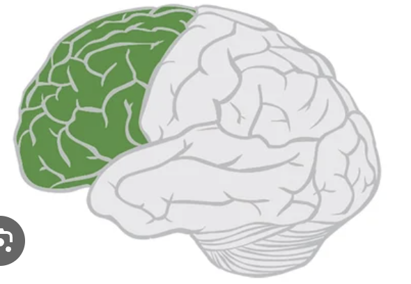
Frontal lobes: Function
Decision making
Problem solving
Concentration
Symptoms of Frontal Lobe Damage
Changes in social behaviour, mood and personality
Weakness of loss of movement in areas of the body (paralysis)
Temporal lobe: location
Located on the side of the head above the ears
Temporal lobe: function
Hearing and understanding sounds
Understanding and creating speech
Symptoms of Temporal Lobe Damage
Difficulty in learning and remembering new information
Difficulty in understanding spoken words
Parietal lobes: location
Near the back and top of the head
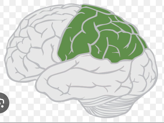
Parietal lobes: function
Sense of touch
Spatial orientation
Symptoms of Parietal Lobe Damage (left side)
Confusion between right and left of the body
Difficulty with writing, reading and mathematics
Symptoms of Parietal Lobe Damage (right side)
Problems with self-care skills
Weakened ability to analyse pictures
Symptoms of Parietal Lobe Damage (both sides)
Problems with visual attention and motor skills
Inability to control your gaze
Occipital lobe: location
The back of the brain
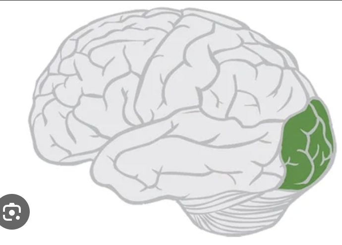
Occipital lobe: function
Visual perception
Distance and depth perception
Symptoms of Occipital Lobe Damage
Difficulty in identifying colours
Problems with vision
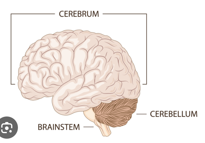
Cerebellum: location
At the back of the head, above and behind where the spinal cord connects to the brain
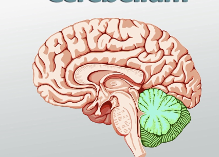
Cerebellum: function
Muscle and balance control
Relays information between body muscles
Symptoms of Cerebellum Damage
Loos of coordination of motor skills (asynergia)
Slurred speech (dysphonia)
Muscle tone
The tension/resistance to movement in muscles, helps us to hold our bodies upright when we are sitting and standing, includes the control, speed and amount of movement we can achieve
Cerebrum
The largest part of the brain which contains all the lobes
Cerebrum: function
Initiates and coordinates movement
Regulates temperature
Lateralisation of function
Each hemisphere of the brain has different functions, some behaviours are controlled more from the left than the right, vise versa
Asymmetrical functions
Both hemispheres are not exactly the same, their structure and their functions vary which makes them asymmetrical despite looking similar (not mirror images), each hemisphere controls the opposite side of the body
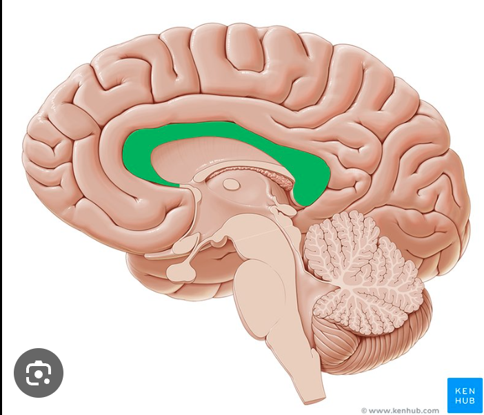
Corpus Callosum
A thick layer of nerve fibres which connects the brain functions for the two hemispheres to communicate and work together
Absence of the Corpus Callosum
Happens at birth and mostly diagnosed in the first two years of life, the disorder can be without apparent symptoms in milder cases
Absence of the Corpus Callosum: possible symptoms
Vision problems
Low muscle tone (hypotonia)
Unusual head shape and facial features
Brain abnormalities
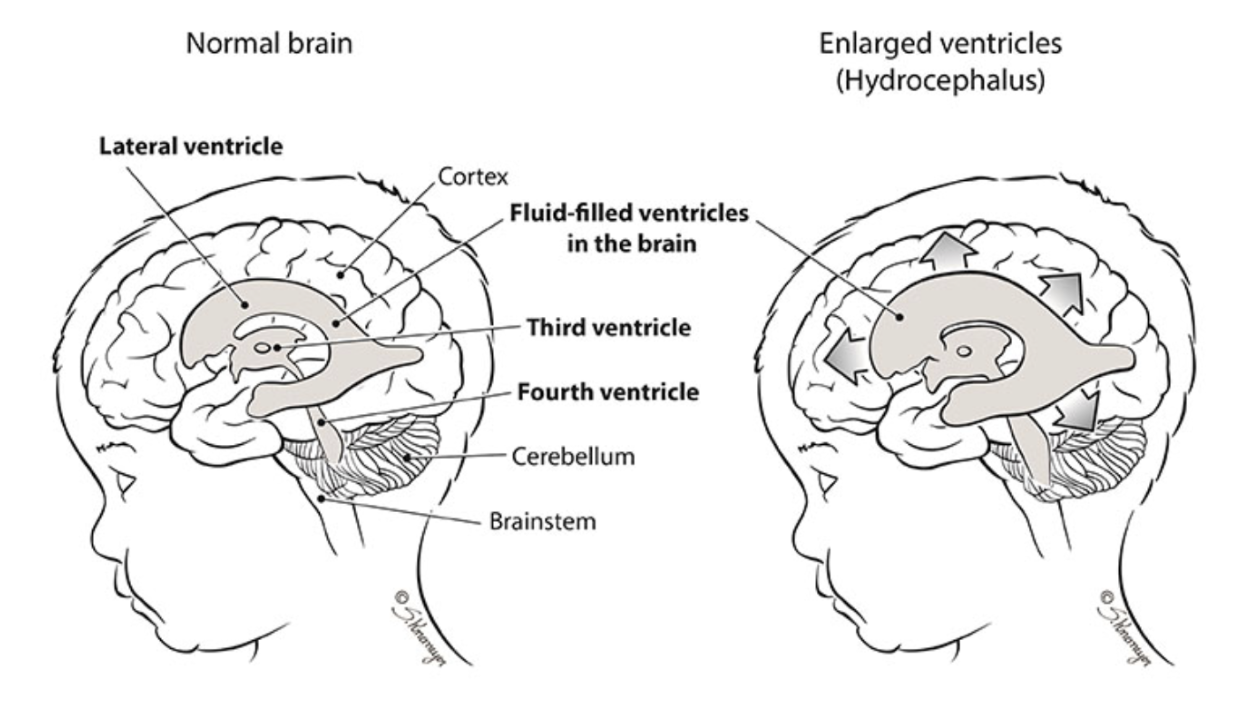
Hydrocephalus
A brain abnormality where there is excess fluid in the brain which puts harmful pressure on the brain’s tissues
Right Hemisphere
R for recognition
Spatial awareness
Ability to recognise and perceive faces
Creativity
Processing music heard
Left hemisphere
L for language
Processing language
Contains the Broca’s Area
Ability to write and understand language
Broca’s Area
Controls production of speech, linked to the control of nerve cells in the face that allow us to speak and process language, located in the frontal lobe

Sex differences
It was always assumed that females were better at language (the left) and males were better at spatial skills (the right)
Sex differences: evidence
Strengths of lateralisation explaining sex differences
There is evidence from a study by Harasty et al. (1997)
Study by Rilea et al. (2005)
Lots of reliable and scientific evidence available
Weaknesses of lateralisation explaining sex differences
Some research by Rilea et al. are weak
Study by Sommer et al. (2004) which suggested no strong evidence
Damasio et al. (1994) Phineas Gage
After a metal pipe pierced through Gage’s skull, Damasio et al. used a 3D model to find out which parts of his brain was damaged for his personality to change drastically
Damage to Phineas Gage’s brain
Damage to both hemispheres of only the frontal lobe
Damage to his ventromedial area of the frontal lobes caused him to be impulsive
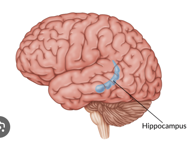
Hippocampus
Responsible for memory and learning, holds short-term memories and transfers them to long-term storage
Alzheimer’s disease
A progressive disease where dementia symptoms gradually intensify, starting from mild memory loss to possible loss of ability to respond to conversations and environments. This happens to patients with a loss of volume in the hippo campus, the more tissues lost, the worse the disease gets.
Neurons
Cells in the nervous system that help with communication within the body, they send electrical and chemical messages to connect organs and glands, they can relation information back into the central nervous system
Neuron composition
Cellbody with nucleus, dendrites, axon, myelin sheath, terminal knobs
Dendrites
Carry electrical charges from one neuron to the next
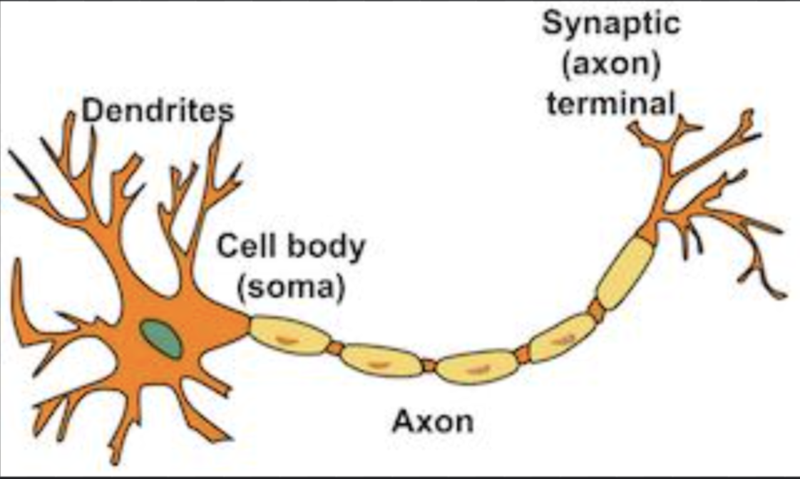
Axon
Carries the charge down the neuron and connects the cell body of a neuron to the terminal button, protected by a fatty myelin sheath
Terminal button
End of a neuron
Neurotransmitters
Chemicals that are released during neurotransmission when a nerve impulse reaches the end of a nerve fibre, passes messages from one neuron to another
Electrical impulse order
Dendrites
Cell body
Axon
Terminal branches
Synaptic vesicles
Synaptic gap
Receptor site
Central Nervous System (CNS)
Helps the brain and body communicate by passing messages back and forth between them. Sensory nerves in the body send messages to the brain through the spinal cord and vise versa
Peripheral Nervous System (PNS)
The spinal cord can activate this system and makes the body do actions the brain is telling it to do. Information is passed around in a fraction of a second.
Synapse
A gap between two neurons that allows messages (in the form of neurotransmitters) to pass from one cell to another
Visual agnosia
Information sent from the eyes to the brain cannot be understood, so the person cannot identify the things they can see. The person can still see the object perfectly well but cannot make sense of this information
Visual agnosia example
Unable to recognise the colour of an object, objects and name them or places they are familiar with
Prosopagnosia (face-blindness)
Unable to recognise faces even though they can be seen, the brain is unable to recognise who the face belongs to even if they know the person very well. All faces they see look the same and cannot tell them apart.
Cause of Prosopagnosia
Damage to a part of the brain (called the fusiform face area (FFA)) near the back of the temporal lobe, next to the occipital lobe