Topic 9: Cerebral Circulation
1/22
There's no tags or description
Looks like no tags are added yet.
Name | Mastery | Learn | Test | Matching | Spaced |
|---|
No study sessions yet.
23 Terms
Hypoxia
deprived of an adequate oxygen supply
role of the cerebral circulation
• to maintain an uninterrupted delivery of oxygen to the brain
• Local neuronal activity increases metabolic rate and the cerebral circulation must be capable of regional adjustment of blood flow (active areas of the brain receive more oxygen)
Measuring regional adjustment of blood flow
Positron Emission Tomography (PET) - Radiotracers, such as oxygen-15 (H215O) and fluorine-18 (18F) are injected into the participant’s bloodstream in order for changes in blood flow in particular brain regions to be directly measured
Blood brain barrier
• Nowhere in the body is there more need for homeostasis (eqm) than in the brain
• The mechanism for maintaining this barrier function lies in the capillary network supplying blood to the brain
• Ion concentration levels in plasma may fluctuate abruptly
• The blood-brain barrier protects the brain against surging fluctuations in ion concentrations.
Arterial supply to the brain
• Brain tissue is supplied by two pairs of arteries: the internal carotid (ICA) and vertebral arteries (VA)
• The ‘Circle of Willis’ is an anastomosis (connection) between the internal carotid and vertebral systems
•
Internal Carotid Artery
• The paired ICA’s arise at the bifurcation of the common carotid arteries in the neck
• It enters the cranium through the carotid canal in the temporal bone
• Branches of the ICA:
o Ophthalmic artery
o Posterior communicating artery
o Choroidal arteries
o Anterior cerebral artery (ACA)
o Middle cerebral artery (MCA)
Vertebral Arteries
• The VA branches from the subclavian artery
• Ascends the neck by passing though a foramen in the cervical vertebrae
• The paired VA’s join to form the basilar artery (BA)
Basilar Artery and branches
• At the upper border of the pons the basilar divides into two posterior cerebral arteries
• Branches of the BA:
o Pontine
o Labyrinthine
o Anterior inferior cerebellar
o Superior cerebellar
o Posterior cerebral artery (PCA) – remember this one
Circle of Willis – base of brain
• The circle of Willis is formed by an anastomosis between the two ICA’s and the two VA’s
• This arrangement of vessels can potentially maintain brain perfusion even when one ICA becomes blocked
• From this circle other arteries – ACA, MCA, PCA - arise and travel to all parts of the brain.
• Evolutionary adaptation to ensure blood flow to the brain (even if an artery is blocked)
• Perfusion = delivery of blood to a capillary bed
Arterial Branches
• Cortical branches - surface of the brain, supply cortical grey matter
• Choroidal branches - supply the choroid plexus (Cerebrospinal fluid derives from blood, choroid plexus produces this fluid, therefore this area needs blood supply)
• Central branches - penetrate into the brain substance to supply fibre tracts and sub-cortical grey matter
• Branches from the BA supply the brainstem
Central branches further detail
Deep branches arising from major arteries (particularly the MCA and PCA) supply basal ganglia and internal capsule
Basal ganglia - a group of nuclei located deep within the brain that are crucial for motor control, including the initiation and suppression of movement – issues involved in Parkinsons.
Internal capsule - a crucial V-shaped bundle of white matter tracts in the brain's cerebral hemispheres that connects the cerebral cortex to the brainstem and spinal cord, transmitting motor and sensory information.
Lenticulostriate Arteries
• small perforating arteries arising from the MCA and supplying deep structures in the cerebrum, including the internal capsule and basal ganglia
• terminal arteries without anastomoses, making them more susceptible to ischaemia (restriction in blood supply)
• Strokes in these vessels are common and can cause extensive damage
Anterior Cerebral Artery
• Cortical branches supply all of the medial surface of the cortex and a strip of cortex on the lateral surface
• The ACA supplies the leg area of the motor and sensory cortex
• Central branches supply parts of the basal ganglia and parts of the anterior visual pathway
Middle Cerebral Artery
• largest branch of the ICA
• Cortical branches supply the large part of the superolateral surface (including motor and sensory cortex and speech areas on the dominant hemisphere)
• The MCA supplies a large part of the visual pathway
• The medial and lateral striate branches supply the medial parts of the basal ganglia and internal capsule
Posterior Cerebral Artery
• supplies the posterior lobe and part of the temporal lobe
• supplies the posterior part of the visual pathway
• Central branches supply a large proportion of the thalamus (which relays sensory and motor signals)
Venous drainage
• Blood drains from deep in the brain substance into superficial veins which drain into large venous sinuses in the dura mater
• Venous sinuses drain into the internal jugular vein
• The cavernous sinus receives blood from the anterior part of the base of the brain.
Cerebrovascular Disease
• any disease affecting an artery within the brain, or supplying blood to the brain.
• most common is atherosclerosis - plaques (fatty deposits) form, leading to narrowing of the arteries.
• There may also be a defect or weakness in a blood vessel in the brain which can cause an aneurysm (ballooning of an artery)
• Cerebrovascular disease makes it more likely that a stroke will occur, when there is a sudden blockage or rupture of a blood vessel within the brain.
• Blockage may be due to a blood clot forming in the cerebral arteries (a thrombosis) or by a fragment of material (blood clot, piece of tissue, cholesterol or various other substances) travelling in the blood stream (an embolism)
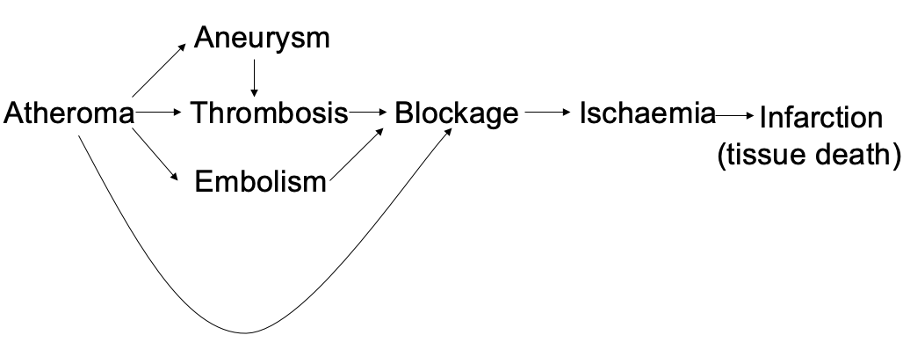
Atheroma
• characterised by the deposition of fibrous growths (plaques) within the walls of arteries, narrowing them
• Consequences of atheroma:
o Smaller vessels may become blocked
o Plaques may become the site of a thrombosis (blood clot forming over the atheroma part)
o A piece of plaque becomes detached from an atheroma and form an embolism – other things such as fat, bits of tumour or air bubbles can form emboli.
o A plaque can be the site of an aneurysm – common in the brain, can be clipped or filled with endovascular platinum coils under direct x-ray visualisation (seal off balloon so it doesn’t burst).
o Aneurysms are often the site of thrombosis
Cerebrovascular disease risk factors - non-modifiable
Non-Modifiable Risk Factors for Stroke:
• Age
• Sex
• Race/ethnicity
• Family history
• Beginning at age 55 years, the rate of stroke doubles every decade. Men are more likely than women to experience early stroke
Cerebrovascular disease risk factors - modifiable
Hypertension
Diabetes
Smoking
Hyperlipidemia
Carotid stenosis (narrowing of carotid artery)
Atrial Fibrillation (atrial beating faster than ventricles)
Ischemic vs haemorrhagic
• Ischemic stroke is caused by a blocked blood vessel,
haemorrhagic stroke is caused by a blood vessel that ruptures and bleeds.
Cerebral Ischemia symptoms
sudden weakness, numbness, or tingling on one side of the body.
Sudden difficulty speaking or understanding others.
Sudden vision problems.
Dizziness or a loss of balance.
A severe, sudden headache.
Confusion.
Aphasia
Diagnosis and treatment
Thrombolysis - dissolves blood clots
CT - haemmoraghic stroke
MRI - fast, clear image
Carotid duplex sonography
Aspirin - ischemic
Thrombectomy - removal of a thrombus (blood clot) under image guidance