Physiology Lecture 1
 Call Kai
 Learn
Learn Practice Test
 Spaced Repetition
 Match
Match Flashcards
 Knowt Play
1/26

There's no tags or description
Looks like no tags are added yet.
Name | Mastery | Learn | Test | Matching | Spaced | Call with Kai | Chat |
|---|
No analytics yet
Send a link to your students to track their progress
27 Terms
Cell physiology
structure of cells and their interaction
Cell
Structural and functional unit
Smallest unit that can carry on life processes
Cell components - Eukaryotic cells
Plasma membrane
Cell organelles
Interior of the cell: nucleus (genetic material) and cytoplasm (organelles suspended in cytosol)
Biological Membrane Composition
Double layer of lipid molecules with embedded proteins
May have different ratios of lipids and proteins based on function
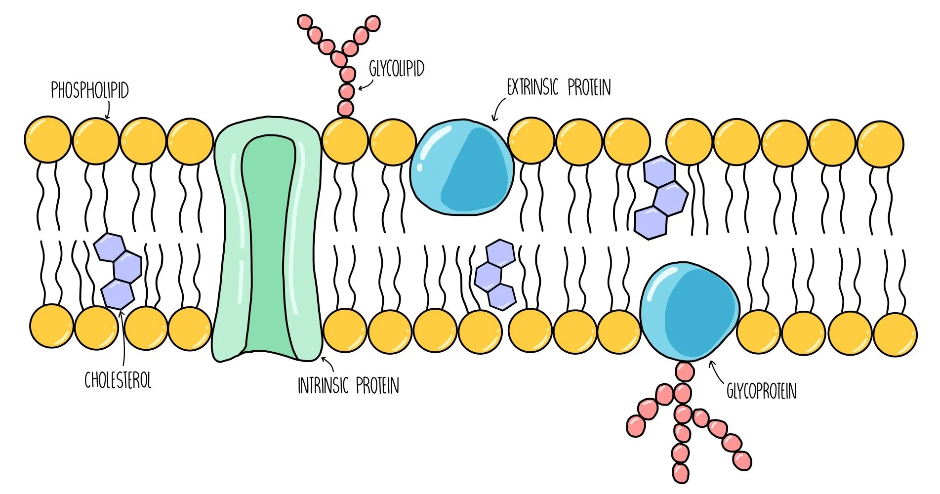
Lipids
Primarily carbon and hydrogens, some oxygen and phosphates
lipids are amphipathic: have polar and nonpolar regions
Phospholipids, steroids, and glycolipids
Phospholipids
Polar heads face out, nonpolar tails face eachother
Polar head group: phosphate attached to glycerol, nitrogen containing R group, glycerol backbone
Hydrophilic (loving)
Nonpolar tail: 2 fatty acids chains attached to flycerol backbone, made up of carbon and hydrogen
may be saturated or unsaturated (causes kinks/bends)
Hydrophobic (fearing)
Steroids
Cholesterol
Amphipathic (single OH group)
Sits between FA chains to maintain membrane fluidity
Glycolipids
lipids with short CHO chains attached
Part of the outer leaflet of the plasma membrane
Amphipathic
Short chain of monosaccharides extend from cell surface in ECF forming glycocalyx
Membrane proteins
Distributed throughout the membrane unequally, are responsible for selective permeability
Can be:
Integral/intrinsic
Peripheral/extrinsic
Glycoproteins
Integral/intrinsic membrane proteins
Amphipathic
May partially span membrane or fully span (transmembrane)
Peripheral/extrinsic membrane proteins
Not amphipathic
Only associate with polar regions on inner or outer surface of membrane
Influence cell shape and motility
Membrane proteins - glycoproteins
proteins with attached carbs
Found on extracellular surface forming glycocalyx
Specialized junctions
stabilize interaction and promote communication between cells
Desmosomes
adhering, anchor cells together
found in tissues subject to stretching
Formed from
plaques: proteins on cytoplasmic membrane that are anchoring points for cadherins
cadherins: transmembrane proteins that link cells
intermediate filaments: linked to plaque and anchor cytoplasmic surface to interior of cell for support

Tight junctions
link cells together and limit movement of molecules through spaces between cells, forces substance to cross plasma membrane to move through tissues
Composed of: occludins which attach to adjacent cells forming impermeable junctions
Found in: epithelial tissues specialized for molecular transport like the kidneys or intestines

Gap junctions
used for fast communication, transmembrane channels link cytoplasm of adjacent cells
Composed of: protein channels called connexons that link to connexons of other cells
Function: electrically and metabolically couple cells allowing small molecules and ions to move between
Found in: heart muscle to allow simultaneous contraction of cells

Nucleus
Function: store/transmit genetic information
Nuclear envelope: double layered membrane (2 phospholipid bilayers) with pores to allow selective movement
Nucleolus: site of ribosomal RNA synthesis; dense region, no membrane
Chromatin: consists of DNA and associated proteins found in the nucleus and condensed to chromosomes during division
Ribosomes
Non-membrane bound
Function: protein synthesis; AAs → proteins
Consist of 2 subunits (small and large) composed of protein and ribosomal RNA
separate in cytoplasm when not in use, functional when joined together
2 types:
Free in cytoplasm
Bound to ER
Endoplasmic Reticulum
Fluid filled membranous system; distributed throughout cytoplasm
2 types:
Rough ER: appears granular due to bound ribosomes; flattened sac in layers
Function: synthesis of proteins with bound functional ribosomes and post-translation modification in the RER
Smooth ER: appears agranular; branched tubular structure
Function: synthesizes lipids (FAs and steroids), has additional functions in specialized cells (ex. stores calcium in muscle cells, drug detox in liver cells)
Golgi Apparatus
Composed of 3-8 cisternae - sets of flattened and slightly curved membrane bound sacs stacked in layers
Associated with many vesicles
Function: further post-translational modification of proteins form RER
Sorts and packages proteins into vesicles
Lysosomes
Small oval sphere
Contain digestive enzymes: hydrolyze enzymes and breakdown polymners into small subunits
Can only function at pH of 5 (phospholipid membrane allows acidity without changing pH of entire cell)
Function: degrade extra/intracellular debris which are then removed by exocytosis or recycled
Peroxisomes
Small spherical
Contain oxidative enxymes: use oxygen to remove hydrogen from molecules and breakdown FAs, alcohol, and drugs (mainly liver and kidney)
Produces H2O2 which can be toxic so they also contain catalase which can breaks down H2O2 into water and oxygen products
Mitochondria
Converts energy from chemical bonds to ATP (usable form)
Double membrane: inner is folded into cristae to increase SA
Matrix contains products for cellular respiration
Have their own DNA - double stranded, circular, and have instructions for making proteins for cellular respiration
Cytoskeleton
Non-membrane bound
Made of protein filaments that are anchored to membrane and dispersed through cytoplasm; dynamic and can dissemble/assemble
Functions: maintain cell shape, organelle position, cell/organelle motility
Cytoskeletal filaments: Actin / Microfilaments
Protein subunit: G-actin (globular)
Support, cell shape, and movement
Cytoskeletal filaments: Intermediate filaments
Protein subunit: several (ex. keratin)
Support, mechanical strength
Desmosomes anchor intermediate filaments of neighbouring cells together
Cytoskeletal filaments: Microtubules
Protein subunit: tubulin, forms large hollow tubes
Support, acts as a track for organelles and vesicles to move along