BLOOD SUPPLY TO THE BRAIN
1/66
There's no tags or description
Looks like no tags are added yet.
Name | Mastery | Learn | Test | Matching | Spaced | Call with Kai |
|---|
No study sessions yet.
67 Terms
what are the cranial meninges
three layers of thin tissue that cover and protect the brain and spinal cord
how many layers does the cranial meninges consist of
three layers
name the layers of the cranial meninges from superficial to deep
dura mater
arachnoid mater
pia mater
which meningeal layer is the toughest and which is the thinnest
dura mater: toughest layer
pia mater: thinnest and most delicate of the three layers
what structure is the cranial meninges continuous with
the cranial meninges is continuous with the spinal meninges
how many layers is the dura mater composed of
two layers
what are the two layers of the dura mater and outline them
DURA MATER
outer layer
endosteal layer - tightly bound to the cranium
inner layer
meningeal layer - in contact with the arachnoid mater
outline the structure and function of dural folds
inward foldings/ projections/ reflections of the meningeal layer of the dura mater to form double layered folds
these folds help stabilise and protect the brain
what are the main dural folds
falx cerebri
tentorium cerebelli
falx cerebelli
diagram of the dural folds
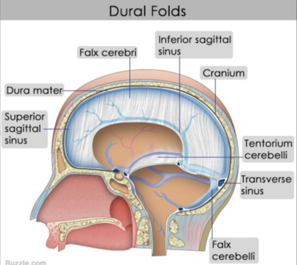
what does the falx cerebri separate
falx cerebri
separates the two cerebral hemispheres
what does the tentorium cerebelli separate
tentorium cerebelli
separates the occipital and temporal lobes
what does the falx cerebelli separate
falx cerebelli
separates the right and left cerebellar hemispheres (cerebellum)
beneath the tentorium cerebelli
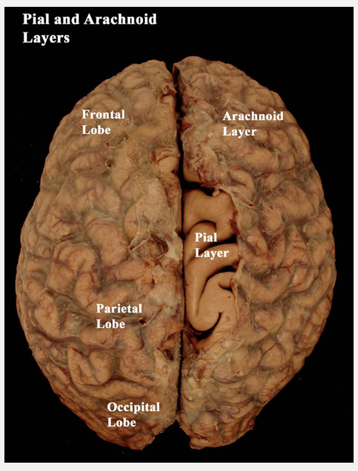
outline the arachnoid mater
ARACHNOID MATER
beneath the dura mater
covers the brain surface
does not follow the brain’s underlying folds
what is the subarachnoid space
extends between the arachnoid and pia mater
fibres extend from the arachnoid to the pia mater to bridge the gap
outline the pia mater
PIA MATER
thin layer tightly adhered to the brain
extends into every fold of the brain
loosely connected to the arachnoid mater via tiny fibres
not distinguishable from the surface of the brain using the naked eye - can be seen histologically
image of the arachnoid and pia mater layers
what is a potential space
a space that is not usually present in the brain of a healthy individual but can appear in disease/ trauma
what is a real space
a space in the brain that is normally present in a healthy individual
what type of space is the extradural space
the extradural space is a potential space between the dura mater and cranium
outline how the extradural space can appear
a potential space
between the dura mater and cranium
—
the meningeal arteries are embedded in the endosteal dura mater
rupture of these vessels results in bleeding into the extradural space
this causes rapid localised increase in intracranial pressure
could lead to coning
what type of space is the subarachnoid space
a real space between the arachnoid and pia mater
outline the subarachnoid space
contains cerebrospinal fluid, arteries to the brain, veins from the brain
large space, therefore a subarachnoid haemorrhage takes time to spread - symptoms are usually 24-48h after injury
increased pressure will eventually cause coning
what is a function of the CSF other than cushioning the brain
the CSF reduces the apparent weight of the brain
1400g in air compared to 50g in CSF
where are venous sinuses located
venous sinuses
spaces between reflections of the meningeal layer of the dura mater
outline venous sinuses
veins containing deoxygenated blood moving away from the brain empty into venous sinuses
venous sinuses drain into the internal jugular veins
what is the function of emissary veins
emissary veins
emissary veins passing through the skull connect venous sinuses with extracranial blood vessels
what does the course of emissary veins increase the risk of
potential spread of infection
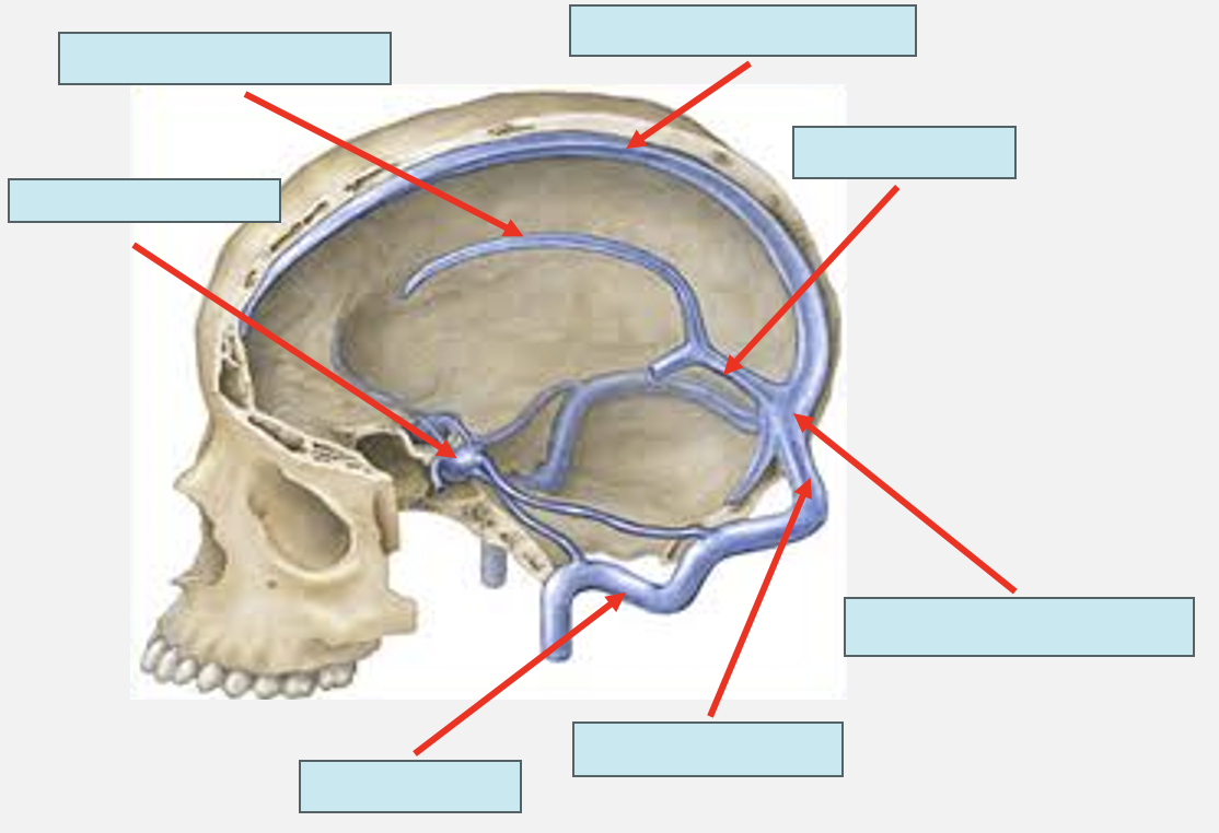
label the venous sinuses
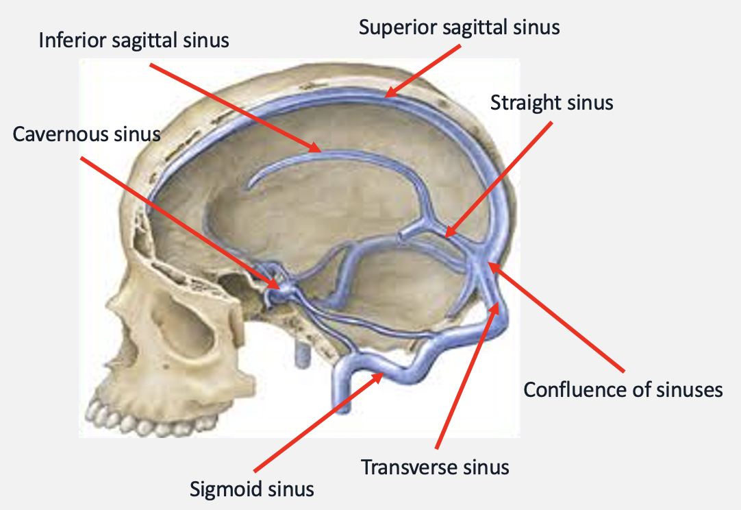
which venous sinuses are paired and which are unpaired
paired
transverse sinus
sigmoid sinus
cavernous sinus
unpaired
superior sagittal sinus
inferior sagittal sinus
straight sinus
what is the ultimate function of venous sinuses
venous drainage - the system of veins that collect deoxygenated blood from tissues and organs and returns it to the heart
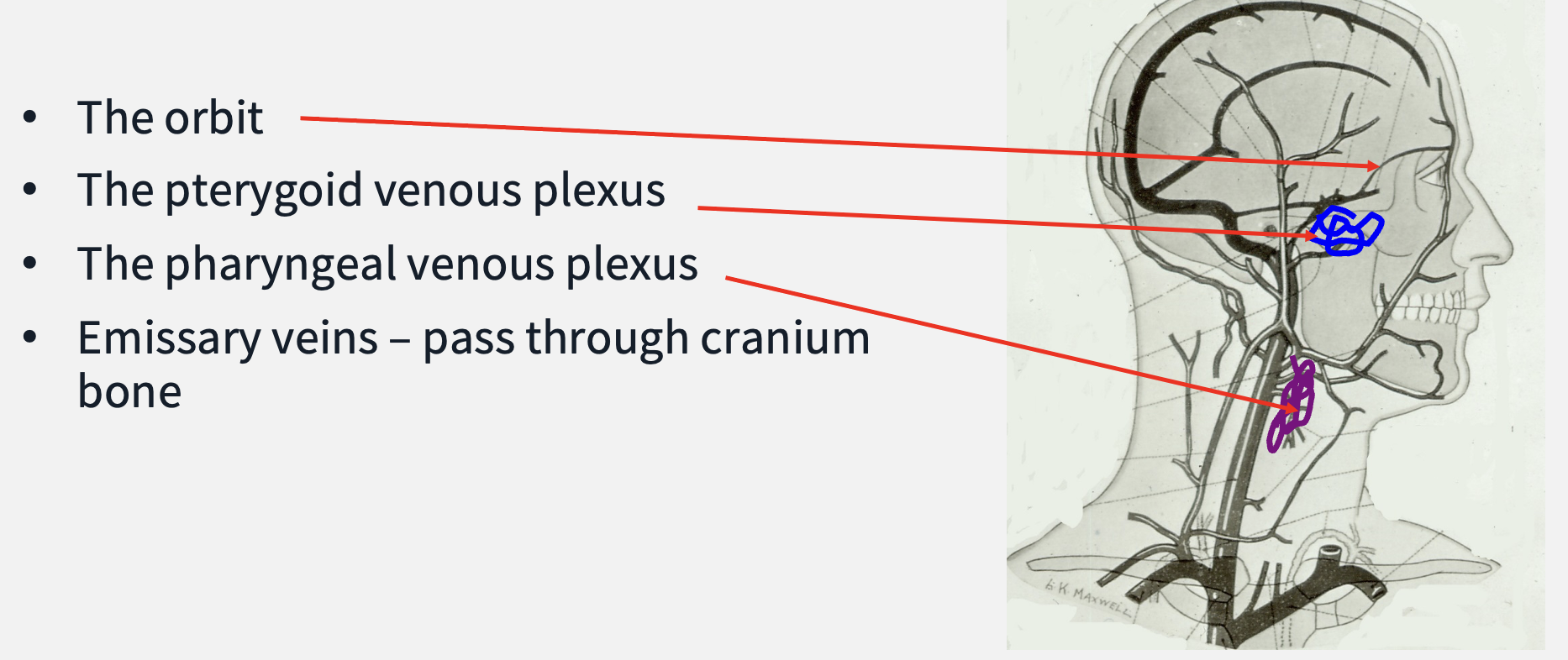
through which structures do venous sinuses connect with superficial and deep veins
the orbit
the pterygoid venous plexus - drains into maxillary vein then heart
the pharyngeal venous plexus - drains into internal jugular vein then heart
emissary veins - pass through the cranium bone
diagram of location of pterygoid and pharyngeal venous plexuses
the brain is highly _____________ active but has no _________ reserves
the brain is highly metabolically active but has no metabolic reserves
which ONE nutrient is the brain dependent on
glucose
what is the term for a lack of glucose
hypoglycaemia
what other element is the brain dependent on
oxygen
what does the figure ‘≈ 2 minutes’ refer to
within how long oxygen deprivation to the brain can cause irreversible damage
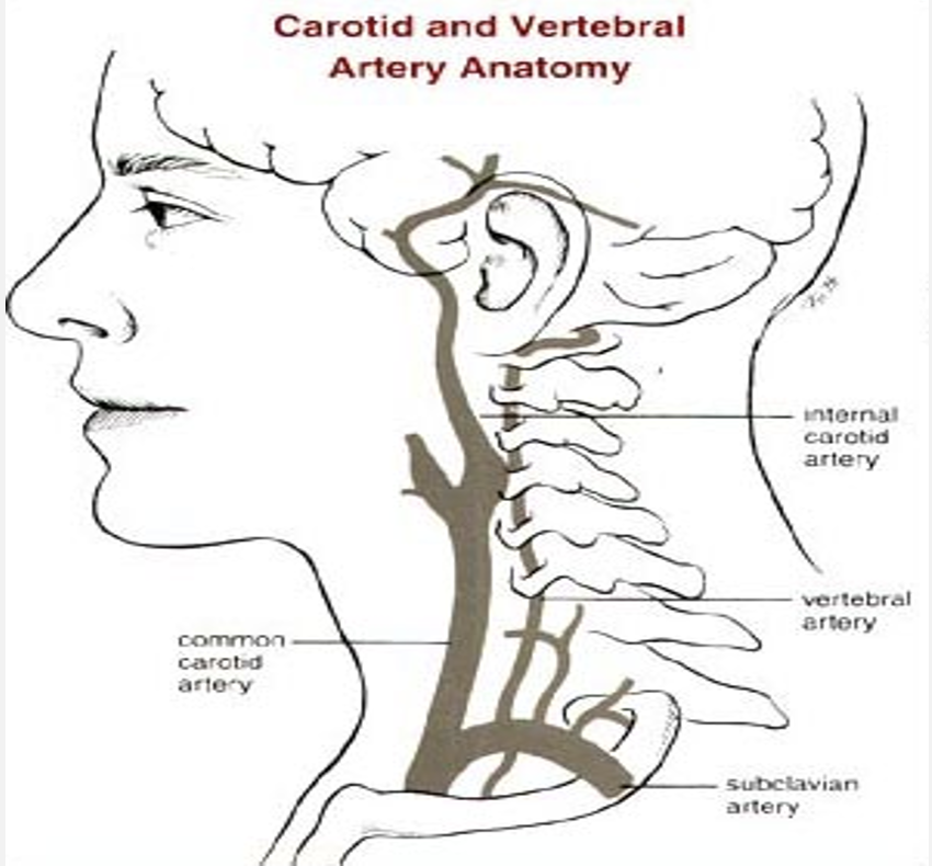
where does the brain receive its arterial supply from
internal carotid arteries - enters brain via carotid canals
vertebral arteries - branches of the subclavian arteries
what is the collective term given to the arteries supplying the brain i.e. the internal carotid and vertebral and what does it mean
true end arteries: one artery supplies a specific area and there is no alternative blood supply
if the artery is damaged or diseased the area will be deprived of nutrients and oxygen and will die
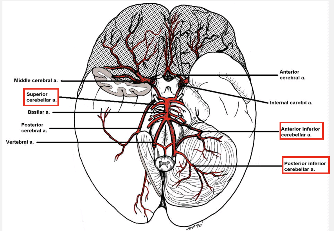
carotid and vertebral artery anatomy diagram
outline the vertebral arteries
vertebral arteries
run either side of the medulla and join at the level of the pons to form a single basilar artery
vertebrobasilar system
as they travel across the pons the basilar artery gives off short medullary and pontine arteries
where do the cerebellar arteries arise from and what do they supply
cerebellar arteries
arise from the vertebrobasilar system
supply the cerebellum and lateral aspects of the brainstem
how many pairs of cerebellar arteries are there
there are three pairs of cerebellar arteries
outline damage to the cerebellar arteries
parts of the brainstem supplied by the cerebellar arteries contain the cranial nerve nuclei
damage to the cerebellar arteries therefore results in:
cerebellar ataxia
cranial nerve signs and symptoms depending on the level of brainstem that is affected
what is lateral medullary syndrome
loss of blood supply to the lateral part of the medulla
how does lateral medullary syndrome come about
manifestation of blockage of posterior inferior cerebellar artery (PICA) or the vertebral artery
can result in cerebellar ataxia
which cranial nerve nuclei can be damaged as a result of lateral medullary syndrome and what are the consequences
damage to nuclei of CN IX (glossopharyngeal)
no sensation to pharynx
no swallowing reflex » dysphagia
damage to nuclei of CN X (vagus)
no sensation to larynx - no cough reflex
no motor innervation to pharyngeal constrictor muscles » dysphagia
to motor innervation to larynx » dysphonia
which arteries supply the cerebral hemispheres and how many pairs are there
cerebral arteries - there are three pairs
which artery supplies the majority of the cerebral hemispheres
the internal carotid arteries
outline the internal carotid arteries
internal carotid arteries
paired
when they enter the cranium through the base of the skull they almost instantly divide into:
anterior cerebral arteries (paired)
middle cerebral arteries (paired)
where do the posterior cerebral arteries arise from
the vertebrobasilar system
the basilar artery eventually bifurcates and gives rise to the posterior cerebral arteries
diagram of the cerebral arteries
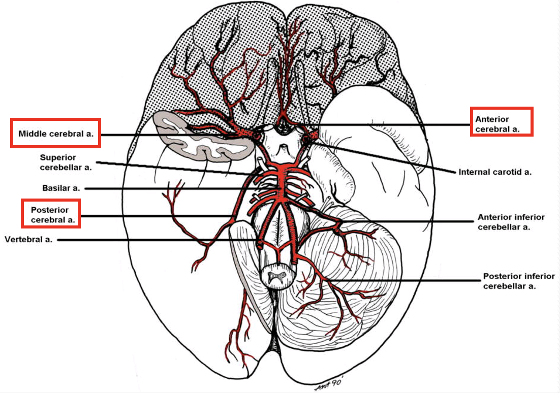
diagram showing the supply of cerebral arteries to the corresponding area of the cerebral hemisphere
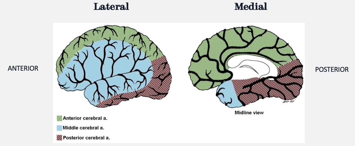
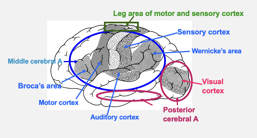
functional areas of the cortex supplied by the anterior cerebral artery
leg area of motor and sensory cortex
loss of blood flow would affect the contralateral side of the body

functional areas of the brain supplied by the middle cerebral artery
Broca’s area - Broca’s aphasia if blood flow is lost
Wernicke’s area - Wernicke’s aphasia if blood flow is lost
sensory cortex
motor cortex
auditory cortex

functional areas of the brain functional areas of the brain supplied by the posterior cerebral artery
visual cortex
loss of blood flow would affect vision
what is the medical term for a stroke
cerebrovascular accident (CVA)

what are the types of stroke and outline them
ischemic stroke
due to a blood clot that blocks the flow of blood
haemorrhagic stroke
due to a burst blood vessel (aneurysm)
can also be due to trauma (tearing)
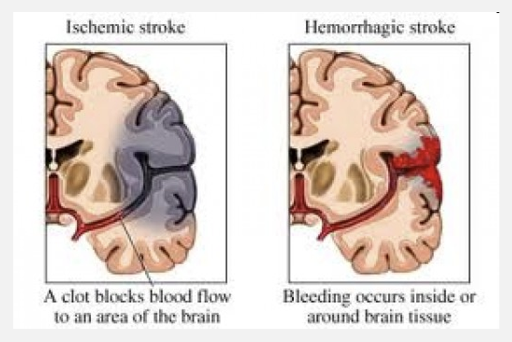
consequences of an ischemic stroke
brain function may be lost in areas where blood supply is permanently absent
not much resolution

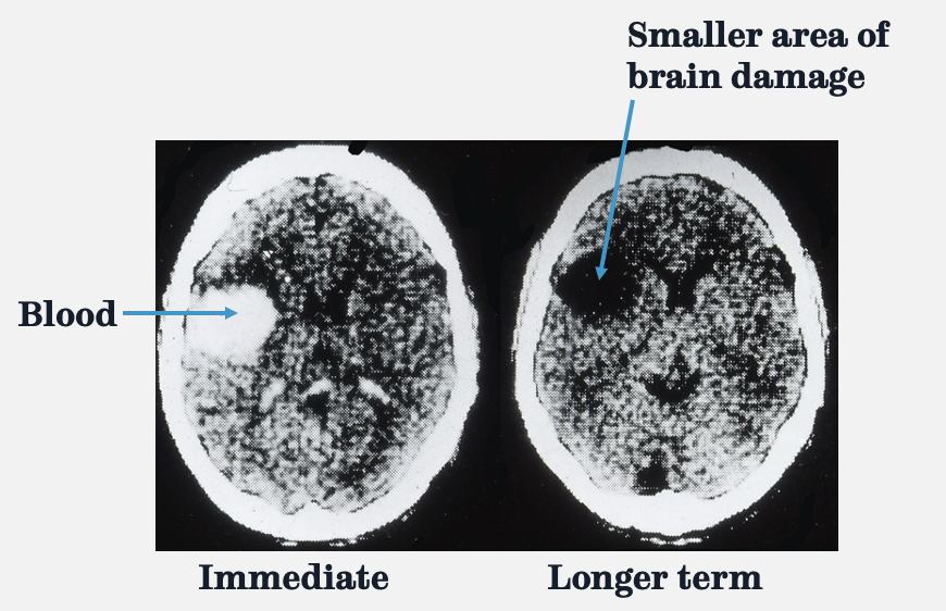
consequences of a haemorrhagic stroke
short term effects are worse than long term
as the mass of the hematoma decreases, function may return to the affected area
brain scan showing damage
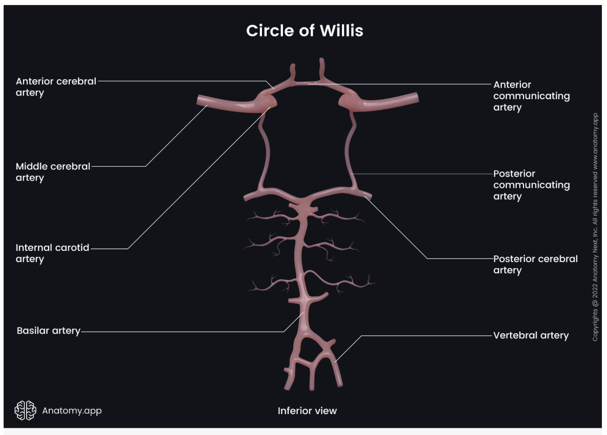
what is the Circle of Willis (CoW)
Circle of Willis
a ring shaped network of arteries at the base of the brain
connects the internal carotid arteries with the vertebrobasilar system
function of the CoW
can act as an alternative blood supply if major vessels are damaged
in what % of people is the CoW complete in
≈ 60% of people
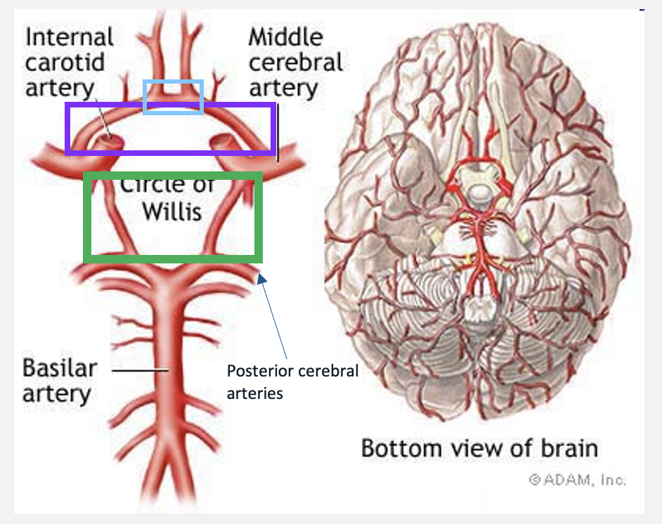
label the coloured boxes
blue rectangle: anterior communicating artery (unpaired)
purple rectangle: anterior cerebral arteries
green rectangle: posterior communicating arteries
complete CoW diagram