Chapter 31: Vascular Technology
1/15
There's no tags or description
Looks like no tags are added yet.
Name | Mastery | Learn | Test | Matching | Spaced | Call with Kai | Chat |
|---|
No analytics yet
Send a link to your students to track their progress
16 Terms
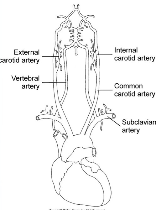
Extracranial Cerebrovascular System
Consists of common carotid arteries (CCA), internal carotid arteries (ICA), external carotid arteries (ECA), and vertebral arteries (VA); right side: innominate artery branches into CCA and subclavian arteries; left side: CCA and subclavian arteries arise separately from aortic arch
Common Carotid Artery (CCA)
Part of extracranial cerebrovascular system; courses in anterolateral aspect of neck and lateral to trachea, esophagus, larynx, and pharynx; bifurcates into ECA and ICA usually at level of superior thyroid cartilage - ICA is posterior and lateral to ECA in most patients; bulb-dilation of vessel is usually at origin of ICA; diameter should be 0.5-0.6cm
Internal Carotid Artery (ICA)
Part of extracranial cerebrovascular system; no extracranial branches; intracranially (feeds brain), give rise to ophthalmic artery; is divided into 4 major segments:
cervical
Petrous
cavernous
Cerebral
diamter should be 0.4-0.5cm

External Carotid Artery (ECA)
Part of extracranial cerebrovascular system; demonstrates extracranial branches, including superior thyroid, ascending pharyngeal, lingual, facial, occipital, posterior auricular, superficial temporal, and maxillary arteries (feeds face); diameter should be 0.3-0.4cm

Vertebral Arteries (VA’s)
Part of extracranial cerebrovascular system; first branch of subclavian arteries; pass cranially through foramina of transverse processes of upper 6 cervical vertebrae; 2 vertebral arteries join to form basilar artery; has antegrade flow; diameter should be 0.2-0.3cm
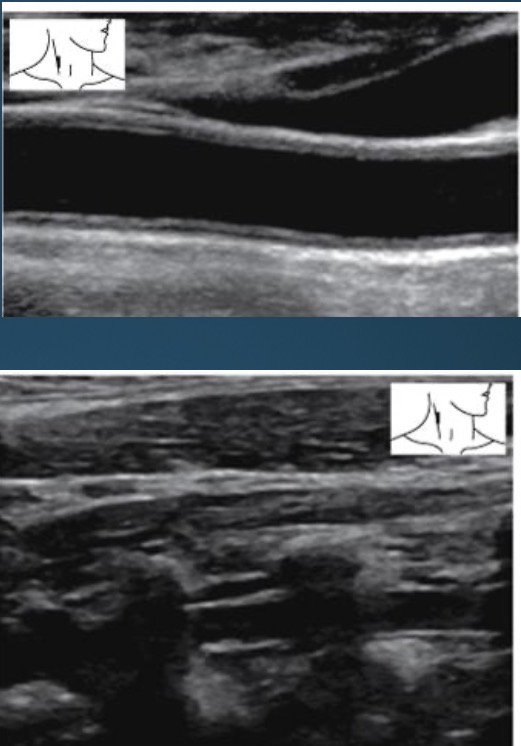
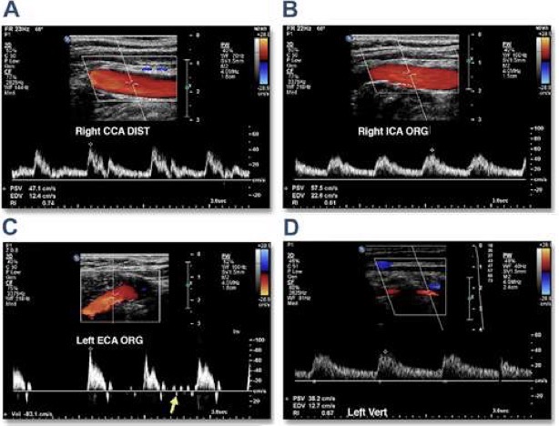
Sonographic Appearance of Extracranial System
anechoic lumen; thin hyperechoic walls; vertebral arteries course through fossae of transverse vertebra process
Normal Variants of Extracranial System
Absence of innominate artery with right subclavian and common carotid originating from arch
common origin of innominate and left common carotid arteries
presence of a left innominate artery
aorta may arch to the right with normal arterial arrangements reversed
absence of CCA, with ICA and ECA arising from arch
absence of carotid bifurcation
agenesis of either ICA or ECA
Hemodynamics
CCA, ICA, VA: low resistance vessels (arteries that supply organs that need constant forward blood flow or perfusion)
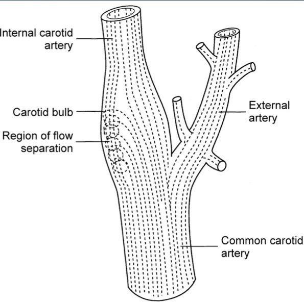
Carotid Bulb: increase size results in pressure flow gradient
boundary layer separation
flow entering ICA and flow reversing towards posterolateral wall
ECA: high resistance vessel (arteries with low or reversed flow in diastole that supply organs not in constant demand for blood flow
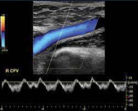
Color Flow Doppler: used to evaluate lumen for narrowing, plaque, or abnormal flow; direction of flow determined
Spectral Doppler: provides flow velocity measurements used to evaluate for stenosis; PSV and EDV using 60 degree angle or less
used to document areas of flow disturbance seen with stenosis, occlusion or trickle flow
Extremity Venous System
Veins of upper and lower extremity are divided into: deep, superficial, and perforating; deep veins accompany arteries and share same names
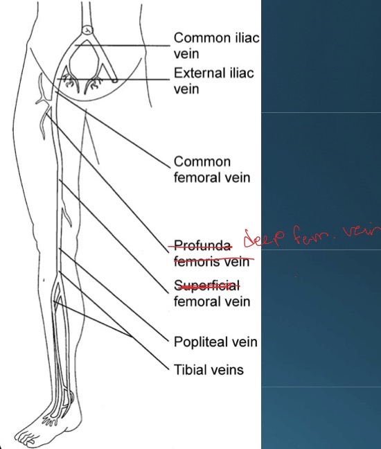
Deep Lower Extremity Venous System
Deep System: anterior tibial veins, posterior tibial veins, peroneal veins, popliteal vein, femoral vein, common femoral vein, deep femoral vein, external iliac vein, common iliac vein; most common anomalies are duplication of popliteal and/or femoral veins, duplication of distal segment of femoral vein, subsequently uniting to form single vein in mid proximal thigh, or presence of 3 or more tibial, popliteal, or femoral veins
left common iliac vein courses posterior to right iliac artery, accounting for higher incidence of left lower extremity deep vein thrombosis and swelling; normally, there’s no valves in common iliac veins
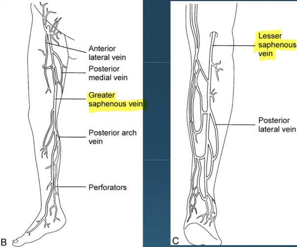
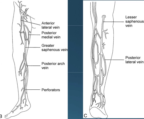
Superficial Lower Extremity Venous System
greater saphenous vein (longest vein in body), and small saphenous vein
Perforating veins in Lower Extremity Venous System
In the calf, there are 8 perforating veins
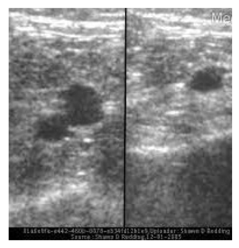
Sonographic Appearance of Lower Extremity Venous System
Lumen is anechoic and walls of vein will coapt entirely with gentile transducer pressure
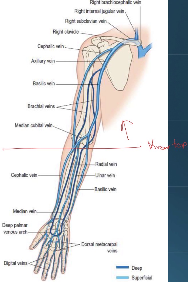
Upper Extremity Venous System
Deep: brachial veins (paired), axillary vein, and subclavian vein
Superficial: Basilic and cephalic veins; they travel from wrist to shoulder and communicate with each other at antecubital fossa via the median cubital vein
Venous Hemodynamics
Able to undergo large volume changes with little change in transmural pressure (veins ability to change shape allows it to increase blood volume without changing pressure gradient); hydrostatic pressure increases when standing, decreases or negligible supine; calf muscle pump/competent venous valves; changes in intrathoracic pressure
Color Doppler: assist with vessel location and flow disturbance
Spectral Doppler: spontaneous, phasicity, augmentation, competency, and pulsatility
Common Diagnostic Tests
Vascular angiography
Computed tomography angiography
Magnetic resonance angiography