BIO130 second half
 Call Kai
 Learn
Learn Practice Test
 Spaced Repetition
 Match
Match Flashcards
 Knowt Play
1/207

There's no tags or description
Looks like no tags are added yet.
Name | Mastery | Learn | Test | Matching | Spaced | Call with Kai | Chat |
|---|
No analytics yet
Send a link to your students to track their progress
208 Terms
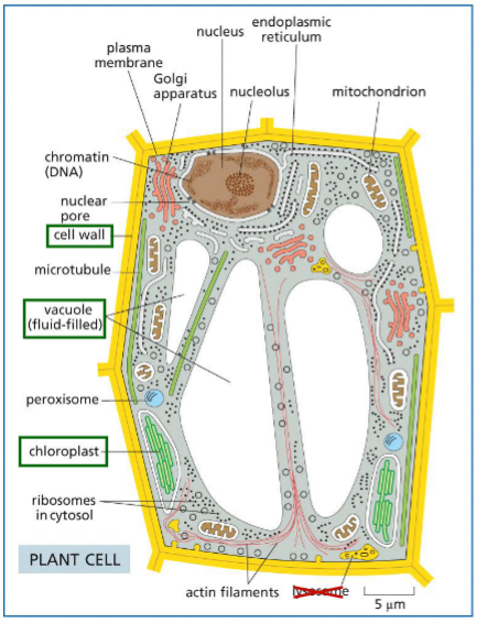
Lec 13: membrane structure: animal vs plant cell specialties
Animal cell:
Extracellular matrix
Lysosomes — degradation of cellular components
.
Plant cell:
Cell wall — shape and anti-stress
Vacuoles — degradation (like lysosome) and storage
Chloroplast — photosynthesis
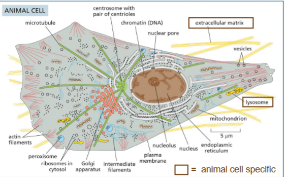
Cytoplasm, Cytosol, and Lumen
Cytoplasm
contents of the cell outside the nucleus (cytosol + organelles)
.
Cytosol
Aqueous part of the cytoplasm (no organelles)
.
Lumen
Inside of organelles
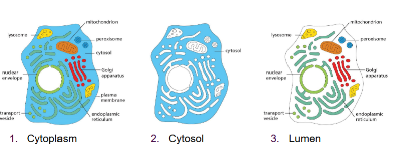
Cellular functions at membranes
Divide cell into compartments
Scaffold for biochemical activities (e.g. mitochondria)
Selectively permeable barrier + transport proteins regulate transport
Receptors respond to external signals
Proteins for interactions between cells
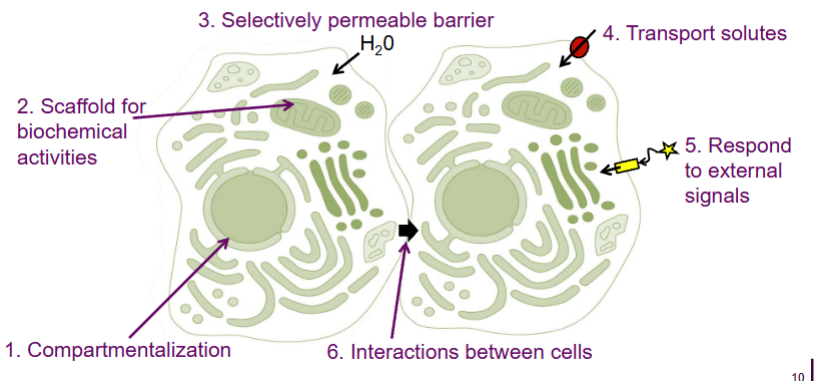
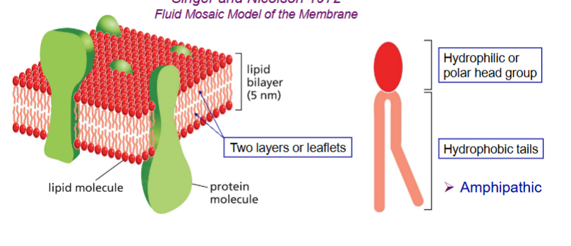
Membrane bilayers
Bilayer
composed of amphipathic molecules (hydrophilic and hydrophobic parts)
hydrophilic head
hydrophobic tail
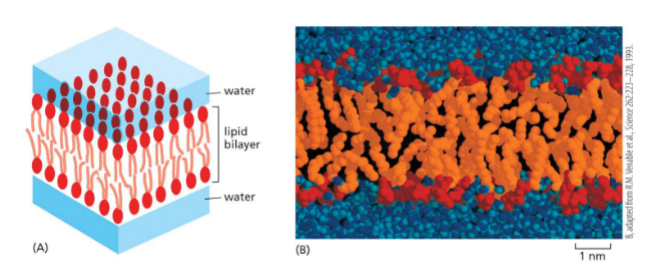
spontaneously assemble into bilayer
hydrophilic head interacts with aqueous outside
hydrophobic tails interact with eachother on inside
proteins and other stuff inside —→ fluid mosaic model
Types of membrane lipids
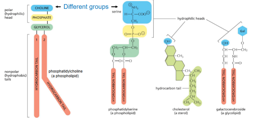
Phospholipids, sterols, glycolipids
Different groups on the molecule
All are amphipathic
Phospholipids
Many types of phospholipids
Hydrophilic head:
Different groups
Phosphate
Glycerol
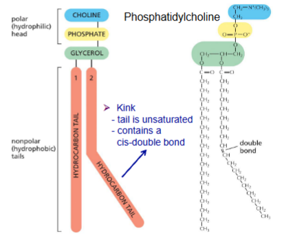
Hydrocarbon tail
length: 14-24
saturated/unsaturated
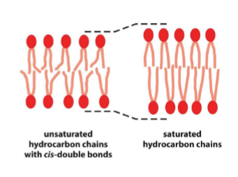
cis double bond leads to kink
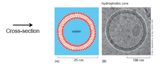
Sealed compartments of bilayers & Liposomes
After assembly into bilayer, form into a sphere
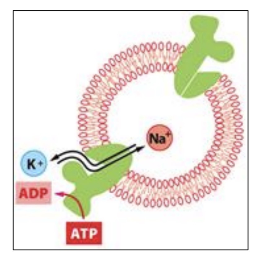
Liposomes: artificial lipid bilayers
Used for
studying lipid movement
protein properties
drug delivery into cells
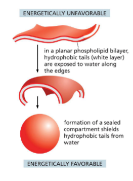
Cell membranes are fluid
Can be deformed without damage
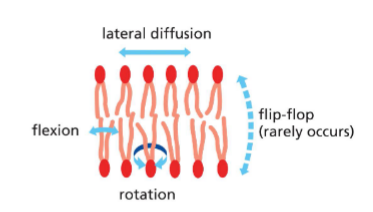
Phospholipid movement
(leaflet: one side of bilayer)
Phospholipids in one leaflet can
move laterally
rotate
flex
Rarely..
move from one leaflet to another (flip flop) on their own
hydrophilic head has to move through hydrophobic core
not energetically favourable
.
Membrane fluidity regulated to keep it consistent
Membrane has many proteins
Factors affecting membrane fluidity
Temperature
lower temp — less fluid
more important for single-celled
Composition
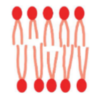
phospholipid saturation — unsaturated more fluid (kinks give space)
tail length — some shorter more fluid
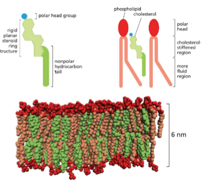
lipid composition — cholesterol stiffens, less permeable to water
Sterols — Cholesterol
Animals — mainly cholesterol
Plants — plant sterols, some cholesterol
.
There can be up to 1:1 ratio of cholesterol and phospholipids
decreases mobility of tails
membrane less permeable to polar molecule
thickness of bilayer increases
Lipid movement to other leaflet — scramblase
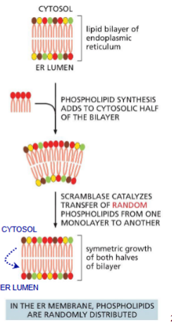
Enzyme in ER membrane
catalyzes random flip-flops
Why?
phospholipids are synthesized in cytosolic leaflet of ER
to maintain symmetry
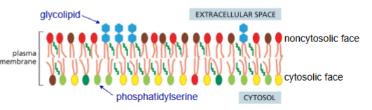
Asymmetry of the lipid bilayer
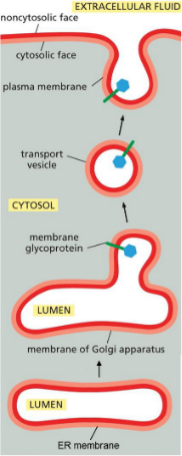
Membranes always have same orientation
cytosolic leaflet
noncytosolic leaflet
Membrane proteins also retain orientation
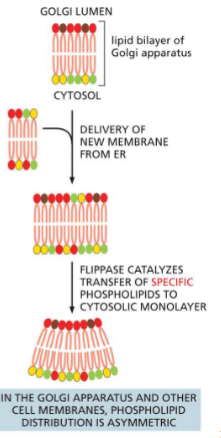
Lipid movement to other leaflet — Flippases (in? to?)
Enzyme in golgi membrane
flip-flop of specific phospholipids to cytosolic leaflet
some can bind cytosolic proteins at plasma membrane (e.g. protein kinase C)
Asymmetry of the lipid bilayer — glycolipids and glycoproteins (in? to?)
Formed by adding sugar groups to lipids/proteins on luminal face of golgi
Ends up on plasma membrane or other organelles on noncytosolic face
Protects membrane from harsh environments
(extracellular, lysosomes, etc)
Lec 14: membrane proteins types overview
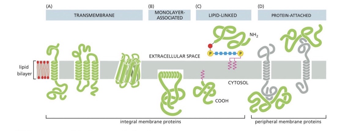
Carry out functions in the membrane
Integral
directly attached to bilayer
transmembrane/monolayer/lipid covalent attached
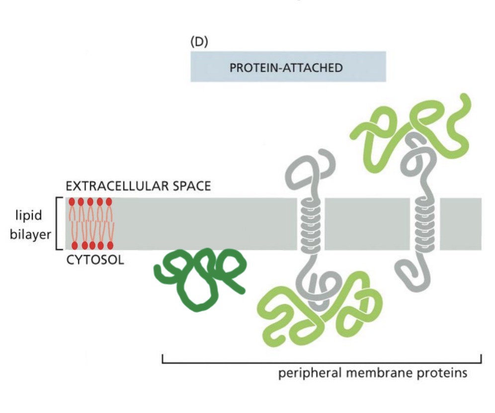
Peripheral
protein attached
Non-covalent interactions
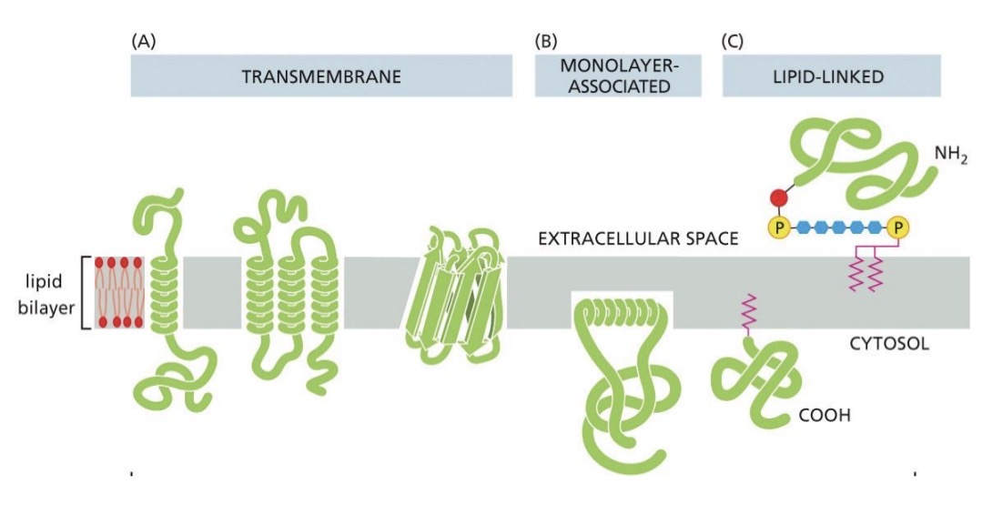
Integral membrane proteins + extraction methods
Proteins directly attached to lipid bilayer
inserted into bilayer
covalently attached to lipid inserted into bilayer
Extraction methods use detergents to denature bilayer
Peripheral membrane proteins
Not inserted into membrane, on either face of membrane (non-covalent interactions)
bound to other proteins
bound to lipids
Gentle extraction methods used, lipid bilayer intact
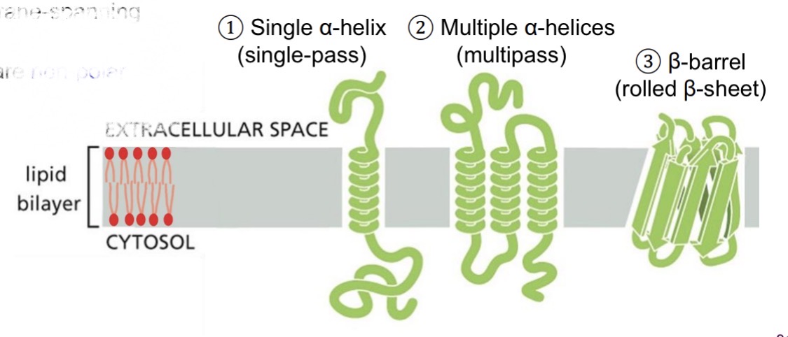
Integral—transmembrane proteins/membrane-spanning proteins
Amphipathic proteins
Hydrophobic membrane-spanning domains (non-polar side chains)
Hydrophilic domains in aqueous outside (polar side chains)
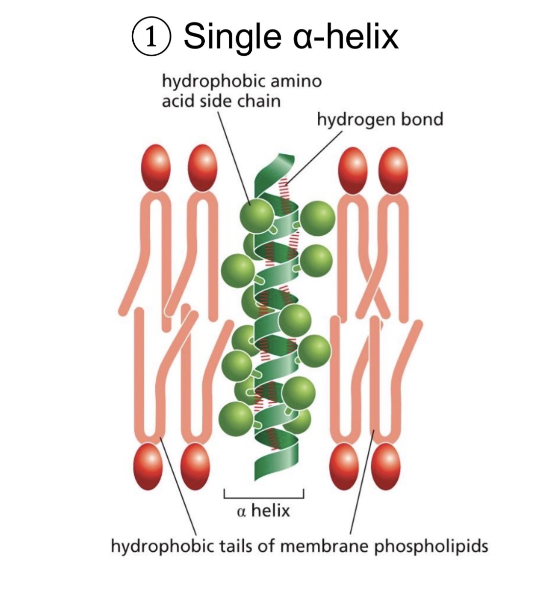
Transmembrane protein—single a-helix (single pass)
Membrane spanning helix: ~20 hydrophobic amino acids
hydrophobic side chains in the membrane
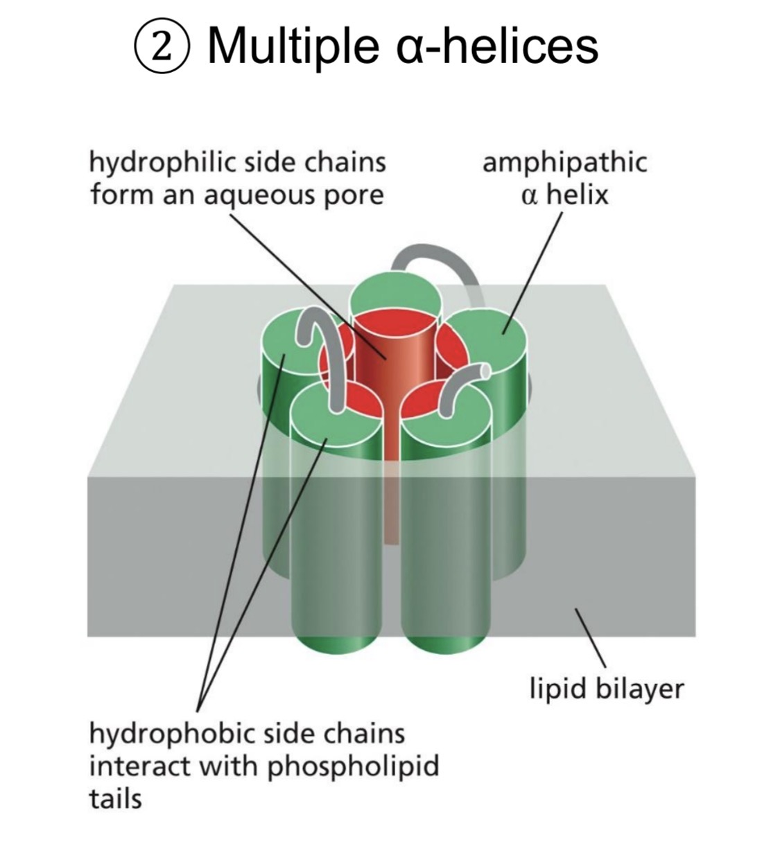
Transmembrane protein—multiple a-helices (multipass)
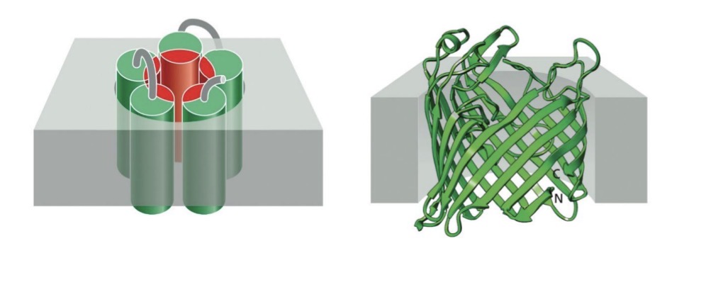
Multiple a-helices
hydrophobic side chains on outside of channel
Hydrophilic side chains on inside
Forms aqueous channel
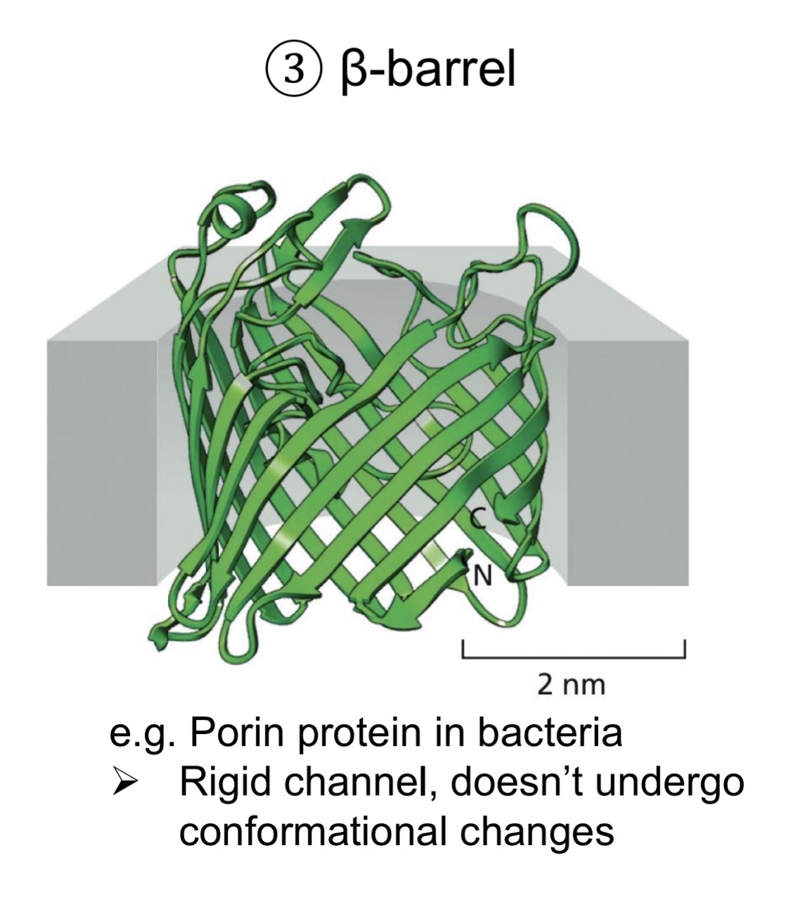
Transmembrane proteins—beta-barrel (rolled beta sheet)
Rigid channel, no conformational changes
hydrophobic side chains on outside
Hydrophilic side chains on inside
Forms aqueous channel
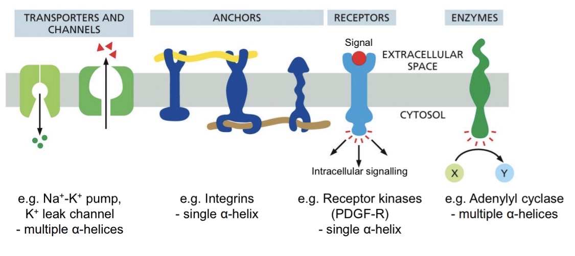
Overview of transmembrane protein functions
Each protein has a specific orientation—essential for function
transporters and channels (sodium/potassium pump)
anchors (integrins)
receptors (receptor kinases)
enzymes (adenylyl cyclase)
How are transmembrane protein structures identified?
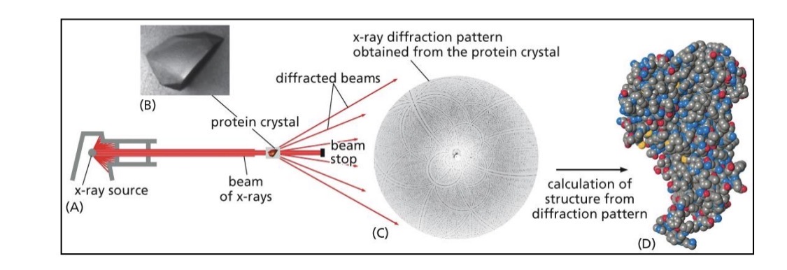
X-ray crystallography
purify protein crystal
shine x-rays
diffraction pattern
computer program gives structure
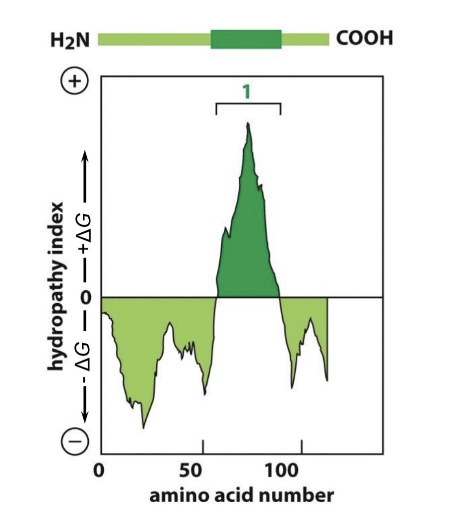
Hydrophobicity plots
find sections of hydrophobic/hydrophilic amino acids
from N-terminus to C-terminus
above is hydrophobic, below is hydrophilic
look for peaks 20-30 amino acids wide
how many peaks is how many transmembrane domains
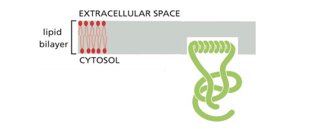
Integral—monolayer-associated proteins (a-helix)
Only attached to one layer/leaflet
amphipathic a-helix
hydrophobic side chains in leaflet
hydrophilic side chains on outside
specialized functions
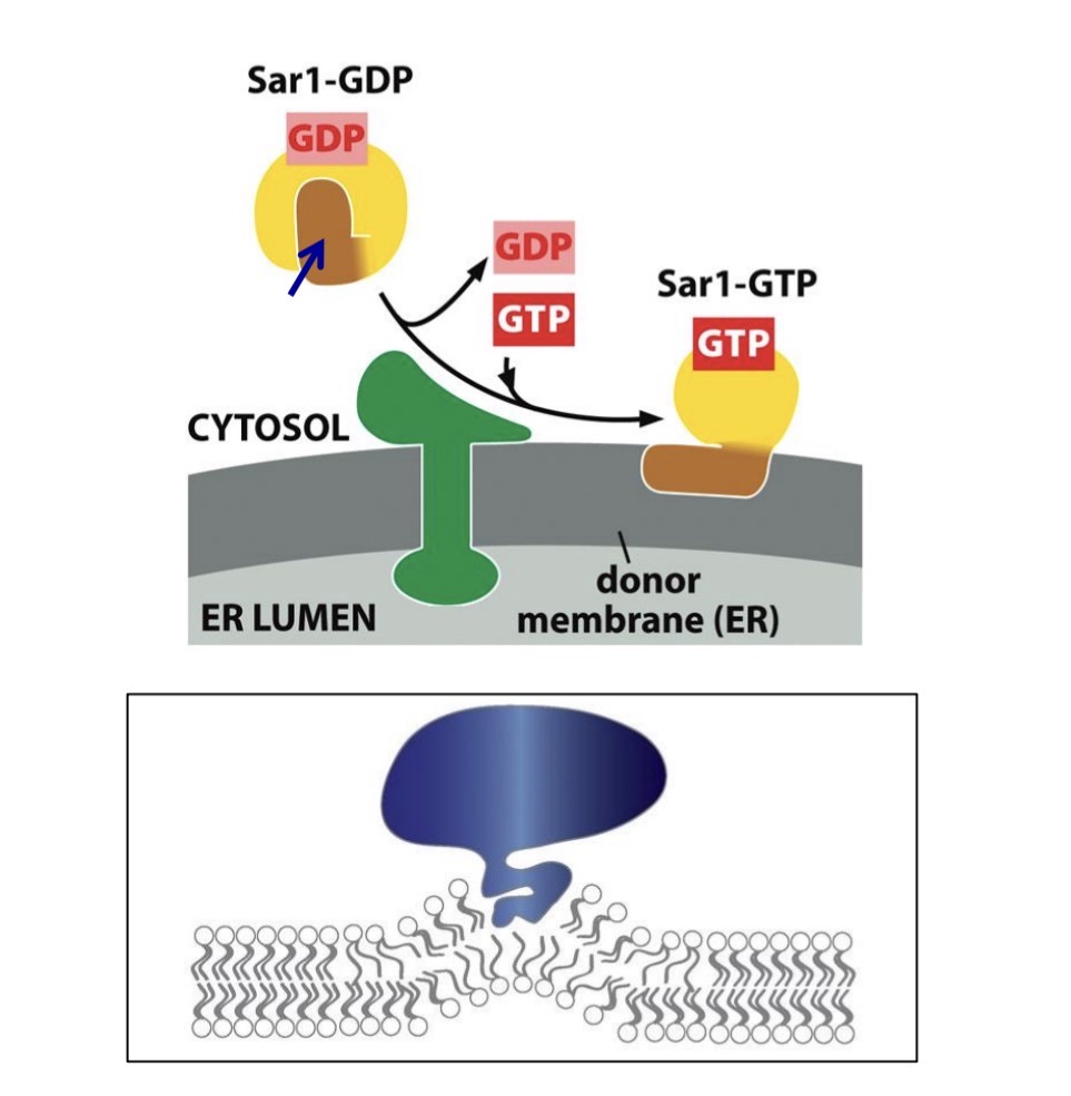
Integral—monolayer-associated proteins: Sar1 example
Protein in membrane bending to form vesicle
inserts into cytosolic leaflet
causes bump in the membrane
vesicle budding at the ER
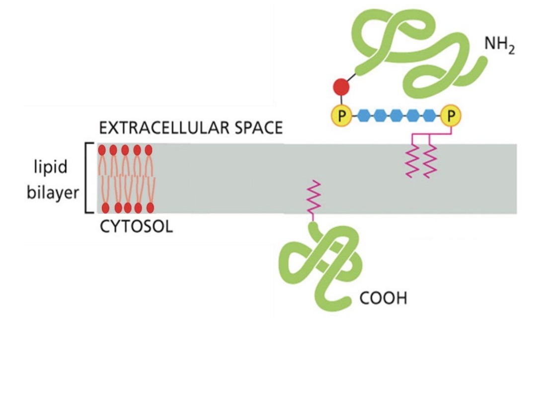
Integral—lipid-linked membrane proteins (GPI & lipid anchored)
For specific protein functions/roles, need to be on specific leaflet
.
Protein with GPI anchor
synthesis in ER lumen
attach to non-cytosolic face
end up on outside of cell
.
Protein with lipid anchor
add to cytosolic face
end up on inside of cell
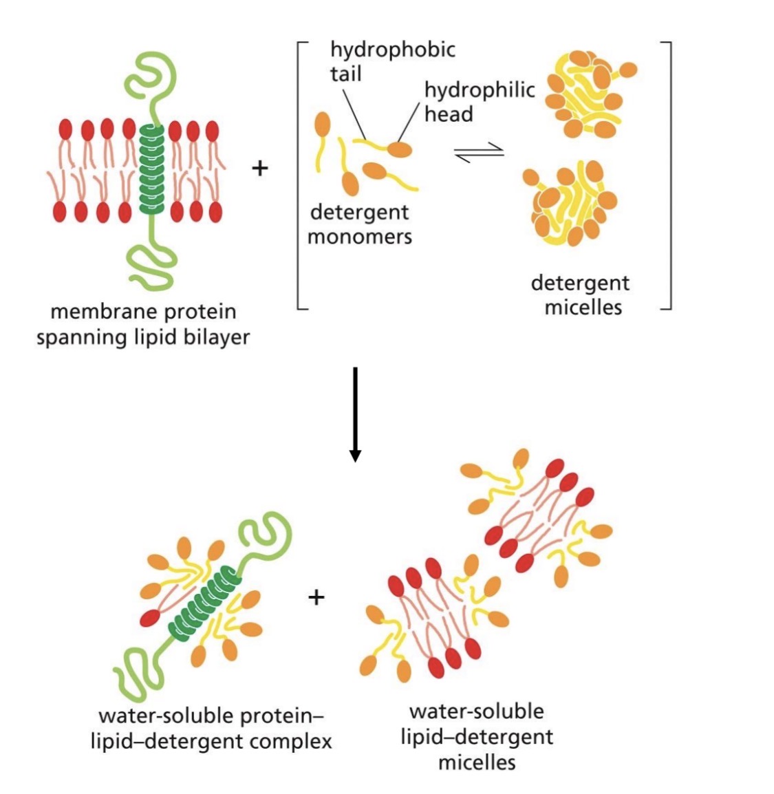
Techniques: Extraction of membrane proteins
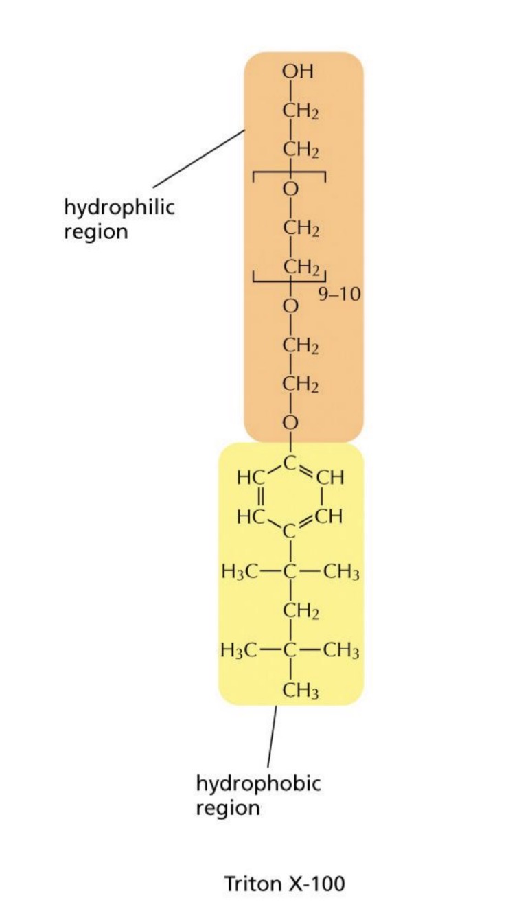
Mild detergent: denatured bilayer but not protein
hydrophilic head and hydrophobic tail
forms micelles in aqueous solution
.
forms water soluble complexes with amphipathic protein and membrane lipids
Techniques: Studying the properties — liposomes
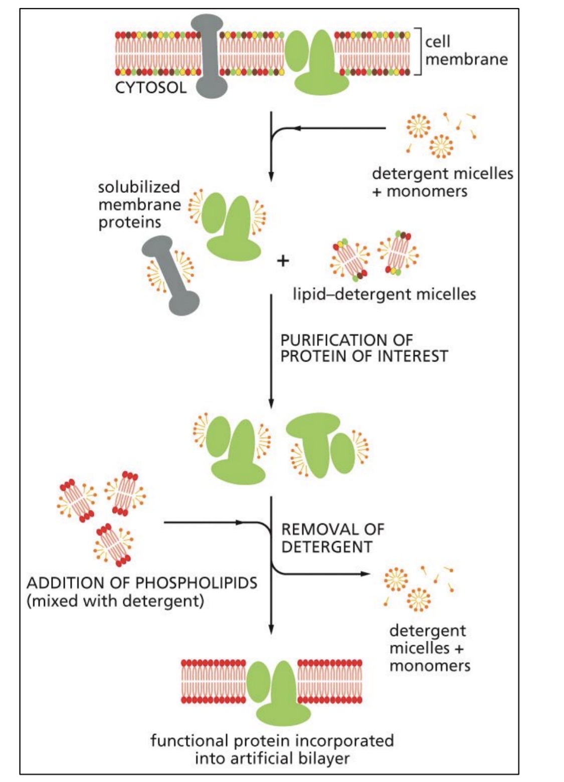
detergent to denature bilayer
purify protein
add artificial lipids
remove detergent
form liposome
study protein
Techniques: lateral diffusion of membrane proteins (GFP)
Some proteins need to move, some need to be immobile (~range)
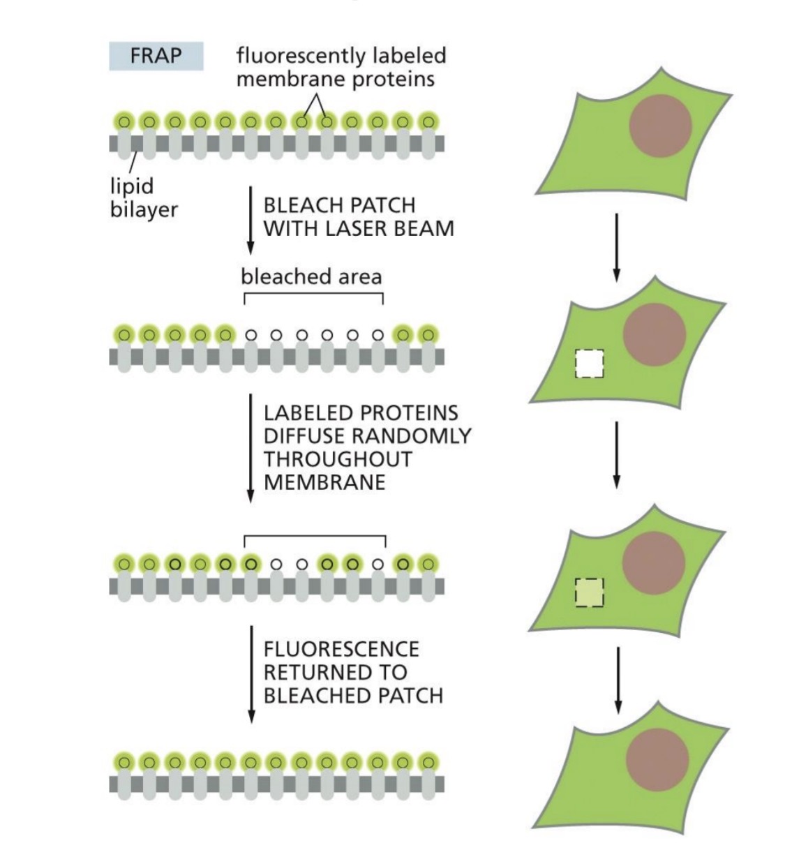
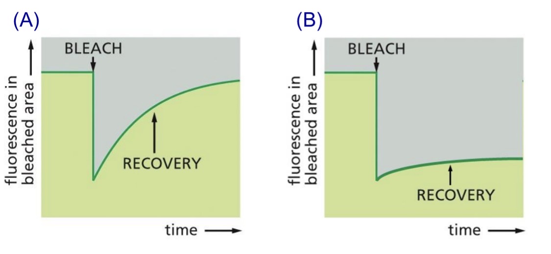
Study lateral diffusion — Fluorescence recovery after photobleaching
Protein fused to GFP (green-fluorescent protein) or labelled with fluorescent antibody
Photobleach area
Recovery: Measure rate of diffusion/rate of fluorescence recovery
.
Not anchored, mobile — recovery
Anchored, immobile — no recovery
Lec 15: Permeability of the lipid bilayer overview (can control…)
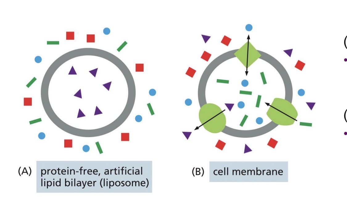
Artificial bilayer
impermeable to most water soluble molecules
Cell membrane
membrane transport proteins to transfer specific molecules — facilitated transport
can control selectivity and directionality
Movement across the bilayer — without proteins
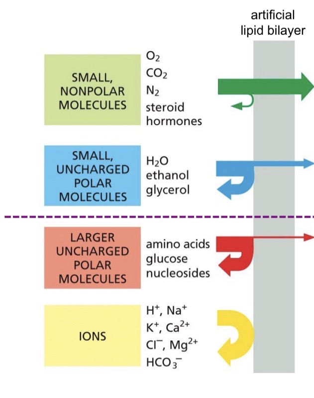
Permeable (simple diffusion down the concentration gradient)
small non polar molecules very good, fast
small uncharged polar not very well
Impermeable (require membrane proteins)
Larger uncharged polar molecules verrry little
Ions
What do transport proteins transport
Transmembrane transport protein
creates protein-lined path across membrane
transport polar and charged molecules
Transport proteins are selective
transports specific molecules
Different cell membranes have different proteins
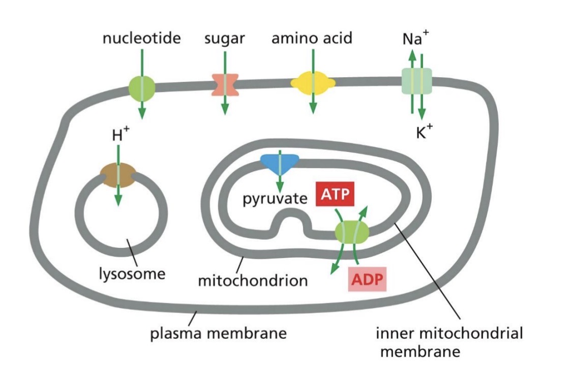
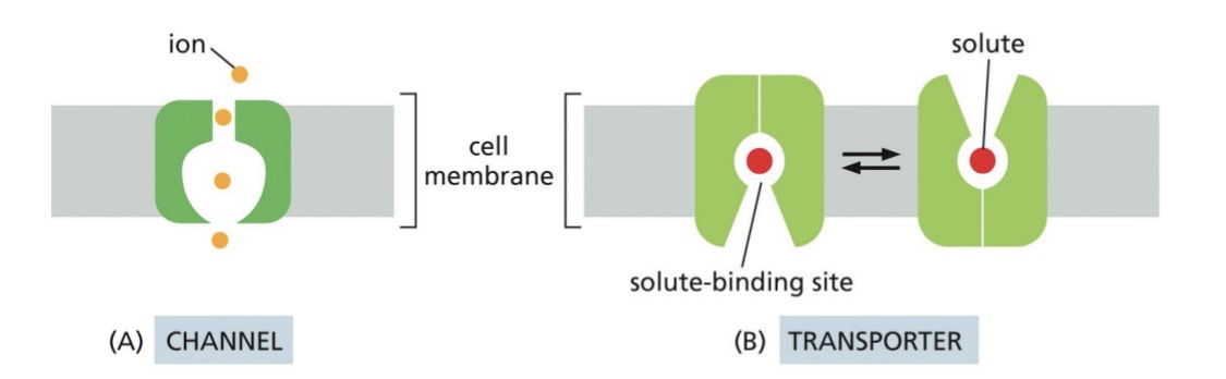
2 main classes of membrane transport proteins
Channel
selective: size and charge of solute matching
passive transport
no conformational changes through open channel
Transporter
selective: solute fits into binding site
passive or active transport
conformational changes for transport
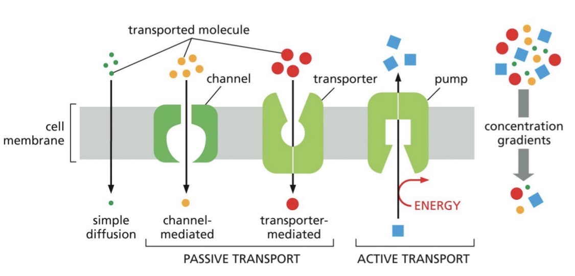
Passive (what kinds?) and active transport
Passive: down concentration gradient (electrochemical gradient if charged)
simple diffusion
channel-mediated
transporter-mediated
Active transport: against gradient
needs energy
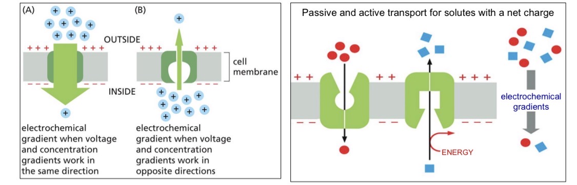
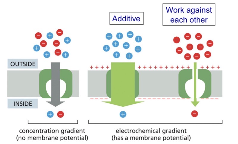
Concentration gradient + Resting membrane potential = Electrochemical gradient (electrical gradient)
Voltage and concentration gradient can work together or against for the net driving force
Additive or work against
MOST of the time direction depends on concentration gradient (rate gets affected)
Channel proteins
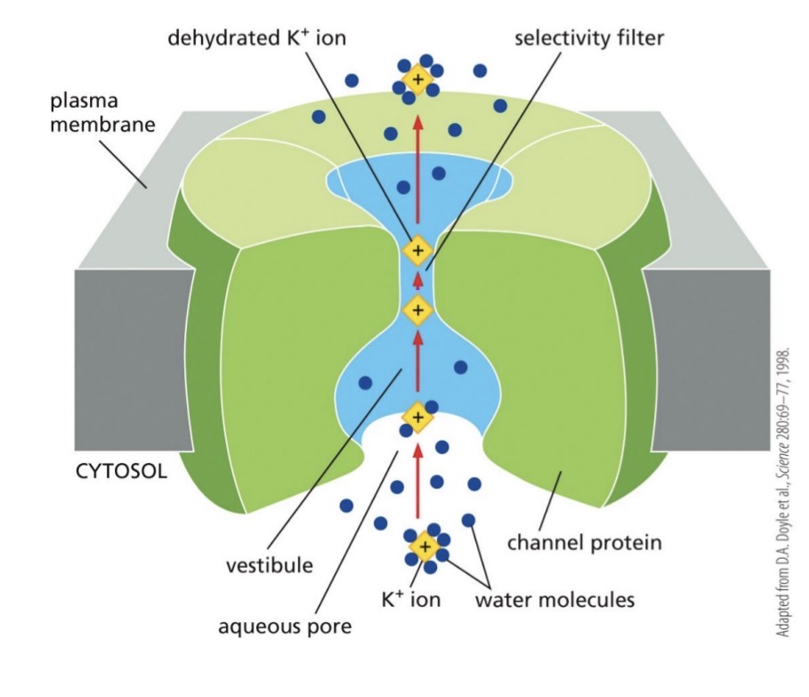
Hydrophilic pore across membrane
Most channel proteins are selective — transient interactions with the channel wall
ALL Passive transport
Faster than transporters
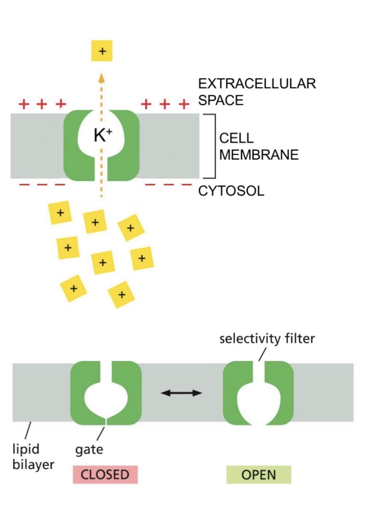
Ion channel example (transient recognition) — non-gated vs gated
Selectivity at narrow part of channel
ion first surrounded by water, then lose
transient recognition by charged AA in channel
narrow to select for size
.
Non-gated ion channels — always open
e.g. K+ leak channels
major role in generating resting membrane potential in animal cells
negative charge on inside to attract ions
but against conc. gradient for transport out of the cell
.
Gated ion channels
need signal to open channel
specific ions transported
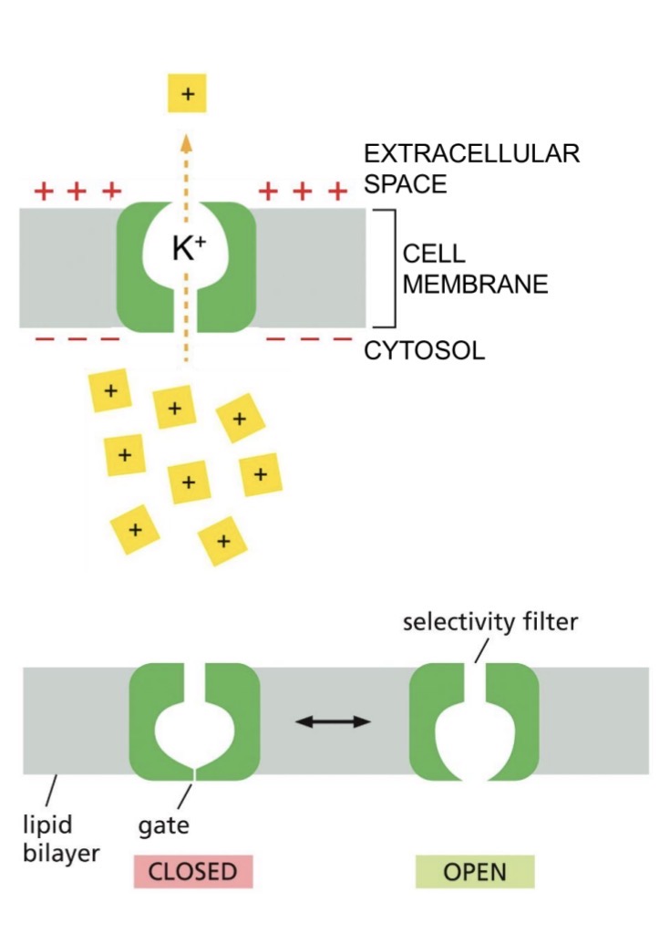
Types of gated ion channel signals
Mechanically-gated: plasma membrane mechanical stress
Ligand-gated (extracellular): ligand signal
Ligand-gated (intracellular): ligand signal
Voltage-gated: change in voltage signal
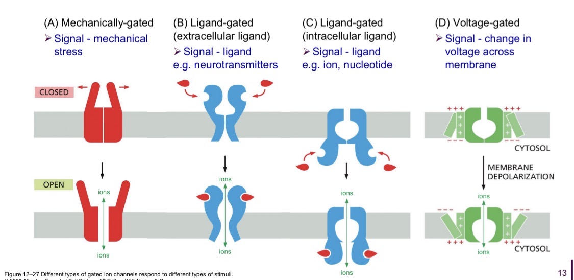
Transporter proteins
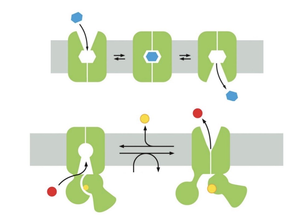
Binds a specific solute
goes through conformational change to transport solute across membrane
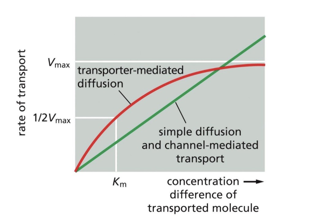
Rate of transport: channel vs transporter
Channel transport rate increases as concentration difference increases
Transporter transport rate has a maximum rate due to all binding sites completely saturated
(channel proteins transport faster)
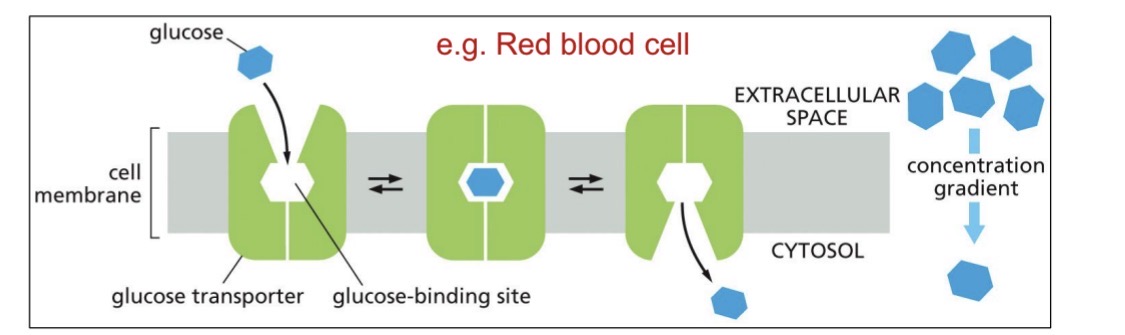
Transporter proteins: Uniport (direction is….)
Uniport: one solute
passive transport down electrochemical gradient
direction of transport is reversible
random conformational changes, just happens to transport
E.g. GLUT uniporter
glucose is the only uncharged
Types of active transport — needs energy (3 kinds)
Gradient driven pump
ATP driven pump (ATPases)
Light-driven pump (bacteria)
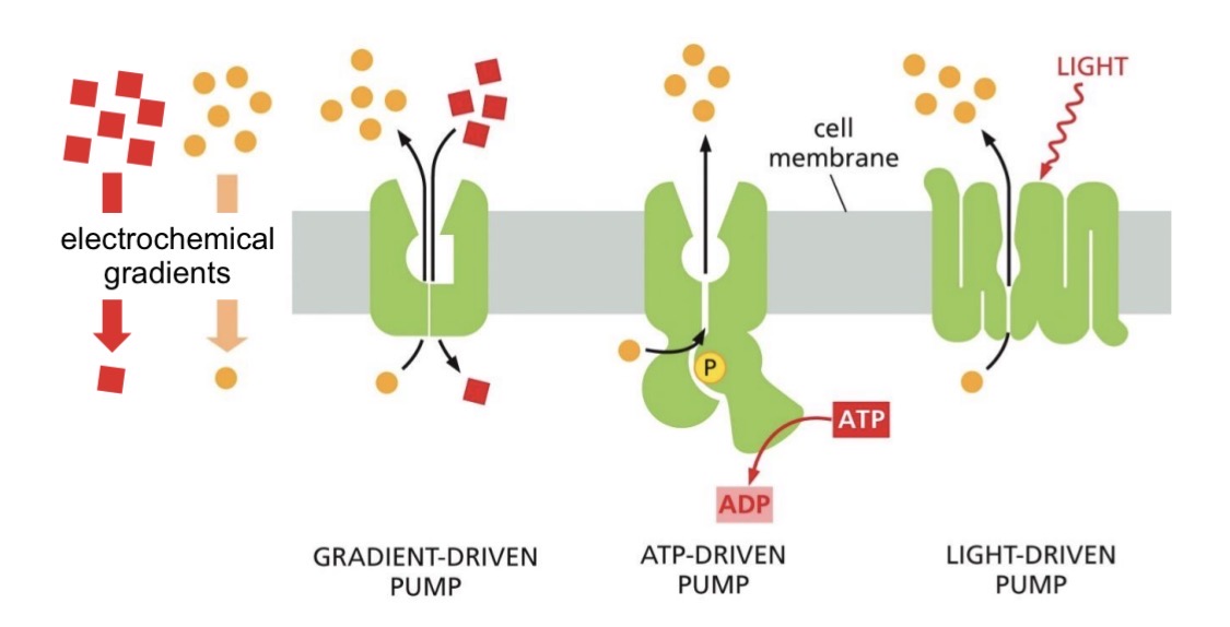
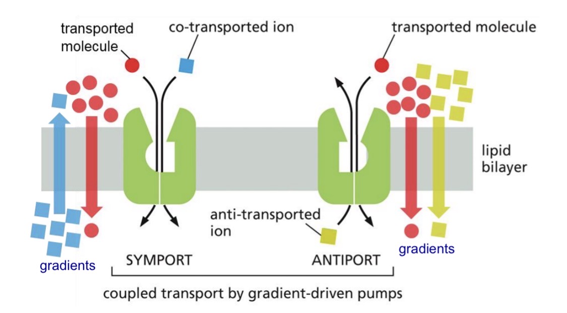
Active transprt: Gradient driven pumps — Symport and anitiport
Symports: two molecules moving in same direction
Antiport: two molecules moving in opposite direction
Free energy from one solute moving down its electrochemical gradient
Powers transport of other solute moving against its electrochemical gradient
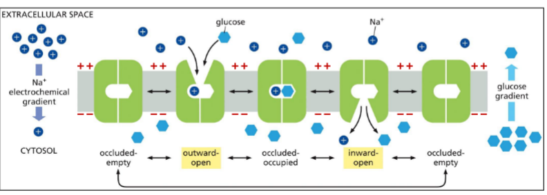
Symport example: Na+ —glucose symporter
Na+ down its electrochemical gradient provides energy to move glucose against concentration gradient
random oscillations between conformations
BUT conformational changes only happen if both sites occupied (cooperative binding) or both sites empty
occluded-empty or occluded-occupied
then open up again randomly
.
Sits open with Na+ until glucose binds
if open to the other side, sodium and glucose released
Antiport example: Na+—H+ exchanger (transported to?)
Na+ down its electrochemical gradient provides energy to move H+ against its
electrochemical gradient
Cytosolic pH needs to be regulated for best enzyme function (pH~7.2)
But excess H+ occurs in cytosol
from acid forming reactions, or leaks out of lysosome (pH~5)
Transporters maintain cytosolic pH
.
e.g. Na+ — H+ exchanger in the plasma membrane
Excess H+ in cytosol
transporter activity increases
H+ transported to extracellular space

How is the Na+ electrochemical gradient maintained?
Symport, Antiport needs Na+ electrochemical gradient to provide energy
but continued action could equalize Na+ gradient
How is it maintained in animal cells? Na+ K+ pump (plasma membrane ATP driven pump)
ATP-driven pumps
Active transport, use energy from ATP hydrolysis to transport solutes

Types of ATP-driven pumps — P type pump (example?)
Use ATP
Phosphorylated during pumping cycle
Many types (transport ions)
AND flippases (transport phospholipids)

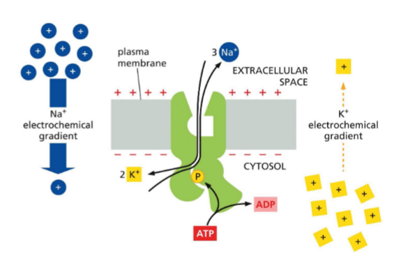
ATP-driven pumps: ① P-type pump (Na/K pump) (extracellular and cytosol conc?)
Animal plasma membrane
Na+ and K+ moved against their electrochemical gradients
3 Na+ out
2 K+ in
more positive out than in
(Na+ gradient available for gradient driven transport)
.
Na+ high in extracellular
K+ high in cytosol
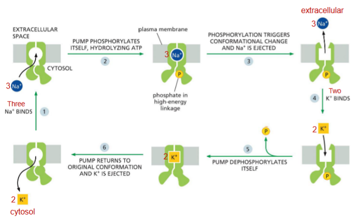
Pumping cycle of the Na/K pump
Has to go through these sequences
3 sodium ions bind to pump (open to cytosol)
ATP hydrolysis —→ phosphorylation —→ change comformation, sodium out
2 potassium ions bind (open to extracellular
dephosphorylate —→ change conformation back, potassium out
(then start again)
(phosphorylation and dephosphorylation powers the pump)
if any stage was blocked, stop working

P-type pumps and electrochemical gradients — animal vs plant
Function in generating and maintaining electrochemical gradients
.
Animal — Na+/K+ pump, animal cell plasma membrane
.
Plant — H+ pump, plant cell plasma membrane (doesnt have Na/K pump)

Types of ATP driven pumps — other than P (what does V do?)
ABC transporter
use 2 ATP hydrolysis, pump small molecules
pump toxins out of the cell (bad for cancer treatment)
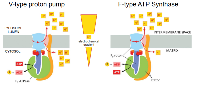
V-type proton pump
pumps H+ into organelles to acidify the lumen

F-type ATP synthase
Structurally related to V-type proton pump, but opposite mode of action
uses H+ gradient to drive the synthesis of ATP
in mitochondria, chloroplasts, bacteria

Transport proteins regulate critical cellular processes (Transporter can work together)
Transcellular transport of glucose by transporters
Generation of membrane potentials
e.g. GLUT uniporter (passive), Na+ glucose symporter, Na+K+ pump

Transporters work together to transfer glucose from the intestine to the bloodstream
Glucose taken up from gut
Na+/glucose symporter on the apical domain villi
(against gradient, ACTIVE TRANSPORT)
Transported through cell
Transported to bloodstream via GLUT uniporter
on basalateral domain
(down gradient, PASSIVE TRANSPORT)
Na+ K+ pump on basal domain to maintain membrane potential
.
(low glucose in gut, high in cytosol of epithelial cell, low in extracellular fluid)
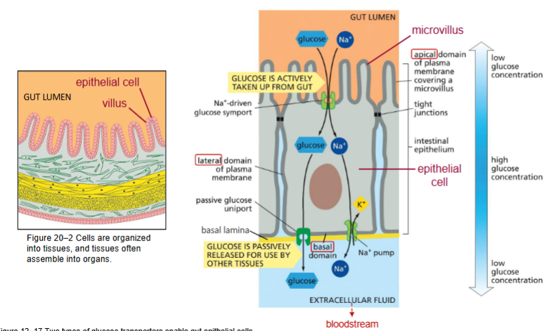
Transcellular Transport of Glucose Requires the Asymmetric Distribution of Membrane Proteins
Intestinal epithelial cells
Transport proteins restricted to certain places by tight junctions (creates boundary, seals)
Apical membrane — Na+-glucose symporter
Basolateral plasma membrane — GLUT2 uniporter
Na+-K+ pump
Membrane potential (pumps and?)
Difference in electrical charge on two sides of membrane
Used by gradient-driven pumps to carry out active transport (Symport, Antiport)
Electrical signaling
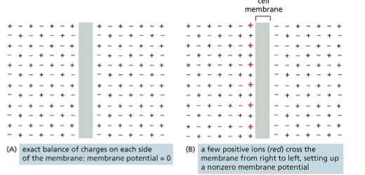

Generation of Membrane Potential (animal cells)
K+ Leak channel (to extracellular)
major role in membrane potential
passive transporter
voltage gradient opposite to concentration gradient, creates positive charge
will reach equilibrium — no net movement
.
Na+-K+ pump (to extracellular)
~10% of membrane potential
Na+ gradient with low cytosolic [Na+]
K+ gradient with high cytosolic [K+]
3Na:2K, Net 1+ ion out
![<ol><li><p>K+ Leak channel (to extracellular)</p><ul><li><p>major role in membrane potential</p></li><li><p>passive transporter</p></li><li><p>voltage gradient opposite to concentration gradient, creates positive charge</p></li><li><p>will reach equilibrium — no net movement</p></li></ul></li></ol><p>.</p><ol><li><p>Na+-K+ pump (to extracellular)</p><ul><li><p>~10% of membrane potential</p></li><li><p>Na+ gradient with low cytosolic [Na+]</p></li><li><p>K+ gradient with high cytosolic [K+]</p></li><li><p>3Na:2K, Net 1+ ion out</p></li></ul></li></ol><p></p>](https://assets.knowt.com/user-attachments/60263466-22d2-4d67-8645-61323526494b.png)

Generation of Membrane Potential (animal cells) — what is resting membrane potential?
Net result:
Bit more positive on outside (Na+, K+)
Bit more negative on inside (Cl- and fixed anions)
Equilibrium = resting membrane potential
(animal cells: vary from -20 mV to -200 mV)
Generation of Membrane Potential (plant cells) (used for?)
Plasma membrane P-type pump (to extracellular or lumen)
H+ pump
generates H+ electrochemical gradient = membrane potential
-120 to -160 mV (in cytosol)
.
Used for:
Used by gradient-driven pumps to carry out
active transport (e.g. H+ driven symport)
Electrical signaling
Regulate pH

Lec 17: Intracellular compartments
Mitochondrion, Golgi, ER, vesicles, endosome, peroxisomes…
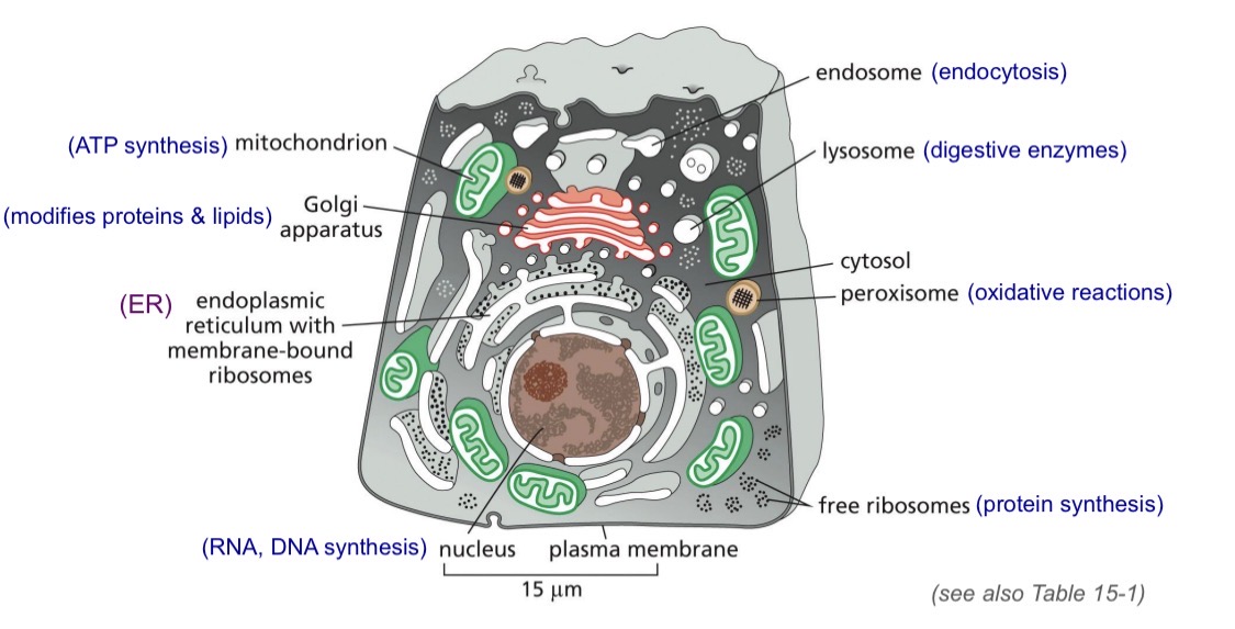
Intracellular Compartments - volumes
Cytosol takes up half the cell volume
Volumes will differ for different cell types
Intracellular compartments — membranes
Plasma membrane very small portion of the cell membranes
Rough and smooth ER take up 50-60%
Rough ER — membrane bound ribosomes, synthesis of soluble and transmembrane proteins
Smooth ER — phospholipid synthesis, detox
Organelle definition
A discrete structure or subcompartment of the a eukaryotic cell that is specialized to do a particular function
Organelle types
Membrane-enclosed
Nucleus
ER
Golgi
Not membrane-bound
Nucleolus
Centrosome
biomolecular condensates
Protein sorting — simple
Proteins are nuclear encoded (made from genes)
mRNA arrives in cytoplasm — translation starts on cytosolic ribosomes
Cytosolic protein: no sorting signal, defualt location is cytosol
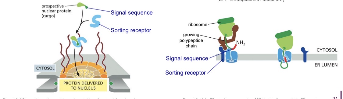
Sorted proteins: have sorting signal, signal sequence
(can be modified experimentally to identify the types of signals)
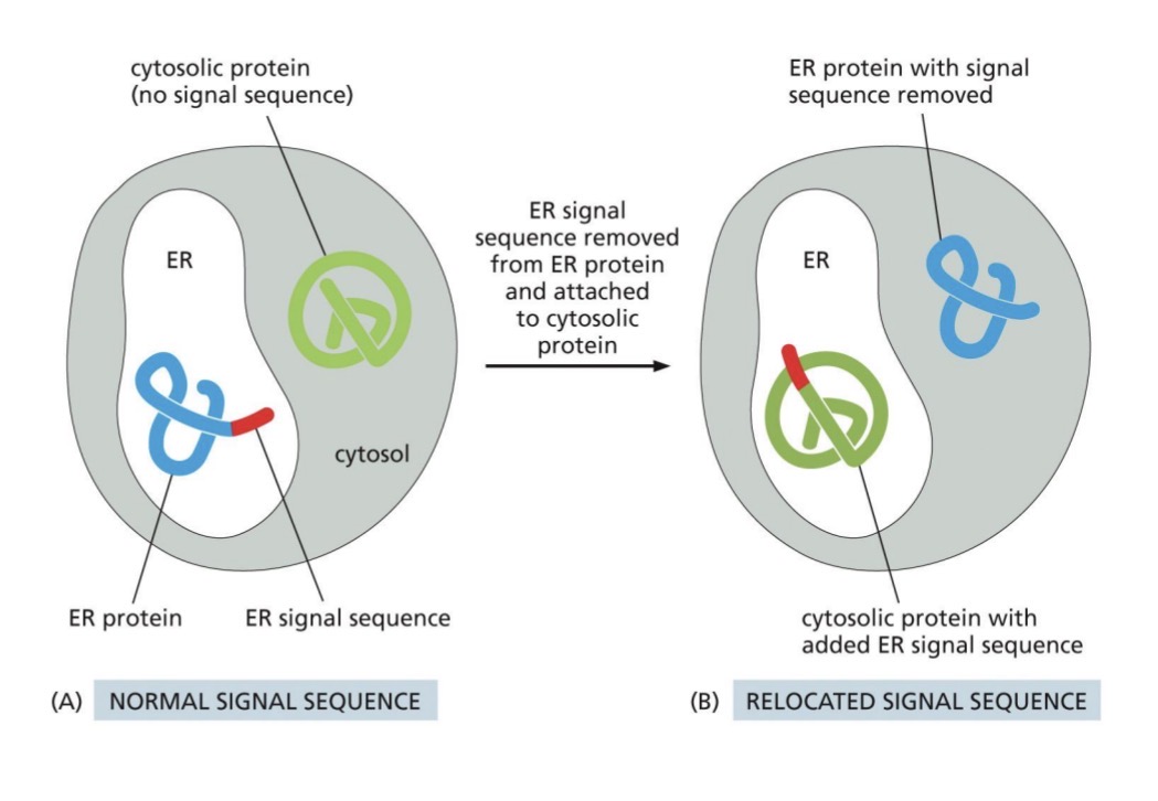
How are specific proteins sorted to different organelles? (signal sequence, sorting receptors)
Sorted by signal sequence
Part of AA sequence
Specifies desination — nucleus, mitochondria, ER, peroxisomes,
etc
Recognized by sorting receptors that take proteins to the destination
Signals are very different
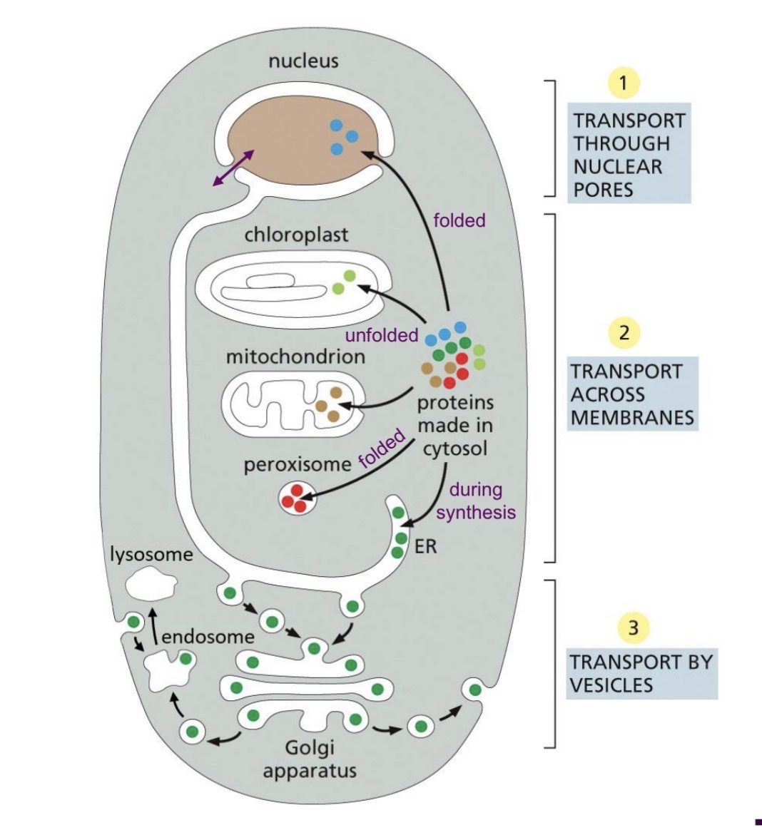
Protein sorting: post vs co-translational
Post-translational
Proteins nuclear-encoded
Fully synthesized in cytosol before sorting
Can be folded or unfolded
Co-translational sorting
Proteins nuclear-encoded
ER signal sequence
associated with ER during protein synthesis in the cytosol
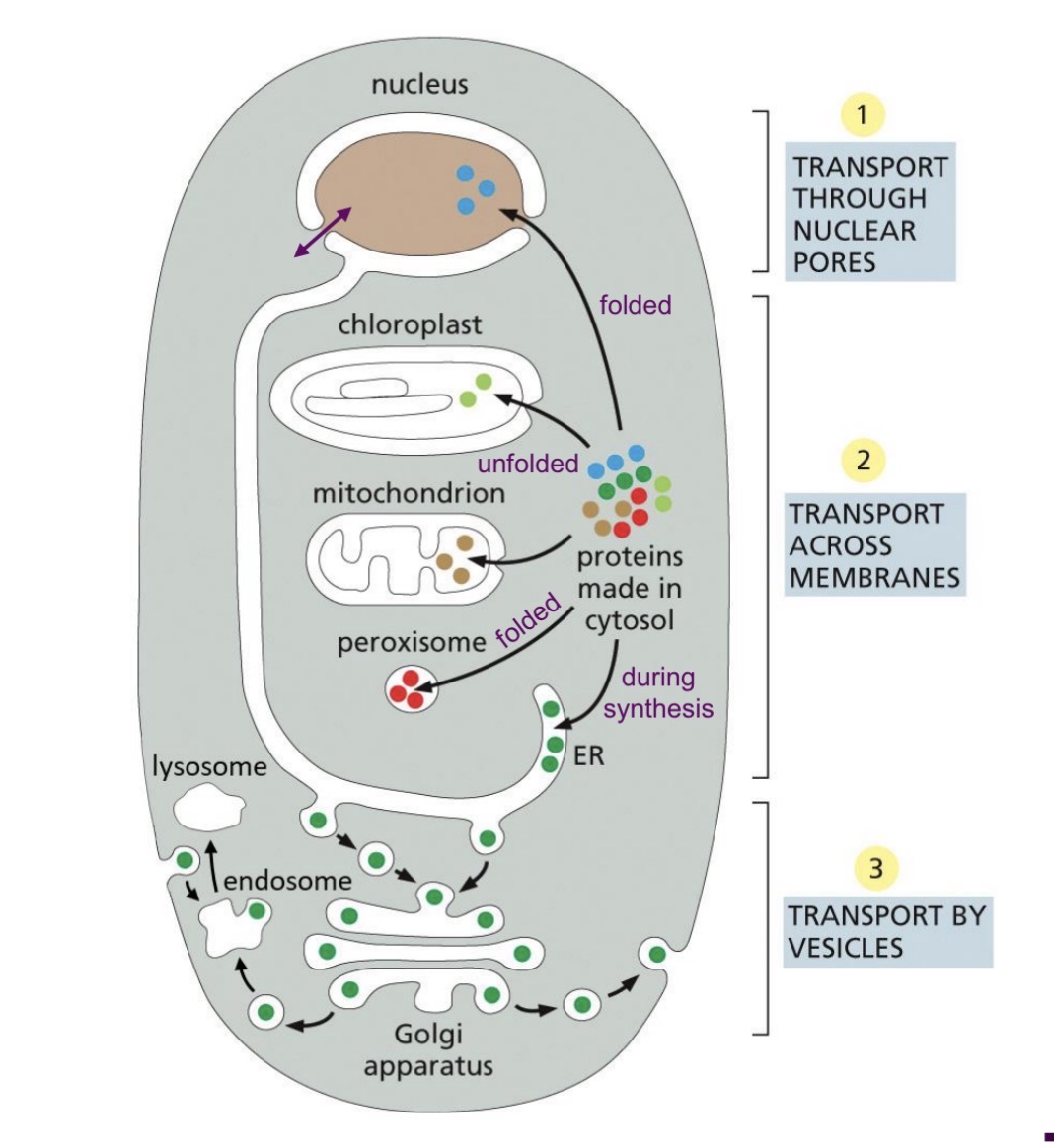
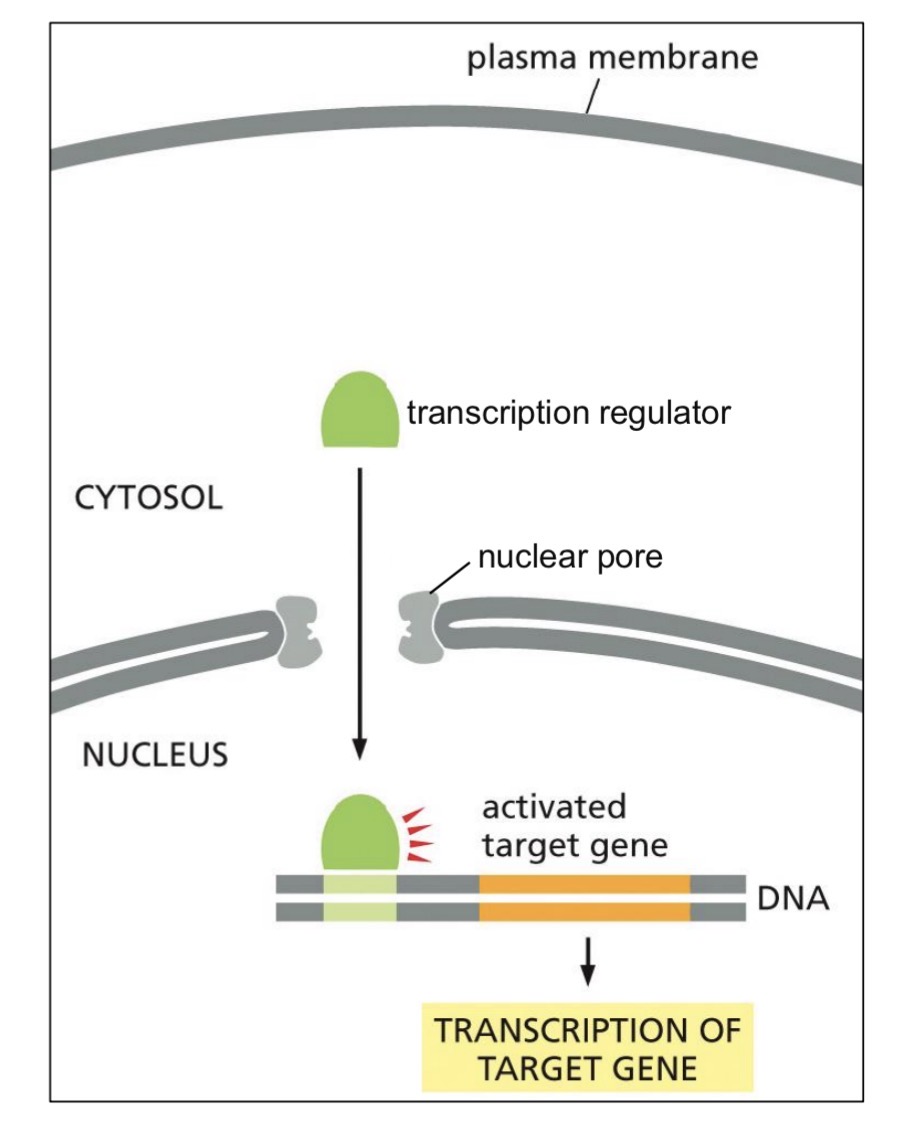
Proteins for nucleus (whats the signal called?)
Post-translational
Nuclear-encoded
Fully synthesized
Folded
.
Targeted by signal sequence for import into nucleus (nuclear localization signal)
Recognized by sorting receptor
Transported through nuclear pore
Detached
Example
transcription factors required in the nucleus
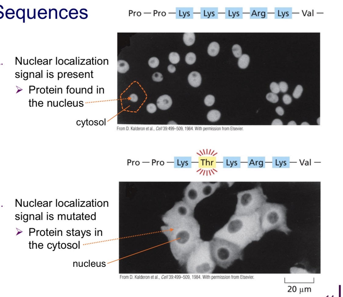
Mutation of nuclear localization signal
If mutated, not recognized by sorting receptor — stays in the cytosol
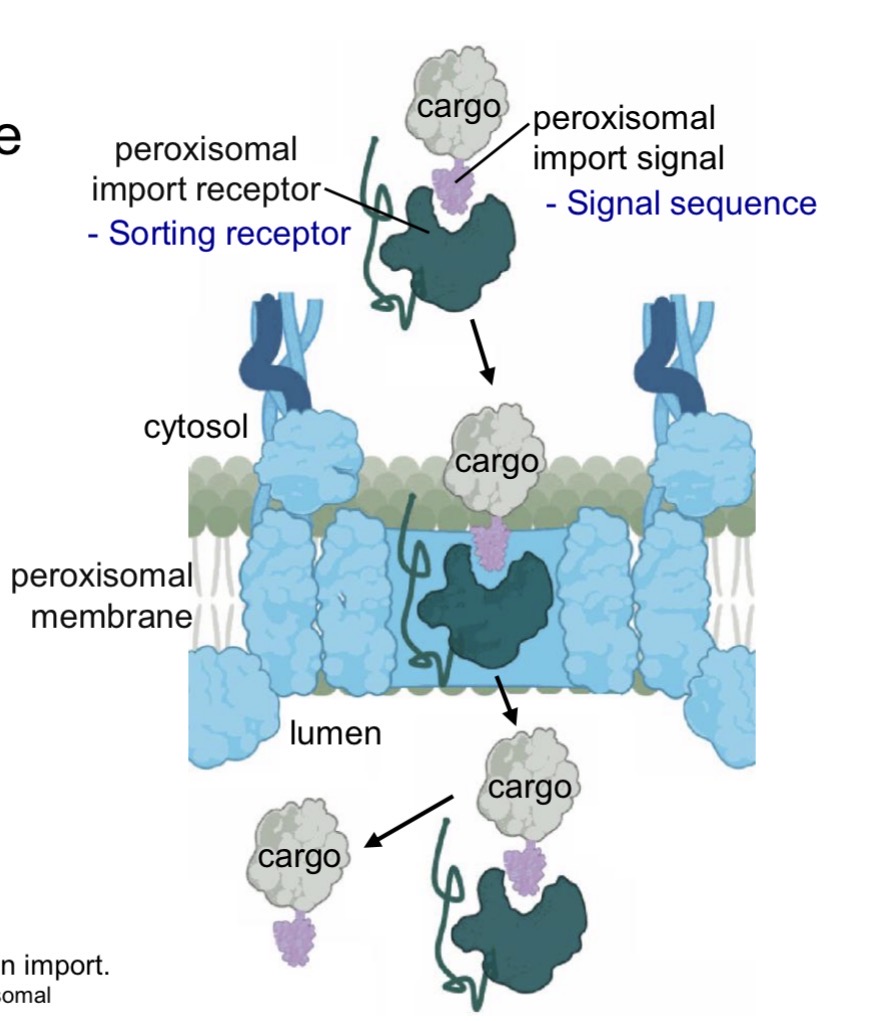
Proteins for Peroxisomes (through the?)
Post-translational sorting
Nuclear-encoded
Fully synthesized
Folded
.
Targeted by sorting receptor for import into peroxisome
Transported into peroxisomes through transmembrane protein complex
.
e.g Enzymes for oxidative reactions
Proteins for Mitochondria & chloroplast
Post-translational sorting
Nuclear encoded
Fully synthesized
Unfolded
.
Targeted by signal sequence
Proteins for Mitochondria & chloroplast — continued
Mitochondria and chloroplasts have own genomes and ribosomes
But most proteins are nuclear encoded
Unfolded to transport through transmembrane proteins
Asociated with cytosolic hsp70 chaperone proteins
In matrix — mitochondrial hsp70 fold protein into shape
Signal cleaved
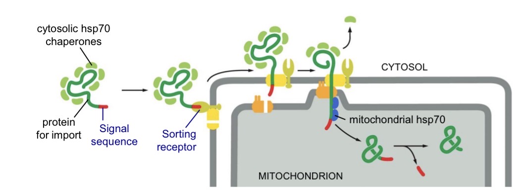
Proteins for the ER (signal sequence is hydro?)
Co-translational sorting
Nuclear encoded
ER signal sequence — hydrophobic
associated with ER during protein synthesis in the cytosol
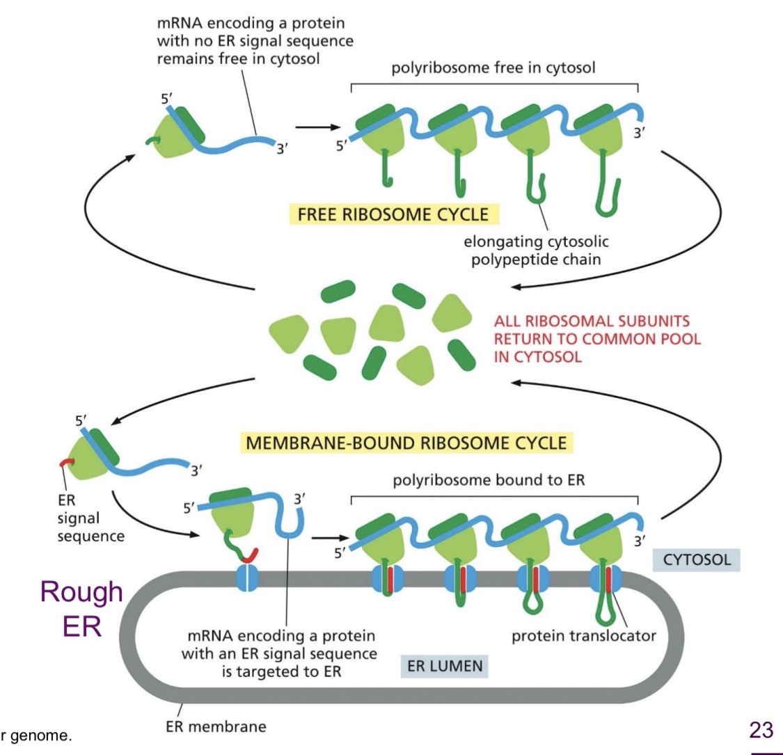
Proteins for the ER — Why sort to ER?
Entry point to the endomembrane system
Endoplasmic Reticulum (soluble and transmembrane proteins vs phospholipids)
Rough ER (with membrane-bound ribosomes)
• synthesis of soluble and transmembrane proteins for the endomembrane
.
Smooth ER
• phospholipid synthesis, detoxification
Sorting Proteins to the ER
mRNA arrives at cytoplasm
Translation starts on ribosomes in cytosol
ER signal sequence
Insertion of protein into ER starts as translation continued
.
Soluble proteins
Transmembrane proteins
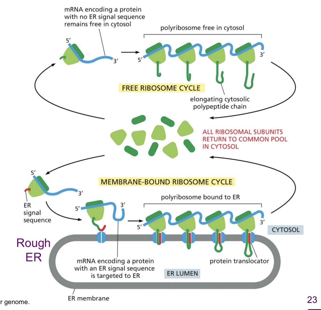
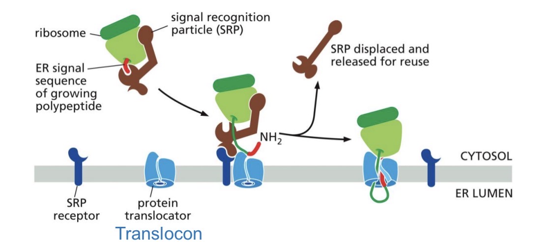
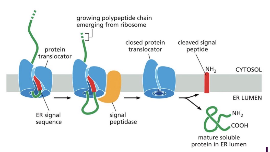
Co-translational Translocation: Soluble protein (SRP, SRP receptor, Translocon, Signal peptidase)
Translation start, N-terminal ER signal sequence emerges
Recognized by SRP, elongation stopped
SRP-ribosome complex — to SRP receptor — to translocon
Translocon opens
Protein synthesis resumes with protein transfer into ER lumen
Signal peptidase cleaves ER signal sequence (hydrophobic — in bilayer)
Protein released into ER lumen
Translocon closes
Destination: lumen of endomembrane organelles or extracellular secretion
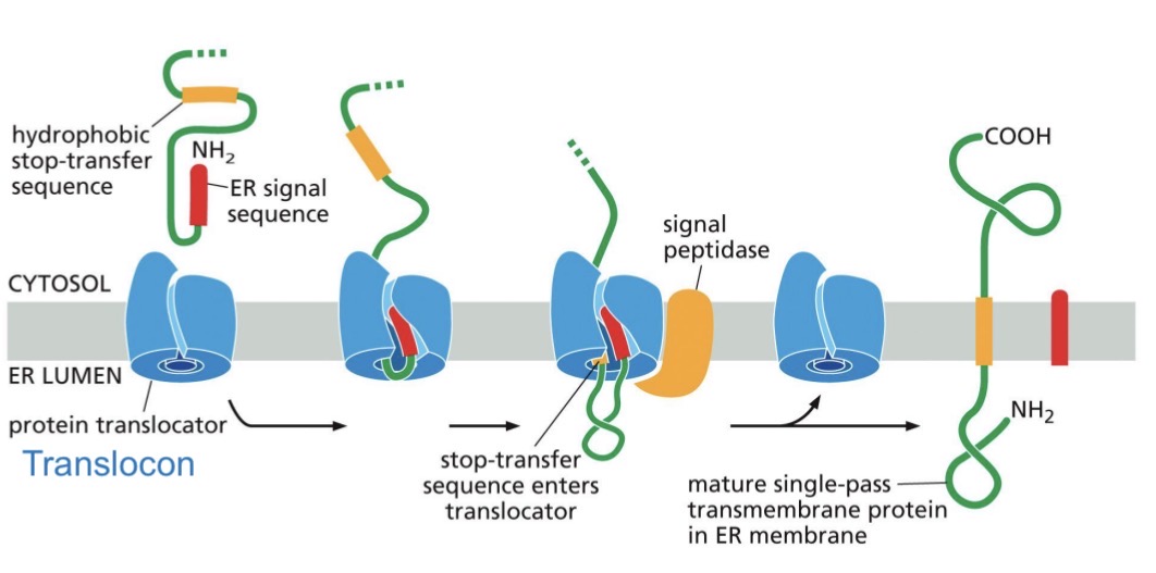
Co-translational Translocation: Transmembrane protein
SRP-ribosome complex
Stop-transfer sequence (hydrophobic a-helix) enters translocon
Protein transfer stops
Transmembrane domain released into bilayer
Signal peptidase cleaves
Translocon closes
protein synthesis completed
Destination: membrane of endomembrane organelle or plasma membrane
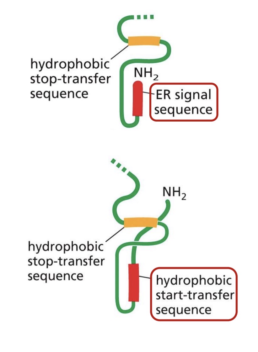
ER signal sequence types (N terminal & internal)
N-terminal ER signal sequence
At N-terminus
hydrophobic AA
removed by signal peptidase
.
Internal ER signal sequence
start-transfer sequence
hydrophobic AA
not removed — becomes membrane spanning a-helix
Co-translational Translocation: Transmembrane protein — multipass
Start-transfer emerges, pause ribosome, move to ER membrane
Transports through membrane until Stop-transfer sequence enters translocon
Both are released into bilayer — membrane spanning a-helices
Translocon closes, translation finishes
Number of transmembrane domains determines whether terminuses are on the same side or opposite sides
Intracellular compartments are dynamic
Endomembrane system
ER
Golgi
Endosomes
Lysosomes
.
Intracellular compartments exchange components
Proteins (soluble and membrane)
Lipids — fused into destination membrane
Pathways of the endomembrane system
Secretory pathway
proteins and lipids from the ER
ER to outside (exocytosis)
ER to lysosomes (endosomes)
.
Endocytic pathway
contents move into cell (endocytosis)
degredation
Retrieval pathway
Retrieval of lipids and selected proteins for reuse
Exocytosis and Endocytosis
Exocytosis:
vesicle contents delivered to extracellular space
vesicle membrane becomes part of the plasma membrane
Endocytosis:
vesicle contents come from extracellular space
vesicle membrane buds from plasma membrane
Vesicular transport (cargo and specificity ddd?)
Vesicle is:
small membrane-enclosed organelle in cytoplasm
Soluble proteins + membrane proteins = cargo
receptors in membrane select cargo
or diffusion into vesicle
receptors can get recycled
Specificity from:
docking proteins bringing vesicle to target membrane
Secretory pathway — constitutive exocytosis pathway (proteins & lipids)
In all eukaryotic cells
continual delivery of proteins (transmembrane and soluble) and lipids to plasma membrane
not regulated
Secretory pathway — Regulated exocytosis pathway
regulated secretion in specialized cells
specialized secretory vesicles
need extracellular signal before vesicle fuses with plasma membrane
e.g. insulin and blood glucose levels
Path of secreted proteins from translation to plasma membrane
Translation starts in cytosol
Co-translational translocation at ER
Secreted protein through secretory pathway
Path of a transmembrane protein from translation to plasma membrane
Translation starts on cytosolic ribosomes
Co-translational translocation at ER
Transmembrane protein through secretory pathway — fused with plasma membrane
Maintenance of membrane protein asymmetry
Membrane has orientation
Protein asymmetry is maintained through vesicular transport
Golgi apparatus (receive….and?) (animal vs plant)
recieves proteins and lipids from ER, modifies them, then dispatches them to other destinations in the cell
Stacks of enclosed sacs
Animal cells has one large golgi
Plant cells have many small golgi
Protein glycosylation
Starts in the ER
single type of oligosaccharide is attached to many proteins on lumen side
.
Complex oligosaccharide processing occurs in golgi apparatus
different enzymes in each cisterna
glycosylation modifications for proteins and lipids
Endocytic pathway: endosomes and lysosomes
Membrane bound organelles containing meterial digested by endocytosis
Endocytic pathway: endosomes and lysosomes: endocytic vesicles
Fuse with early endosomes and with eachother to form early endosomes
ingested material sorted, stays in endosomes or recycled to plasma membrane
Endocytic pathway: endosomes and lysosomes: gradual maturation of early endosomes into late endosomes
moves to nucleus, stops recycling material to plasma membrane
hydrolases and proton pump delivered from golgi by vesicles
protons pumped into endosome — acidification
hydrolases become more active
Maturation of late endosomes into lysosomes
hydrolases and proton pump continue delivered
pH continues to drop, hydrolase digest
late endosomes fuse with each other and with lysosomes
Lysosome - final detination for endocytosed material being digested
other pathways available — autophagy
Lysosomes are the main site of intracellular digestion (enzymes, membrane, transport)
Contains 40 types of hydrolytic enzymes
acid hydrolases
Lysosomes are acidified by proton pump
.
Membrane-bound organelle
protects cell from digestion
glyosylated proteins to protect from proteases
.
Transport proteins in lysosomal membrane
transfer digested monomers to cytosol
Cytoskeleton
Network of protein filaments, Highly dynamic
Many important functions
