TCP Week 5 (Head and Spinal Trauma)
 Call Kai
 Learn
Learn Practice Test
 Spaced Repetition
 Match
Match Flashcards
 Knowt Play
1/108

There's no tags or description
Looks like no tags are added yet.
Name | Mastery | Learn | Test | Matching | Spaced | Call with Kai | Chat |
|---|
No analytics yet
Send a link to your students to track their progress
109 Terms
traumatic brain injury (TBI)
A significant cause of morbidity and mortality in young adults
•Head injuries in: polytrauma, high velocity injuries/fall from height/falls in the elderly
•1º versus 2º TBI
What percent of head injuries are mild?
80%
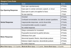
Glasgow Coma Scale
- Readily used for assessment of trauma patients
- GCS + Neurologic assessment is the tenet of care of the brain injured patient
Glasgow coma scale grading
- GCS ≤ 8 = severe head injury
- GCS 9-13 = moderate head injury
- GCS 14-15 = mild head injury
GCS + P = GCS-pupil score
accounting for pupillary changes and supplement GCS adding more information about severity and prognosis -> can lead to change in morbidity/mortality
Neuro exam for TBI
- Systematic approach, using starting with higher function (like sensation to touch etc.) followed by simplest (reflexes)
- Identify areas of tenderness, edema, bruising, spasm, or clonus
- Focus should be on strength testing of extremity muscle groups, muscle tone, sensation of touch, pain, vibration, and proprioception by dermatomes, identification of a sensory level, lateralizing signs, exaggeration, or absence of DTRs, and evaluation of rectal tone
HEENT assessment for TBI
Head:
- bruising
- lacerations
- racoon eyes
- bruising behind the ears
Ears:
- blood behind ear drum
- CSF leak
Eyes:
- pupil size
- equality
- reactivity
- fundoscopy for retinal hemorrhage
Nose:
- CSF leak
- bleeding
exams for neurotrauma assessment include?
- GCS
- Neuro exam
- HEENT
Glasgow Coma Scale pic
Remember, T is used for tube (intubation or trach) with Verbal
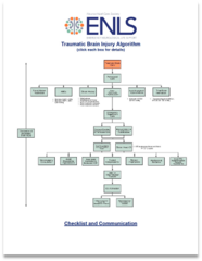
ENLS - Emergency Neurologic Life Support
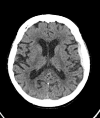
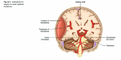
subdural hematoma
blood under the dura but not intracranial (convex)
traumatic injury to bridging veins
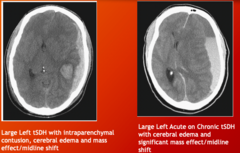
acute versus chronic presentations
who does subdural hematomas occur most in due to what
babies and elderly due to traumatic injury to bridging veins
s/sx of subdural hematoma
- progressive HA
- weakness
- seizure
- alterations of LOC
subdural hematoma imaging
surgery indication for subdural hematoma
>1 cm thickness
AND/OR
>0.5 cm of midline shift (BTF Guidelines)
tx for Subdural hematoma
AEDs—highly epileptogenic in acute phase
Surgery
most commonly seen TBI
traumatic subarachnoid hemorrhage
traumatic subarachnoid hemorrhage
trauma to small capillaries and veins, generally seen in the sulci or close to the surface of the brain (cortex) near traumatic insult:
- direct extravasation of blood from an adjacent cerebral contusion
- arterial dissection
- direct damage to small veins or arteries
- sudden increase in intravascular pressures leading to rupture
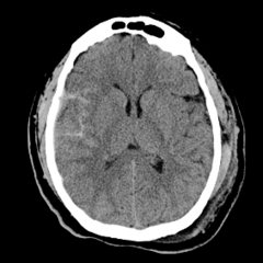
Traumatic subarachnoid hemorrhage tx
- majority are nonsurgical
- doesn't usually require AED
- good prognosis
traumatic subarachnoid hemorrhage notes
- Presentation similar to tSDH, less severe symptoms
- If blood lies closer to the base of the brain/cisterns -> high suspicion of vascular event (e.g. aneurysm SAH or aSAH)
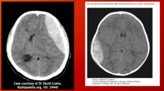
traumatic epidural hematoma epidemiology
less common but higher levels of morbid/mortal if untreated
Younger > Older patients
how do traumatic epidural hematomas occur?
Bleeding outside the dura (epi-above), but trapped between the bone/suture lines:
- False space
- Lens like shape
- Commonly associated with occult skull fracture
traumatic epidural hematoma notes: arterial type bleeding and symptom specific to EDH
arterial type bleeding: rupture of parent or branch of MMA
presence of lucid interval ~25% of patients (high velocity trauma, patient improves rapidly, then rapid decline)
tx of traumatic epidural hematoma
- AEDs indicated
- LEV first line (Keppra)
- Often surgical
- Definitive treatment: Craniotomy
traumatic epidural hematomas imaging
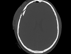
traumatic skull fractures
•High velocity head trauma
•Often treated conservatively
basilar skull fracture s/sx
- more severe mechanism
- battle sign (mastoid/skull base fracture)
- raccoon eyes
traumatic skull fracture tx indication
often treated conservatively but indicated for >1 cm depression (depressed skull fracture): craniotomy with elevation of skull fracture
surgical indications for depressed skull fracture
- > 1cm depression of the inner table of the skull
- open depressed skull fx
- foreign body causing brain irritation
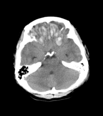
brain contusion and intra cerebral hemorrhage most common location
frontal and anterior temporal lobes
s/sx brain contusion and intra cerebral hemorrhage
- Usually hemorrhage with surrounding edema
- May cause traumatic sub-arachnoid hemorrhage
- Cause broad spectrum of neurological dysfunction
- May cause increased intracranial pressure
neurotrauma definitive care: traumatic intracranial hemorrhage and depressed skull fractures (tx)
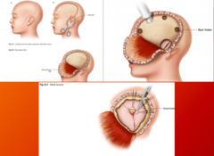
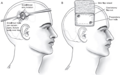
definitive surgical care = craniotomy for large acute trauma (chronic SDH = burr holes)
Large window of bone removed and replaced after hematoma evacuated:
- Craniotomy = replaced
- Craniectomy = removed
focal craniotomy (over hematoma) VS trauma flap (large/wide craniotomy)
surgical acute EDH
image
craniotomy for large acute trauma (chronic SDH = burr holes)
image
surgical indications for traumatic intracranial hemorrhage and depressed skull fractures
Large mass lesion > 1 cm in thickness, or causing >5 mm MLS --> SURGICAL
Depressed skull fractures > 5 mm below inner table of the skull --> SURGICAL
Burr Holes vs craniotomy
- burr holes is less invasive
- often considerations are the surgical lesion, patient age/comorbidities, chronicity and size of SDH
indications for decompressive craniectomy
*same but don't put bone back on
No level 1 recommendations
Level 2 recommendations:
- reduce ICP and improve ICU LOS
- no effect on outcome based on GCS 6 months post injury
TBI Management Pathway (pt 1)
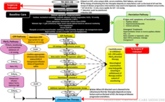
TBI Management Pathway (pt 2)
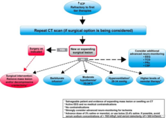
intracranial pressure & what is normal?
the pressure that is maintained within the skull at any given time
normal is 8-12 mmHg
Alterations of ICP fluctuate constantly and cerebral dynamics change to accommodate
Accounting for?
- Blood
- Brain Tissue
- CSF
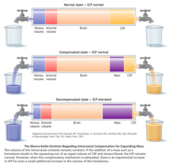
monroe kellie doctrine
when one content in the skull increases, another must decrease to compensate and maintain normal ICP; compensated up until a point
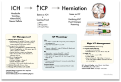
Elevated ICP management
- Elevate HOB
- Prevent hypoxia + protect airway
- Maintain normal PCO2, O2, Temperature, Glucose
- Control Seizure
- CSF Diversion (Ventriculostomy)
- Sedation (Propofol, Midazolam)
- NMB (Cisatricurium)
- Hyperosmolar Therapy
- Osmotic Diuretics (Mannitol)
- Hypertonic Saline
- Barbiturate Coma (Pentobarbital)
IF PERSISTENT ICPs
Surgical Decompression (salvage therapy)
mechanism of traumatic brain injury
-blunt
-penetrating
penetrating head injuries
- gunshot wounds and penetrating sharp objects can penetrate the skull into the brain
- often ballistic traumatic causes widespread traumatic cerebral edema and ICP issues
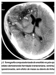
penetrating head injury tx
commonly: emergent decompressive craniectomy
work up for penetrating head injury
- CT
- CTA
- Skull assessment
- Foreign bodies
- Parenchyma and hematomas
indications for CTA for penetrating traumatic injury
used when there is evidence or suspicion of intracranial vascular injury. Projectiles or penetrating injuries that have traversed the base of skull or orbits have a high incidence of vascular injury
CTA for penetrating traumatic injury: what it can show?
- CTA may demonstrate a CTA spot sign indicating active hemorrhage at the time of the scan, as seen in non-traumatic intraparenchymal hemorrhage, which correlates with a worse prognosis
- Vascular injuries include transection, pseudoaneurysm, dissection or thromboembolic occlusion
Penetrating Head Injury- Skull Assessment Work Up
• entry and exit wounds can be identified paying attention to the bone "beveling"
• often there is not an exit wound, especially in incomplete metal jacket bullets (e.g. hollow-point tip) that are designed to fragment and lose kinetic energy along their path 1.
• location and extension of linear fractures as well as any non-penetrating fractures from coexisting blunt head trauma
inward beveling of the inner skull - entry or exit wound?
entry wound
outward beveling of outer skull- entry or exit wound?
exit wound
Penetrating Head Injury- Foreign Body Work Up
Location, number (sometimes difficult if highly fragmented) and density (usually metal in high-velocity injuries) of fragments, particularly in relation to eloquent parts of the brain or vessels
Penetrating Head Injury - Parenchyma and hematomas work up
The direction of tract(s) and the key structures they traverse along with the amount of hemorrhage, particularly if there are sizable hematomas that need evacuation.
This is particularly the case for extra-axial hemorrhages that may occur due to vascular injury.
Complications of penetrating head trauma
infection:
- cerebral abscess
- meningitis
CSF Leak
arterial injury:
- dissection
- transsection
- false aneurysm, etc.
venous injury:
- dural venous sinus thrombosis
Acute infarct/stroke
Traumatic hydrocephalus (delayed or immediate)
Bullet migration/foreign body migration
spine facts/epi
•17,000 new SCI cases each year
•The average age at injury has increased from 29 years during the 1970s to 42 years currently
•Males account for approximately 80% of new SCI cases
•Vehicle crashes are currently the leading cause of injury, followed by falls, acts of violence (primarily gunshot wounds), and sports/recreation activities
spine ligaments
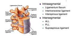
mechanism of injury for spine injuries
- hyperextension
- hyperflexion
- compression
- rotation
- lateral stress
- distraction
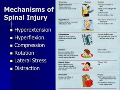
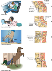
Mechanism of injury may be solely isolated to the spine, or may have secondary injury to the spinal cord
Injury to spinal ligaments without bony fracture may still be?
unstable (not all fx are unstable, if ligament damaged, bones can be unstable)
suspicion of secondary injury is high, with a more significant presentation (e.g. high velocity MVA, fall of significant height)
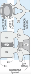
3 column model
- Three columns of stability to the spine
- Theory that an injury to 2 or more columns dictates an unstable injury and thus likely requires surgical fixation
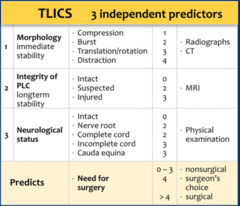
Thoracolumbar injury classification score (TLICS)
1. morphology
2. integrity of PLC
3. Neurologic status
helps to stratify injury and support decision making in spine injured patients
>4 = surgery
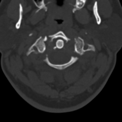
Notable spine fractures: Jefferson fracture
Burst fracture at C1
Bilateral anterior/posterior fractures of the ring of C1
Most commonly associated with diving into shallow water
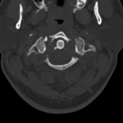
tx of Jefferson Fracture
- Collar
- if ADI/TL disrupted fusion vs external orthosis
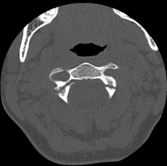
Hangman's fracture ("hangee's fracture")
traumatic spondylolisthesis of axis, or C2
bilateral pars fracture; hyperextension and distraction injury (e.g. MVA)
~25% have neurologic injury
tx of hangman's fracture
most commonly collar, may be fused
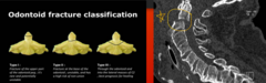
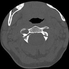
Dens/odontoid fracture
Fracture of the dens, or peg at C2
3 subtypes, treated differently depending on location of fracture
• ex: TII = surgical bc cut off blood supply
facet dislocation/subluxation
Anterior traumatic displacement of one vertebrae on another
Oftentimes associated with high axial forces, and disruption of ligaments:
- Can be in the absence of fracture
Subluxed facet -> Perched Facet -> Locked Facet
subluxed facet joint
- mildest form in which the ligamentous injury leads to partial uncovering of facet joint
- results in mild anterior displacement of one vertebral body on another
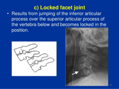
locked facet joint
results from jumping of the inferior articular process over the superior articular process of the vertebra below and becomes locked in the position
basic management of spinal fractures
- Immobilization
- Orthosis (internal or external brace)
- Neurologic/radiographic/clinical assessment
- Operative management (if applicable)
- Rehabilitation considerations
- Management of spinal fracture complications
Prior to the complete evaluation of a spinal fracture, it is paramount to suspect the most significant injury and treat as such (what are some of the most significant injuries?)
high velocity mechanisms:
- MVA
- fall from significant height
- poly-trauma
all have a high spine injury rate even if its not clear
what are precautions the PA can take to prevent re-injury or secondary spinal trauma (often done pre-hospital)
- inline cervical stabilization
- log-roll
this is usually done pre-hospital, however spinal trauma can occur in house, or be missed during initial eval
management with orthoses
essentially bracing, includes both
external bracing
- collar
- brace
- splint
- halo
- tongs
internal bracing (fusion)
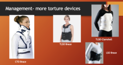
example picture of external cervical orthoses
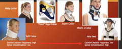
"more torture devices"
internal orthosis
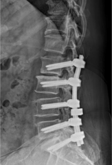
internal brace or fusion provides a rigid construct to stabilize the spine in trauma
fusion definition
the surgical fixation of two or more spinal elements to create immobile segments to aid bone growth
what are the indications of fusion?
- Stability (unstable fracture treatment)
- Correction of deformity
- Decompression of Neural Elements (requiring bone removal/laminectomy)
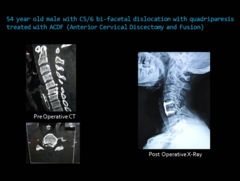
anterior cervical discectomy and fusion (ACDF)
approach through?
risks/complications?
approach through neck, well tolerated in most patients
- post operative dysphagia
- risk of laryngeal injury
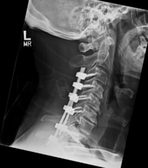
Posterior Cervical Decompression and Fusion (PCDF)
- more morbid than anterior, usually requiring decompression of cord
- adjacent segments fused
spinal shock is due to injury at
devastating injury at or above T6 resulting in a variety of symptoms
*sympathetic chain controlling autonomic nervous system lies above T6, this is what makes that location significant
s/sx of spinal shock
- loss of reflexes
- loss of sensation and flaccid paralysis below area of lesion
- loss of rectal and bladder tone
- hypotension and bradycardia
*inappropriate response to crisis due to unopposed innervation
Recovery dependent on injury; complete spinal cord lesion with poorest prognosis
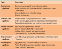
central cord syndrome
common in elderly , hyperextension injury (edema in central spinal cord)
motor dysfunction in UE > LE (legs fine!)
sensory dysfunction below injury
bladder dysfunction
s/sx central cord syndrome
-Increased in elderly population with underlying spinal degenerative problems
-Quadriparesis with loss of pain and temp sensation greater in UE
-Prognosis is good, usually residual loss of fine motor fx of UE
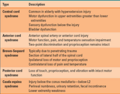
anterior cord syndrome occurs due to
damage to corticospinal, spinothalamic pathways caused by ant. cord compression (flexion of c-spine, thrombosis of ant. cord).
motor function, pain, and temperature sensation impairment distal to lesion
2 point discrimination and proprioception remains intact
tx for anterior cord syndrome
prognosis poor
surgical decompression and stabilization are treatment of choice
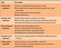
brown-sequard syndrome cause
More frequently caused by penetrating injury causing hemisection of the spinal cord; good prognosis for recovery
s/sx of brown-sequard syndrome
Ipsilateral loss of:
- motor function
- proprioception
- sensation
Contralateral loss of: pain and temp sensation
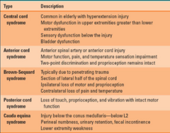
cauda equina syndrome occurs at
the tail end of the spinal cord
s/sx of cauda equina syndrome
- variable motor or sensory loss in LEs
- sciatica
- bladder or bowel dysfunction
- 'saddle anesthesia'
prognosis is good because is considered peripheral nerve injury, regeneration possible
management of a spinal cord injury
• Stabilization of spinal column primary concern
• Referral to neurosurgical services ASAP
• Radiographic studies to evaluate extent of injury
• Intervention to prevent, minimize secondary injury, steroid administration is controversial
surgical intervention of spinal cord injury would be dictated by
Surgical intervention would be dictated by spinal column stability and patient hemostasis:
- Complete Injuries (complete paralysis with loss of motor/sensory below level of injury)
- Incomplete Injuries
ASIA scoring
standardized method of assessing level and motor function of spinal cord injury
level of disability correlating with prognosis and recovery
ASIA A-E
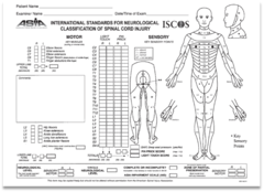
ASIA A
injury is complete spinal cord injury with no sensory or motor function preserved
ASIA B
a sensory incomplete injury with complete motor function loss
e.g. have sensory but no movement
ASIA C
a motor incomplete injury, where there is some movement, but less than half the muscle groups are anti-gravity (can lift up against the force of gravity with a full range of motion)
ASIA D
a motor incomplete injury with more than half of the muscle groups are anti-gravity
ASIA E
normal function
what may have a benefit to improve ASIA score in incomplete spinal cord injuries
Early surgical decompression (injury to 72hrs) may have a benefit to improve ASIA score in incomplete injuries
Continual observation of vital signs, serial neurological observations are essential