Session 3: Caries detection
 Call Kai
 Learn
Learn Practice Test
 Spaced Repetition
 Match
Match Flashcards
 Knowt Play
1/77

There's no tags or description
Looks like no tags are added yet.
Name | Mastery | Learn | Test | Matching | Spaced | Call with Kai | Chat |
|---|
No analytics yet
Send a link to your students to track their progress
78 Terms
dental caries are a (?)-mediated , (?)-modulated process
biofilm-mediated, diet-modulated process that can lead to an imbalance in the cycle of demineralization and remineralization causing localized destruction of tooth structure
microscopic communities of bacteria and slimy matrix
biofilm mediated
in the presence of sugars, the bacteria can produce acid thereby lowering the pH
diet modulated
when is the microbiome in dysbiosis?
in extended low pH

what are some examples of dental caries?
DMFT1938- WHO Classification- standardized method for conducting oral health survey
NYVAD 1999
ICDAS 2005/2011
ADA CCS- convened a group of experts – published in 2015 in JADA
PUFA (pulpal, ulcer, f istula, abscess) 2010
CAST (Caries assessment spectrum and treatment)- instrument2011
which classification system is used at TUSDM?
ADA CCS
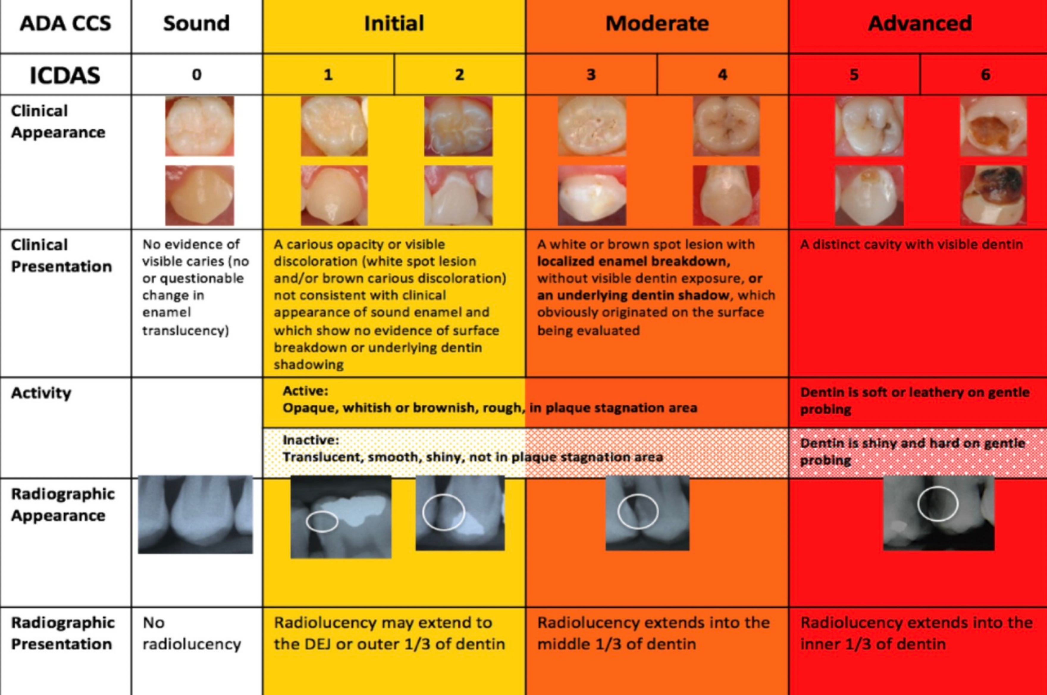
describes how much tooth structure is involved—not whether the lesion is active
severity
ADA Caries Classification System (ADA CCS) categorizes caries lesions according to (what two things?)
extent/severity
activity status
with goal to describe the lesion clinically and support minimally invasive tx decisions
common sites of pit and fissure caries
Occlusal surfaces
Buccal pits of mandibular molars
Lingual pits of maxillary incisors
common sites of smooth surface caries
proximal surfaces
buccal/lingual smooth surfaces
common sites of root surface caries
gingival recession
exposed root surfaces
older adults
smooth and leathery
active
shiny and hard
inactive, arrested
which caries severity/extent clinical appearance:
First visible evidence of mineral loss
White or brown opacity
No cavitation
initial
White spot lesions Early enamel changes
initial

which caries severity/extent clinical appearance:
More advanced enamel breakdown
Localized enamel surface discontinuity
(dentin involvement - not visually - but histologically and radiographically)
moderate
characteristics and management of moderate lesions

which caries severity/extent clinical appearance:
Cavitation with dentin involvement
Structural compromise
Difficult to maintain plaque control
advanced
characteristics and management of advanced lesions

signs of active caries
Chalky/matte appearance
SOFT - WHITHISH/YELLOWISH
Rough surface
Plaque stagnation area Near gingival margin
Recent change in size/appearance
signs of arrested caries
Shiny appearance
BLACKISH / DARKER BROWN
Smooth surface
Hard texture
Not associated with plaque
Smooth Surface Lesions
Whitish/yellowish appearance
Matte/chalky surface
Rough texture
Plaque accumulation
Location associated with stagnation areas
active lesions
Shiny surface
Smooth texture
Hard surface
No plaque stagnation
Darker discoloration may be present
inactive/arrested lesions
what are the tools used for detecting and assessing caries?
visual:
mouth-mirror
reflected light - fiber optic transilluminator for extent, (foti/difoti-microlux)
air-water syringe on a clean tooth surface
WHO probe bc less sharp
AI is decent
radiographs:
BWs bc angulation standardized due to open contacts
what is QLF
quantitative light-induced fluorescence
measures the amnt of fluorescence emitted by tooth structure when exposed to a specific wavelength of light (often blue or violet light)
Healthy enamel emits a measurable amount of fluorescence, while carious lesions emit less
the system quantifies this difference and displays it as a grayscale image, with darker areas indicating higher fluorescence (often sound enamel) and lighter areas indicating lower fluorescence (often carious tissue)
do carious lesions emit more or less fluorescence than healthy enamel?
less
on display the darker areas indicate higher fluorescence
NIRI
Near InfraRed Imaging
an advanced diagnostic tool used primarily in dentistry to detect cavities and monitor dental health without exposing patients to harmful radiation
NIRI technology shines near-infrared light through the teeth. Healthy enamel is transparent to this light, appearing darker on the scan, while dentin and decayed areas scatter the light, making them appear brighter. This contrast helps dentists identify areas of concern effectively

occlusal primary moderate

occlusal primary advanced

occlusal primary initial
chalky white, no E breakdown or microcavitation
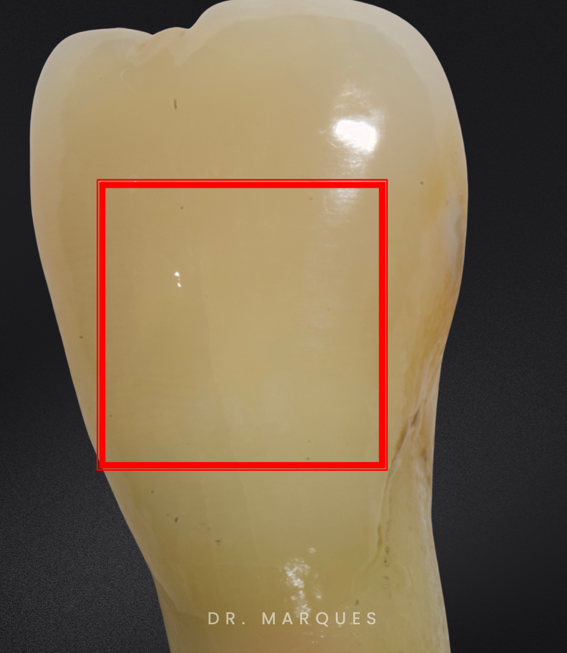
buccal sound
vertical crack not a lesion

occlusal primary moderate
cavitation no D exposed but there is shadowing → E breakdown

occlusal sound

buccal primary advanced
irregular; soft mushy D with plaque buildup

occlusal primary moderate
discolored, cavitation, shadowing, looks shiny and is hard → inactive

occlusal primary advanced

occlusal secondary advanced

occlusal primary initial
no E breakdown but chalky, no microcavitation
apply F for rebalance/mineralization

buccal primary advanced (inactive)
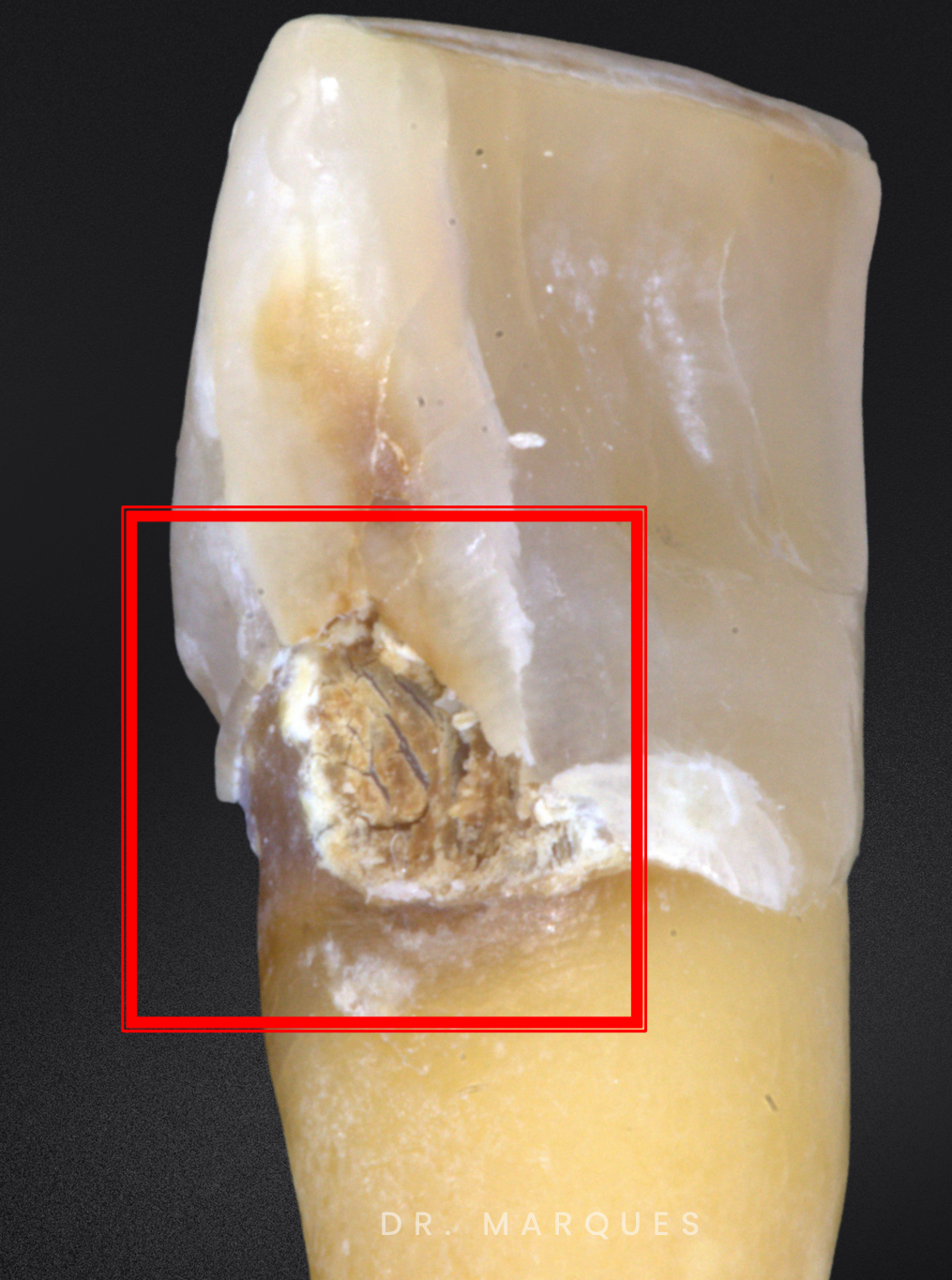
lingual primary advanced
root caries active

occlusal secondary initial (active)
chalky no microcavitation but deep grooves

19 O moderate and B moderate


#5 O moderate

#20 D- advanced O-moderate B-advanced


#29 O moderate

#13 O moderate - shadowing and E breakdown
#12 O initial

27, 28, 29: bucal advanced

#30 O - moderate B - initial
#29 O - initial B -moderate
#28 O - moderate

#19 O and B moderate

#18: Occlusal - moderate B - moderate
#19: Occlusal - moderate

#30: Occlusal - moderate - Buccal - moderate
#31: Occlusal - moderate

#18: Occlusal - moderate
#19: Occlusal - moderate
#21: Occlusal - sound
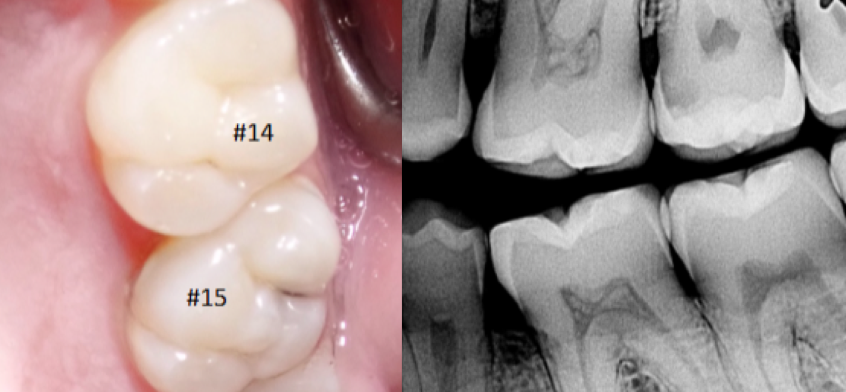
#15: Occlusal - moderate
#14: Occlusal - initial

#11: Facial - initial

occlusal - INITIAL

occlusal - advanced

occlusal - sound

occlusal - moderate
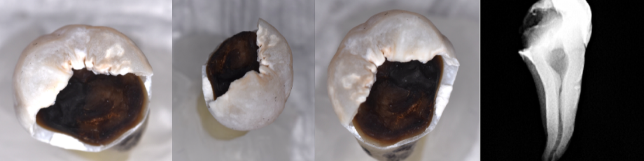
occlusal - ADVANCED

occlusal - ADVANCED

occlusal initial
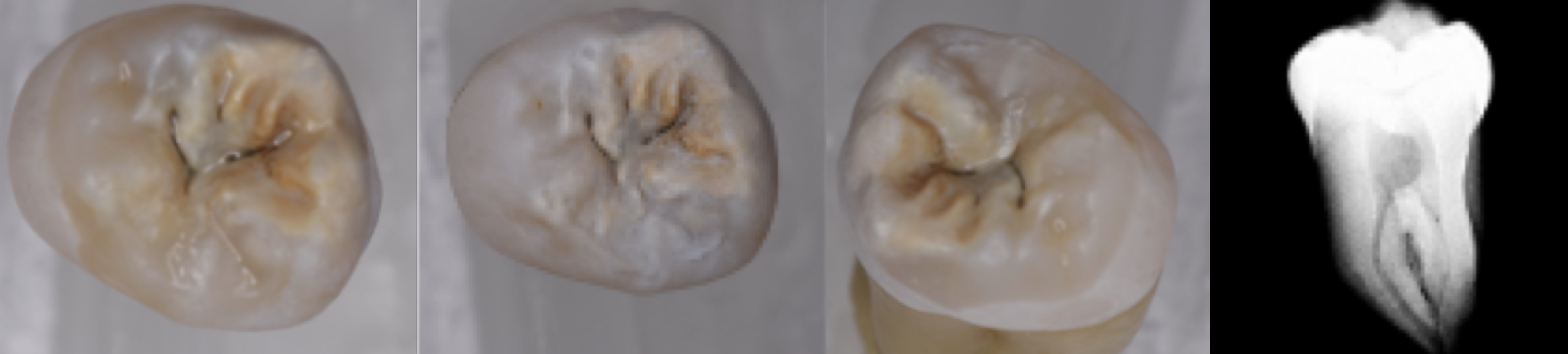
occlusal - moderate
A 2022 publication analyzing GBD 2017 data explicitly notes: • “The Global Burden of Disease (GBD) 2017 study showed that the prevalence of dental caries in permanent teeth ranked (?) among 328 diseases.” PMC
first
untreated caries in permanent teeth had the highest prevalence globally; most common health condition according to the Global Burden of Disease 2021
It is not determined by a single factor but by the (?) between pathological (risk) factors and protective factors
balance
central to the CariesCare International Consensus and modern caries management
The goal of caries management is to (?) pathological factors while (?) protective factors to shift the balance toward oral health
reduce, strengthen
central to the CariesCare International Consensus and modern caries management
CariesCare 4D cycle
determine caries risk, detect and assess, decide on a personalized care plan, do preventative and tooth-preserving care
what are some caries risk factors?
Visible plaque accumulation
Frequent exposure to fermentable carbohydrates
Reduced salivary flow hyposalivation/xerostomia)
Exposed root surfaces
Deep pits and fissures
Orthodontic appliances or other plaque- retentive factors
Frequent consumption of sugary foods or drinks (especially between meals)
Poor oral hygiene
Inadequate fluoride exposure
Irregular dental attendance
Low adherence to preventive recommendations
Medications causing xerostomia
Head and neck radiation therapy
Medical conditions affecting salivary function
Low socioeconomic status (associated with increased disease burden)
Limited access to dental care
Low oral health
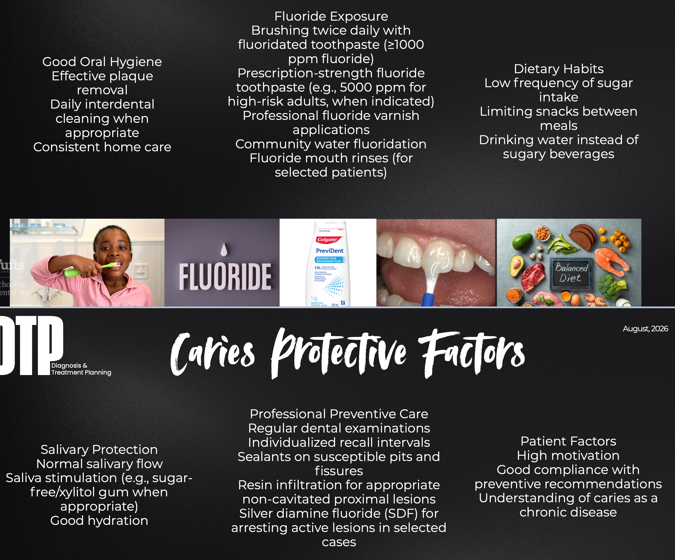
what do caries protective factors do?
reduce the likelihood of caries initiation or progression by enhancing remineralization, reducing demineralization, or modifying the oral environment
what are some caries protective factors?
how much of the tooth structure is involved
caries staging
severity of the lesion
stage
lesion management depends on:
Lesion stage
Lesion activity
Cavitation
Cleansability
Patient caries risk
Patient preferences and clinical circumstances
sound surface tx options
Prevention Routine monitoring
initial arrested lesion tx options
Fluoride varnish
Sealant (occlusal lesions)
Resin infiltration
Active surveillance
initial active lesion tx options
Fluoride therapy
Dietary modification
Oral hygiene improvement
Active surveillance
moderate lesion tx options
Management depends on whether the lesion can be effectively cleaned and controlled
Possible options:
Non-operative care
Micro-invasive treatment
Minimally invasive restoration
advanced/cavitated lesion tx options
When the lesion cannot be adequately cleaned or maintained, operative care is indicated
Goals:
Restore function
Create a cleansable tooth surface
Preserve as much healthy tooth structure as possible
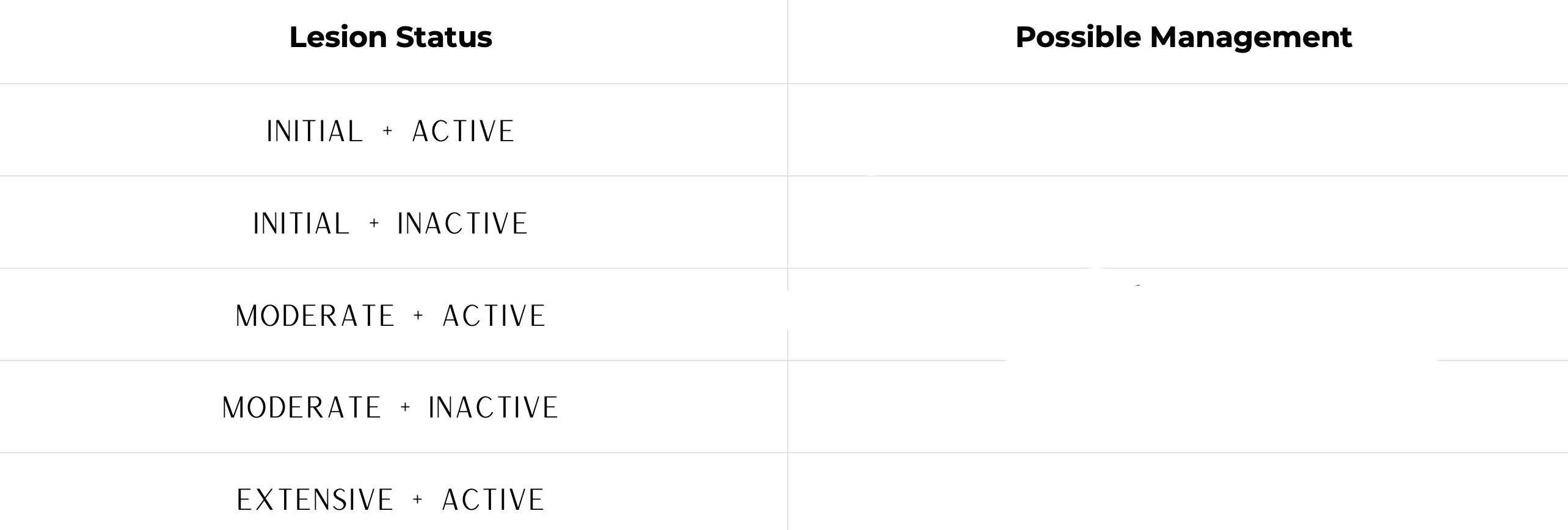
treatment depends on what three things?
pt risk, lesion stage, lesion activity
