Pathology of the Ovary
 Call Kai
 Learn
Learn Practice Test
 Spaced Repetition
 Match
Match Flashcards
 Knowt Play
1/170

Earn XP
Description and Tags
617
Name | Mastery | Learn | Test | Matching | Spaced | Call with Kai | Chat |
|---|
No analytics yet
Send a link to your students to track their progress
171 Terms
Should the Graafian follicle fail to ovulate, it could continue to enlarge and result in a —
follicular cyst
Follicular cysts range in size from —; however, larger cysts have been documented.
3 to 8 cm
surgical intervention or drainage may be warranted because a large cyst increases the risk for —
ovarian torsion
The surgical removal of an ovarian cyst is referred to as —
ovarian cystectomy.
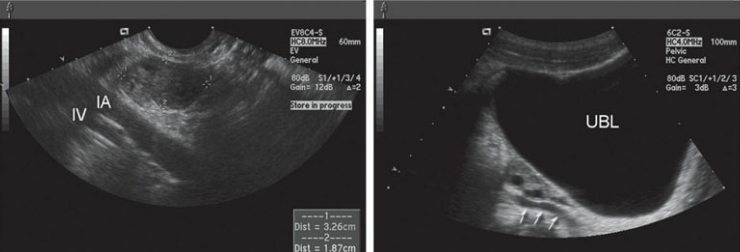
Normal sonographic appearance of the ovary. A. In this longitudinal endovaginal image of the pelvis, the ovary (between calipers) is noted adjacent to the iliac artery (IA) and iliac vein (IV). B. A normal ovary can be seen in this longitudinal transabdominal image of the pelvis between the urinary bladder (UBL) and prominent ureter (arrows).

Transverse image of the ovary on day 5 of the menstrual cycle revealing a high-impedance waveform.
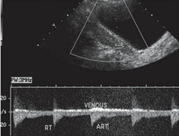
Transverse image of the ovary during day 10 of the menstrual cycle revealing a low-impedance waveform pattern with more diastolic flow. (Color image provided online.)
Early follicular phase
High impedance (resistance) with absent or low end-diastolic velocity
RI = 1.0
Late follicular phase
Low impedance (resistance) with increased end-diastolic flow
RI = 0.5'
Early luteal phase
Low impedance (resistance) with increased end-diastolic flow
RI = 0.5
Late luteal phase
High impedance (resistance) with absent or low end-diastolic velocity
RI = 1.0

Simple ovarian cyst. Coronal image of the right ovary demonstrating a simple ovarian cyst (arrows), surrounded by normal ovarian tissue (arrowheads).
Hyperstimulation of the ovaries, or —, from fertility treatment will also result in the development of multiple, enlarged follicular cysts
ovarian hyperstimulation syndrome
A follicular cyst that contains blood is referred to as a —, and it most often appears complex or completely echogenic depending on the hemorrhagic component present and the stage of lysis
hemorrhagic cyst
The sonographic manifestation of a hemorrhagic cyst may be described as demonstrating a —
weblike or lacy appearance
clinical findings of follicular cysts
aysmptomatic
pain associated with hemorrhage and enlargement of cyst
— is a physiologic (functional) cyst that develops after ovulation has occurred.
The corpus luteum cyst
The corpus luteum is formed as a result of the — and is responsible for producing progesterone
rupture of the Graafian follicle
the corpus luteum will normally regress if— but may rarely be maintained and continue to enlarge
fertilization does not occur
When regression takes place, a small, echogenic structure may be noted within the ovary, representing the —
corpus albicans

Hemorrhagic ovarian cyst
sonographic findings of follicular cyst
Simple cyst–anechoic, thin walled, unilocular, round, posterior enhancement
Hemorrhagic cyst—variable appearances, including complex components or entirely echogenic, depending on the amount of blood and the stage of lysis; may have a weblike or lacy appearance as well
Corpus luteum cysts may reach sizes up to —, with resolution of the cyst taking place within 1 to 2 months in menstruating patients
8 cm
In the presence of a pregnancy, and thus the production of human chorionic gonadotropin (hCG) by the trophoblastic cells of the pregnancy, —
the corpus luteum is preserved
These cysts are considered the most common pelvic masses seen during a first-trimester sonographic examination
corpus luteum of pregnancy
corpus luteum of pregnancy may even reach sizes up to 10 cm, although most often they resolve by 16 weeks gestation and do not exceed —
3 cm
A complex or thick-walled corpus luteum cyst can resemble an —, so precaution to establish the presence of an intrauterine pregnancy should be taken in this regard, as well as careful consideration of the entire clinical picture
ectopic pregnancy
clinical findings of corpus luteum cysts and corpus luteum of pregnancy
asymptomatic
Pain associated with hemorrhage and enlargement of cyst
Corpus luteum of pregnancy accompanies a pregnancy
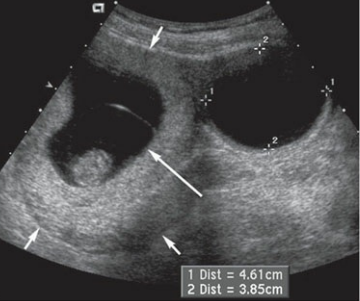
thin-walled unilocular cyst
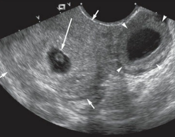
a thick-walled cyst
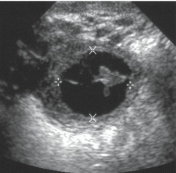
a complex cyst with septations

structure that contains diffuse internal echoes
sonographic findings of corpus luteum cysts and corpus luteum of pregnancy
Simple cyst appearance
May have a thick wall, be completely echogenic, and may be difficult to differentiate from other solid and cystic adnexal masses
Hemorrhagic components may appear complex or have a weblike or lacy appearance depending on the amount of blood and stage of lysis
— are the largest and least common of the functional cysts
Theca lutein cysts
Theca lutein cysts are found in the presence of —, often exceeding 100,000 mIU per mL
elevated levels of hCG
— are common conditions associated with theca lutein cysts
gestational trophoblastic disease (molar pregnancy) and ovarian hyperstimulation syndrome
— would also have higher levels of hCG and therefore increase the likelihood of developing theca lutein cysts
multiple gestations
Theca lutein cysts are large cysts and are frequently —
bilateral and multiloculated, and may reach sizes up to 15 cm
Theca lutein cysts tend to regress after the —
high level of circulating hCG diminishes

Theca lutein cysts. Transabdominal image
clinical findings of theca lutein cysts
Markedly elevated levels of hCG (as seen in cases of gestational trophoblastic disease, ovarian hyperstimulation, and twin gestations)
Nausea and vomiting
Pelvic fullness
Pain associated with hemorrhage, rupture, and ovarian torsion
sonographic findings of paraovarian cysts
Simple cyst located adjacent, but not attached, to the ovary
If hemorrhagic, will appear complex
Parovarian cysts are small cysts located adjacent to the ovary, and most likely arise from the —
fallopian tubes or broad ligaments
paraovarian cysts can range in size from —
1.5 to 19 cm
sonographic findings of theca lutein cysts
Large, bilateral, multiloculated ovarian cystic masses
May contain hemorrhagic components
clinical findings of paraovarian cysts
asymptomatic
If cyst is large, patients may present with pelvic pain and increased lower abdominal girth

Paraovarian cyst. A. A simple appearing cyst (C) is noted adjacent to the ovary (arrows) and posterior to the urinary bladder (B) in this longitudinal image. B. The same patient in transverse.
The most common benign ovarian tumor is the —, also referred to as a dermoid cyst
ovarian cystic teratoma
Dermoids result from the —
retention of an unfertilized ovum that differentiates into the three germ cell layers.
these germ cell tumors are composed of —
ectoderm, mesoderm, and endoderm
As a result of the combination of these germ cells, a cystic teratoma may contain any number of tissues, including —
glandular thyroid components, bone, hair, sebum, fat, cartilage, and digestive elements
Dermoids are commonly found in the — but may also be found in postmenopausal patients
reproductive-aged group
— denotes the sonographic appearance of the mass when only the anterior element of the dermoid cyst is seen, while the greater part of the mass is obscured by shadowing
The “tip of the iceberg” sign
Often, dermoid tumors contain a —
“dermoid plug.”
The dermoid plug contains various tissues that will be a source of —
posterior shadowing
The “dermoid mesh” has been used to describe the visualization of —
hair within the mass.
clinical findings of a cystic teratoma
Often asymptomatic
If torsion or rupture occurs, the patient may present with acute pelvic pain
sonographic findings of a cystic teratoma
Complex, partially cystic mass in the ovary that includes one or more echogenic structures that may shadow
“Tip of the iceberg” sign–only the anterior element of the mass is seen, while the greater part of the mass is obscured by shadowing
Dermoid plug–produces posterior shadowing
Dermoid mesh–produced by hair and will appear as numerous linear interfaces within the cystic area of the mass

Cystic teratoma. A. A coronal image of the female pelvis demonstrating a complex ovarian mass (between large arrows) with several typical sonographic characteristics of a dermoid, including the dermoid plug (small arrows) and the posterior shadowing (between arrowheads) from the plug. B. In the sagittal plane, this dermoid (between arrows) reveals a solid (arrows) shadowing (between arrowheads) structure within its borders.
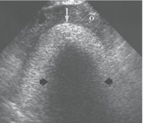
Cystic teratoma and the “tip of the iceberg” sign
— is a benign ovarian sex cord-stromal tumor
A thecoma
Thecomas are most often found in postmenopausal women and may be associated with —
Meigs syndrome
Meigs syndrome describes the condition of a having a —
benign ovarian tumor with ascites and pleural effusion
Thecomas are —; therefore, patients often complain of postmenopausal vaginal bleeding associated with the unconstrained estrogen stimulation upon the endometrium.
estrogen-producing tumors
thecomas are most often — and may appear similar to a pedunculated uterine leiomyoma
unilateral
clinical findings of a thecoma
May be asymptomatic
Postmenopausal vaginal bleeding or abnormal vaginal bleeding secondary to estrogen stimulation
Meigs syndrome (ascites and pleural effusion)
sonographic findings of a thecoma
Hypoechoic, solid mass with posterior attenuation
No posterior enhancement
If large, it may mimic a pedunculated leiomyoma
— is considered to be the most common estrogenic tumor
The granulosa cell tumor, also referred to as the granulose theca cell tumor,
the granulosa cell tumor is also a— like the thecoma
sex cordstromal tumor
the granulosa cell tumors typically occur — but they can also be found in younger patients
unilaterally and are more commonly seen in the postmenopausal female
Because of its estrogen-producing potential, a granulosa cell tumor will present clinically much like the —.
thecoma
As a result of consistent estrogen stimulation, postmenopausal patients with granulosa cell tumors have approximately a 10% to 15% chance of developing —
endometrial carcinoma
Granulosa cell tumors can reach sizes up to — and do have malignant potential.
40 cm
clinical findings of granulosa cell tumors
Adolescence–pseudoprecocious puberty
Reproductive-aged and postmenopausal women will have abnormal vaginal bleeding
sonographic findings of granulosa cell tumors
Solid, hypoechoic mass
Complex or partially cystic mass
In pediatric patients, the granulosa cell tumor is associated with —
pseudoprecocious puberty.

Granulosa cell tumor. Longitudinal image of a 3-year-old girl suffering from premature breast development demonstrating a granulosa cell tumor (between arrows) located posterior to the urinary bladder (B).
An ovarian fibroma is also considered a —
sex cord-stromal tumor
Unlike thecomas and granulosa cell tumors however, fibromas are —
not associated with estrogen production
Fibromas are most often found in —
middle-aged women
fibromas are benign ovarian masses that may be complicated by —
Meigs syndrome as well
Often, fibromas, like thecomas, may mimic —
pedunculated uterine leiomyoma
The ascites and pleural effusions associated with Meigs syndromes usually resolve after —
resection of the tumor
clinical findings of a fibroma
May be asymptomatic
Meigs syndrome (ascites and pleural effusion)
sonographic findings of a fibroma
Hypoechoic, solid mass with posterior attenuation
No posterior enhancement
If large, it may mimic a pedunculated leiomyoma
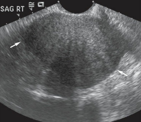
Ovarian fibroma. Sagittal image of the right ovary demonstrating a solid mass (arrows) with a homogeneous echotexture.
Brenner tumors, or —, are most often small, solid, hypoechoic unilateral tumors that may contain calcifications.
transitional cell tumors
brenner tumors are almost always benign, but they can —
undergo malignant degeneration
clinical findings of a brenner tumor
May be asymptomatic
Meigs syndrome (ascites and pleural effusion)
sonographic findings of a brenner tumor
Small, solid, hypoechoic mass
May contain calcifications
— is a benign, blood-containing tumor that is associated with endometriosis and forms from the implantation of ectopic endometrial tissue
An endometrioma
This ectopic endometrial tissue is —
functional
It is this hemorrhage that forms into focal areas of bloody tumors—-
endometriomas
Consequently, they have been nicknamed — because they appear as dark, thick bloody masses during gross examination
“chocolate cysts”
Endometriomas can be located anywhere outside the endometrial cavity, including on any other pelvic organ, such as the bladder and bowel, but are more commonly found on—
the ovary
Most often, these masses are multiple and seen more often in the —
reproductive years.
endometriomas can be found within the — and present as a palpable mass that may change shape throughout the menstrual cycle in relation to hormone alterations
cesarean section scar
clinical findings of endometriomas
Patient may be asymptomatic
pelvic pain
infertility
dysmenorrhea
menorrhagia
dyspareunia
painful bowel movements (dyschezia)
sonographic findings of endometriomas
Predominantly cystic mass with low-level internal echoes (may resemble a hemorrhagic cyst)
Anechoic or complex, mostly cystic mass with posterior enhancement and may have a fluid–fluid level