672 Lab 8/11 Vital Signs
 Call Kai
 Learn
Learn Practice Test
 Spaced Repetition
 Match
Match Flashcards
 Knowt Play
1/74

There's no tags or description
Looks like no tags are added yet.
Name | Mastery | Learn | Test | Matching | Spaced | Call with Kai | Chat |
|---|
No analytics yet
Send a link to your students to track their progress
75 Terms
Pulse Rate
the number of heart beats per minute, the rate at which the ventricular heart contraction pushes a pressure wave of blood throughout the arterial system
Assessing Pulse Rate (in bpm)
often use carotid and radial pulse
never palpate the carotids simultaneously or use the thumb to assess pulses
3 Options for Obtaining Pulse
60 sec
30 sec x 2
15 sec x 4
Normal Range of Pulse for Adults
60-100 bpm
<60 bpm
bradycardia
>100 bpm
tachycadia
Respiratory Rate
the number of breaths a person takes per minute, the rate at which air is drawn into the lungs during inspiration and expelled during expiration when the intercostal muscles and diaphragm relax (allowing the chest wall to recoil)
Obtaining Respiratory Rate in Breaths Per Minute (bpm)
can place arm on shoulder to better feel each breath, easiest to take respiratory rate immediately after pulse, may need to distract patient so they don’t change their breathing pattern
2 Options for Obtaining Respiratory Rate
0 sec
30 sec x 2
Normal Range of Respiratory Rate for Adults
12-20 breaths per min
<12 breaths pm
bradypnea
>20 breaths pm
tachypnea
0 breaths pm
apnea
3 Elements of Pulse Palpation Technique
student palpates carotid pulse bilaterally, one at a time, and reports pulse rate in bpm
student palpates radial pulse bilaterally and reports pulse rate in bpm
student reports if pulse is high, low, or as expected
2 Elements of Reporting Respiration Technique
student obtains and reports respiratory rate in breaths per min
student reports if respiratory rate is high, low, or as expected
Blood Pressure
the force of the blood against the wall of an artery as the ventricles of the heart contract and relax
systolic pressure/diastolic pressure
Systolic Pressure
the force exerted when the ventricles contract
is largely the result of cardiac output, blood volume, and compliance of the arteries
Diastolic Pressure
the force exerted by peripheral vascular resistance when the heart is in the filling or relaxed state, lowest arterial pressure during ventricular filling
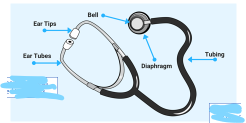
Anatomy of a Stethoscope
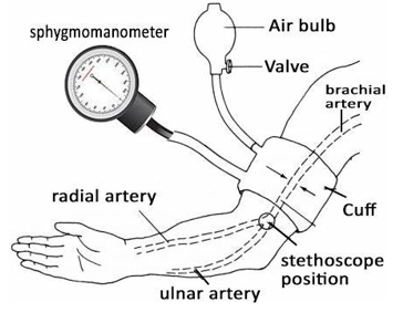
Anatomy of a Sphygmomanometer
How to Place the BP Cuff Correctly
placed approx 1 inch above the antecubital fossa, must be on bare skin, place artery marker over the brachial artery, make sure the artery marker is within the range indicated on the cuff
BP Cuff too Big
underestimated blood pressure
BP Cuff too Small
overestimated blood pressure
Why should the patient’s arm be kept at heart height while BP is being taken?
placing the arm below heart level leads to an overestimation of systolic and diastolic pressures (bp measured is higher than actual bp)
placing the arm above the heart leads to an underestimation (bp measured is lower than actual bp)
How do you ensure the patient’s arm stays at heart height?
either pin it to your side under your arm or rest it on a table/pillow
Taking Systolic BP with Palpation
palpate radial or brachial artery
inflate cuff until you no longer feel a pulse +20-30 mmHg
then deflate the cuff slowly and note when you feel the pulse again (systolic)
document as (SBP)/Palpatory
Obaining BP By Ausculation
place and inflate the cuff same as method used for palpatory
place stethoscope over brachial artery
deflate until you hear the first and last beats (korotkoff sounds)
Normal Adult BP
120/80
can be between <90/<60 and >130/>80
Hypotension
<90/60
Hypertension
>130/80
Korotkoff Sounds
sound made by the turbulent flow of blood in artery
pressure at which the first tapping sound is heard is the systolic bp
pressure at which the last sound is heard is diastolic bp
Orthostatic Hypotension
within 2-5 mins of standing or following a seated or supine position, the systolic bp drops by 20 mmHg+ or the diastolic drops by 10 mmHg+, leading to a “faint” or “lightheaded” feeling upon standing
5 Elements of Blood Pressure Technique
student places cuff appropriately on the patient’s arm
student keeps the arm at or near the level of the patient’s heart
student supports patient’s arm (or rests on table)
student obtains a systolic blood pressure reading by palpation
student obtains BP by auscultation and reports the value
What portion of SOAP do vital signs and comments go in?
objective
3 Components of Physical Exam Used for Vital Signs
inspection
ausculation
palpation
Inspection
note general appearance, symmetry, presence of edema, deformities, color, and presence of any skin lesions always comparing bilaterally
Auscultation
listen to body sounds with and without the stethoscope, use the diaphragm for high-frequency sounds and the bell for low-frequency sounds
Palpation
identify landmarks that are related to the region being examined
note presence of heat, deformities, edema, effusion, tenderness, crepitus, and pulses
4 Vital Signs
temp
heart rate/pulses
respiratory rate
blood pressure
How is body temp maintained and regulated?
controlled by the hypothalamus, set point can be raised or lowered in response to external and internal stimuli
Vasoconstriction (for temp)
increases body temp by reducing heat loss through the skin
Vasodilation (for temp)
decreases body temp by promoting heat loss through the skin
3 Ways Body Temp is Taken
oral or rectal thermometry
infrared thermometry
smart phones
Oral or Rectal Thermometry
preferred for body temp, central thermometry (bladder, rectal) or peripheral thermometry (oral, ear, axilla, forehead)
Infrared Thermometry
measure the amount of heat produced, accuracy may be affected by sweating or vascular changes, can be higher or lower than rectal
Smart Phones Temp
apps that track fever and require an external sensor with accuracy commensurate, not recommended
Normal Body Temp
97.2-99.9 F or 36.2-37.5 C
average 98.6 F or 37 C
How does body temp vary?
varies with age, time of day, level of activity, phase of menstrual cycle (temp rises 0.9 F during ovulation)
highest in evening and lowest in morning
certain non-infectious disease can cause high or low temp
Serial Temperature Measurements
can be used in hospitalized patients to provide a clue to the severity of the disease
>100.4 F or>38.0 C
fever
Persistent Fever
does not fluctuate by >1.8 F in a 24 hour period
Remittent Fever
oscillates by 3.6 F during the day
Intermittent/Spiking Fever
present for several hours at a time before returning to normal
Geriatric Patient Temp
a normal temp is commonly observed in patients with severe infections due to blunted response from the hypothalamus
Arterial Pulse
rhythmic wave produced by the ejection of blood from the left ventricle into the aorta, the pressure causes a smooth and rapid upstroke that can be palpated in arteries, venous pulse is not palpated because it is a low-pressure system
3 Ways Pulse is Evaluated
rate and rhythm of heart
contour of the pulse
amplitude of the pulse
Contour of the Pulse
the shape of the pulse wave
described by the speed of the upward slope, downward slope, and duration of wave
Amplitue of the Pulse
the maximal force of the pressure wave
described as normal, increased, diminished, or double-peaked
5 Types of Irregular Pulse
anacrotic
waterhammer
bisferiens
alternans
paradoxical
Anacrotic
type of irregular pulse, caused by aortic stenosis
small, slow rising, delayed pulse with a notch or shoulder on the scending limb
Waterhammer
type of irregular pulse, caused by aortic regurgitation
rapid and sudden systolic expansion
Bisferiens
type of irregular pulse, caused by aortic regurgitation
double-peaked pulse with a midsystolic dip
Alternans
type of irregular pulse, caused by congestive heart failure
alternating amplitude of pulse pressure
Paradoxical
type of irregular pulse, caused by tamponade, constrictive pericarditis, and chronic obstructive heart disease
detected by blood pressure assessment, and exaggerated drop in systolic pressure during inspiration
6 Arteries to Palpate
carotid
radial
femoral
popliteal
dorsalis pedis
posterior tibial
Adult Athlete Heart Rate
50-60 resting
Children Heart Rates
newborn = 120-170 bpm
toddler = 80-120 bpm
school age 70-110 bpm
Pulse Oximetry
measures the percentage of oxygen-saturated hemoglobin in SpO2, normal is >97%
oxygen saturation is a percentage of the maximum amount of the oxygen the blood can carry
Respiratory Rate
number of breaths per minute, one respiration is start of inspiration to end of expiration
Child Respiratory Rate
newborn = 40-60 bpm
toddler = 20-30 bpm
school age = 16-20 bpm
White Coat Syndrome
isolated elevation of blood pressure measurements in the healthcare setting
Direct vs Indirect Blood Pressure
direct → intra-arterial catheter connected to a pressure sensor
indirect → blood pressure cuff
Width and Length of BP Cuff Bladder Should Be
width → 1/3 to ½ circumference of limp
length → 2x width or 80% of limb circumference
5 Phases of Korotkoff Sounds
phase 1 - sharp thuds, two sounds for systolic
phase 2 - thuds become more blowing or swishing
phase 3 - softer thuds, still crisp less blowing sound
phase 4 - soft thuds become soft blowing that disappears
phase 5 - silence, last sound is diastolic
15 Full Steps of Measuring Blood Pressure
Patient is at rest; sitting or lying down
Palpate the brachial artery
Wrap the cuff around the patients arm with the “artery” indicator towards the brachial artery
Support the patients arm atop your arm at heart level
Hold the inflator bulb in your opposite hand
While palpating the radial artery, inflate the bladder until the pulse is no longer felt
Slowly deflate the cuff by 2 – 3 mmHg per second until you feel two beats of the pulse
Deflate the cuff completely
Record the reading as “Systolic pressure by Palpation” e.g. “120/P”
Wait 30 seconds before inflating cuff again
Inflate the cuff to 20 – 30 mmHg above the systolic pressure determined by palpation
Place the diaphragm of the stethoscope over the brachial artery
Listen for the Korotkoff sounds
Deflate the cuff completely
Document your findings