Other Gram -ve NON fermenting part 1
 Call Kai
 Learn
Learn Practice Test
 Spaced Repetition
 Match
Match Flashcards
 Knowt Play
1/93

There's no tags or description
Looks like no tags are added yet.
Name | Mastery | Learn | Test | Matching | Spaced | Call with Kai | Chat |
|---|
No analytics yet
Send a link to your students to track their progress
94 Terms
Genus Pseudomonas characteristics
Gram-negative medium-sized rods
Arrangement of Pseudomonas
Pairs
Oxygen requirement of Pseudomonas
Obligate aerobes
Pseudomonas are fermenters or nonfermenters?
Nonfermenters
Oxidase test of Pseudomonas
Positive
Catalase test of Pseudomonas
Positive
Most important species of Pseudomonas
Pseudomonas aeruginosa
Habitat of Pseudomonas aeruginosa
Soil water skin and mucous membranes
Characteristic odor of Pseudomonas aeruginosa colonies
Fruity/grape-like odor
Characteristic colony appearance of Pseudomonas aeruginosa
Mucoid colonies
Serotyping of Pseudomonas aeruginosa is based on
O (somatic) antigens
Blue-green pigment produced by Pseudomonas aeruginosa
Pyocyanin
Function of pyocyanin
Produces reactive oxygen species and damages host tissues
Yellow-green fluorescent pigment produced by Pseudomonas aeruginosa
Pyoverdin
Function of pyoverdin
Siderophore that acquires iron
Dark red pigment produced by Pseudomonas aeruginosa
Pyorubin
Brown-black pigment produced by Pseudomonas aeruginosa
Pyomelanin
Major virulence factors of Pseudomonas aeruginosa
Fimbriae capsule pyocyanin pyoverdin exotoxin A elastase hemolysin leukocidin and drug resistance
Function of fimbriae
Adhesion
Function of capsule
Resists phagocytosis
Most important toxin of Pseudomonas aeruginosa
Exotoxin A
Mechanism of exotoxin A
Inhibits host cell protein synthesis
Result of exotoxin A activity
Cell death
Function of elastase
Destroys elastin causing tissue and lung damage
Function of hemolysin
Lyses red blood cells
Function of leukocidin
Kills leukocytes
Pseudomonas aeruginosa is primarily what type of pathogen?
An opportunistic pathogen
Animals at greatest risk of Pseudomonas infection
Immunocompromised animals
Other major predisposing factors for Pseudomonas infection
Burns and concurrent infections
Pseudomonas aeruginosa commonly causes
Urinary tract infections, ear and wound infections
Pseudomonas aeruginosa commonly causes
Ear infections
Pseudomonas aeruginosa commonly causes
Wound infections
Characteristic color of Pseudomonas wound infections
Blue-green
Pseudomonas aeruginosa commonly causes (2)
Pneumonia and mastitis
Pseudomonas aeruginosa commonly causes
Mastitis
Pseudomonas aeruginosa commonly causes in sheep
Fleece rot (green wool)
Pseudomonas aeruginosa commonly causes in horses
Ulcerative keratitis
Characteristic appearance of fleece rot
Green discoloration of wool
Why is Pseudomonas difficult to treat?
High antimicrobial resistance due to porin-mediated drug resistance
Best clues for identifying Pseudomonas aeruginosa
Oxidase positive obligate aerobe blue-green pigment fruity odor and nonfermenter
Genus Burkholderia characteristics
Gram-negative rods
Oxygen requirement of Burkholderia
Obligate aerobes
Major virulence factor of Burkholderia
Polysaccharide capsule
Burkholderia mallei causes
Glanders
Primary host of Burkholderia mallei
Horses and other equids
Other susceptible hosts of Burkholderia mallei
Humans cats and dogs
Transmission of Burkholderia mallei
Ingestion of contaminated food or water
Other routes of Burkholderia mallei transmission
Inhalation and skin wounds
Burkholderia mallei primarily causes
Chronic disease
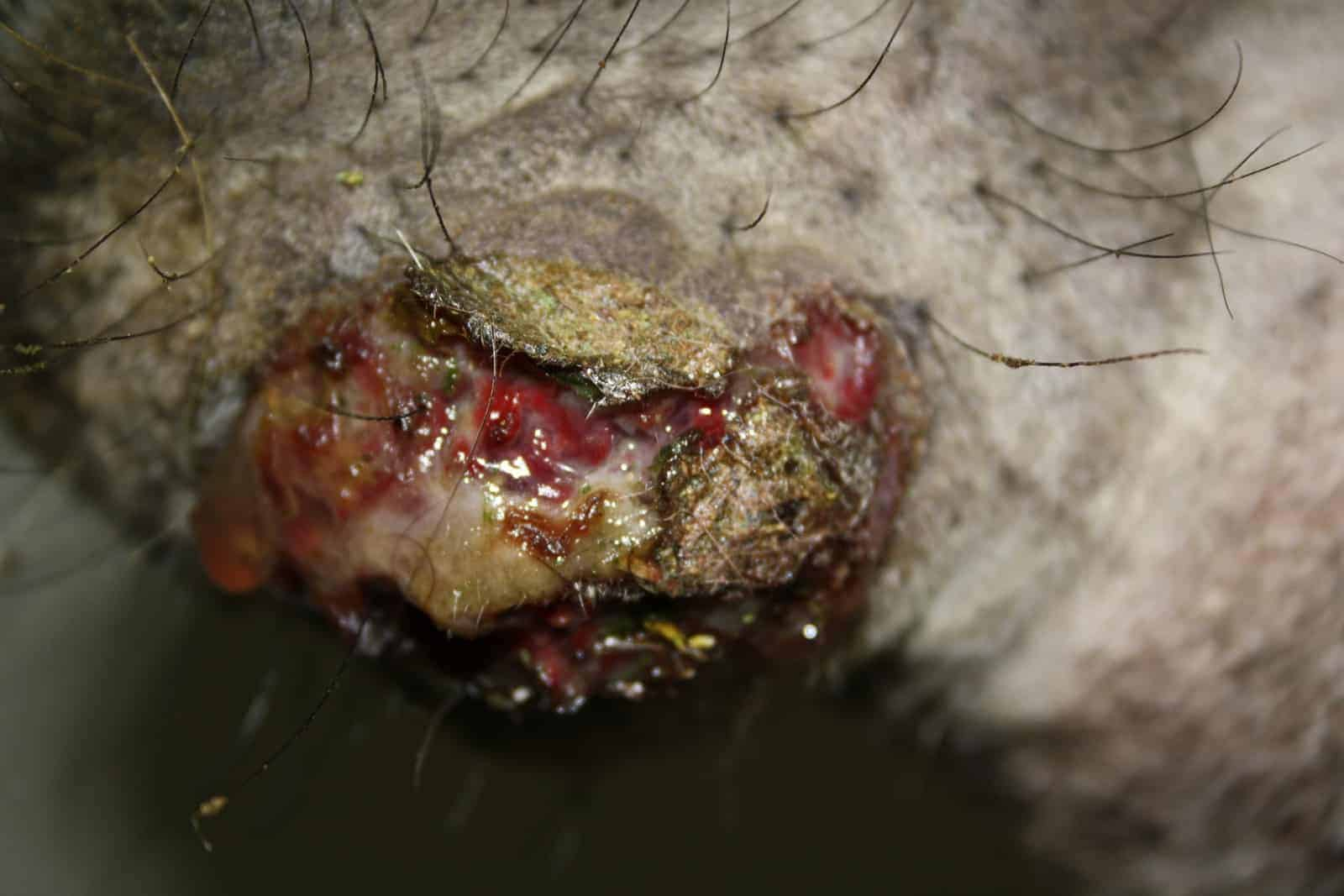
Characteristic lesion of glanders
Caseous abscesses
Burkholderia mallei induces
Delayed hypersensitivity
Three forms of glanders
Nasal pulmonary and cutaneous (farcy)
Nasal form of glanders causes
Nodular lesions and ulcers on the nasal septum
Pulmonary form of glanders causes
Nodular lesions and abscesses in the lungs
Cutaneous form of glanders is called
Farcy
Cutaneous glanders is characterized by
Lymphangitis with discharging abscesses along lymphatic vessels
Diagnostic skin test for glanders
Mallein test
Mallein test is based on
Delayed hypersensitivity reaction
Mallein test is performed by
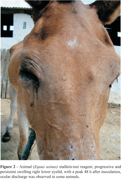
Intrapalpebral injection of mallein
Positive mallein test
Eyelid swelling with purulent ocular discharge
Time to read a mallein test
More than 24 hours after injection
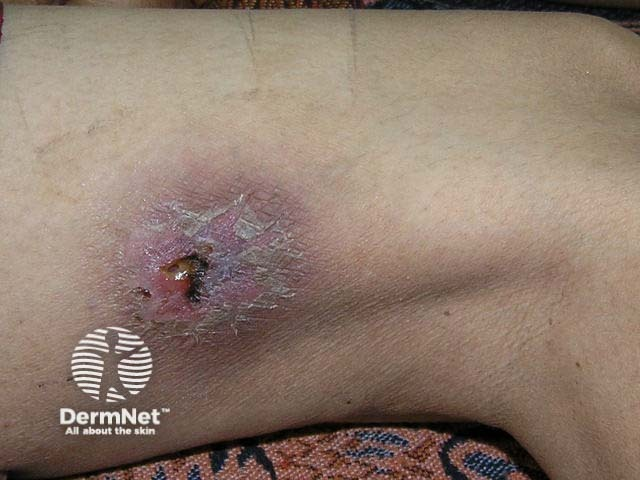
Burkholderia pseudomallei causes
Melioidosis (Pseudoglanders)
PS: because it looks and acts almost exactly like an ancient, highly contagious veterinary disease called glanders, but it is caused by a different, "pseudo" (false) version of the bacteria
Habitat of Burkholderia pseudomallei
Soil water and vegetation
Transmission of Burkholderia pseudomallei
Inhalation ingestion or skin wounds
Acute form of melioidosis causes
Septicemia with abscesses in multiple organs
Chronic melioidosis resembles
Glanders
Lawsonia intracellularis characteristics
Slender curved Gram-negative rod
Oxygen requirement of Lawsonia intracellularis
Microaerophilic (require low levels of oxygen to survive)
Lifestyle of Lawsonia intracellularis
Obligate intracellular bacterium
Lawsonia intracellularis can only be grown in
Cell culture
Lawsonia intracellularis causes
Proliferative enteropathy (contagious GIT disease)
Primary host of Lawsonia intracellularis
Pigs
Most susceptible pigs
Post-weaning and young growing pigs
Lawsonia intracellularis replicates in
Ileal crypt epithelial cells
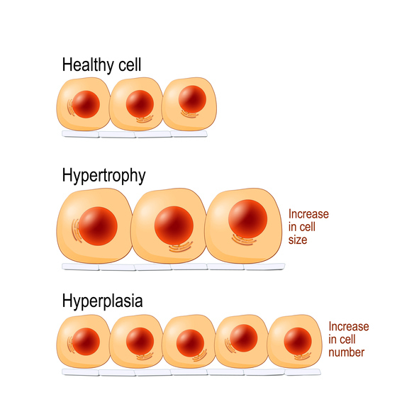
Effect of Lawsonia intracellularis on crypt cells
Hyperplasia (proliferation) (an abnormal increase in the number of cells in an organ or tissue, which causes it to enlarge)
Most severely affected intestinal region
Terminal ileum
Clinical signs of proliferative enteropathy
Diarrhea dehydration abdominal pain and arched back
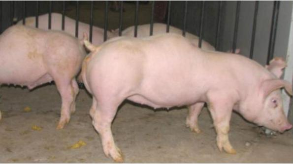
Characteristic intestinal lesion
Thickened intestinal mucosa
Proliferative hemorrhagic enteropathy occurs most commonly in
Older pigs
Characteristic lesion of proliferative hemorrhagic enteropathy
Thickened intestinal wall with hemorrhage into the lumen
Case fatality rate of proliferative hemorrhagic enteropathy
Approximately 50%
Porcine intestinal adenomatosis occurs in
Pigs younger than 12 weeks
Lesions of porcine intestinal adenomatosis
Thickened mucosa of terminal ileum and upper large intestine
Clinical signs of porcine intestinal adenomatosis
Anorexia wasting and recovery
Necrotic enteritis caused by Lawsonia intracellularis occurs in
Pigs younger than 12 weeks
Characteristic lesion of necrotic enteritis
Necrosis of proliferative intestinal mucosa
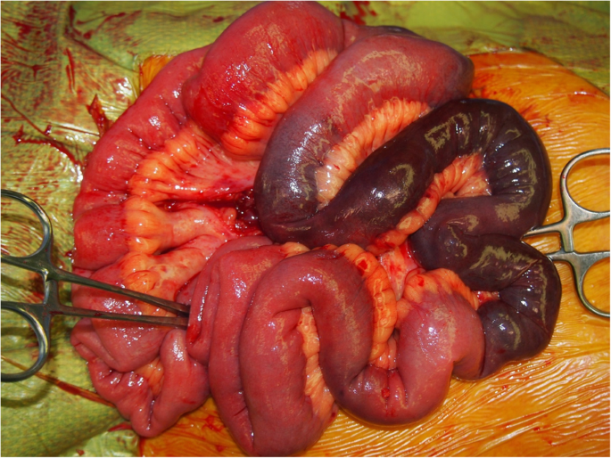
Clinical outcome of necrotic enteritis
Sudden death
Regional ileitis occurs in
Pigs younger than 12 weeks
Nickname for regional ileitis
Hosepipe gut
Why is it called hosepipe gut?
Thick rigid intestine due to muscular hypertrophy
Lesions of regional ileitis
Muscular hypertrophy with inflammation of the submucosa and lamina propria
Serious complication of regional ileitis
Intestinal rupture and peritonitis
Clinical outcome of intestinal rupture
Sudden death
Best clue for Lawsonia intracellularis on an exam
Intracellular curved Gram-negative rod causing proliferative enteropathy with thickened terminal ileum in post-weaning pigs