PATH 3810 Cell Issues
 Call Kai
 Learn
Learn Practice Test
 Spaced Repetition
 Match
Match Flashcards
 Knowt Play
1/71

There's no tags or description
Looks like no tags are added yet.
Name | Mastery | Learn | Test | Matching | Spaced | Call with Kai | Chat |
|---|
No analytics yet
Send a link to your students to track their progress
72 Terms
physiologic adaptations
response to normal stimulation by hormones or
endogenous chemical mediators
pathologic adaptations
responses to stresses that allow cells to modulate
structure and function and escape injury
hypertrophy
adaptive response of cells with limited capacity to divide. caused by increased functional demand, eventually reaches point of no return. may progress to degenerative changes in cells/cell injury
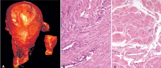
physiologic hypertrophy of the uterus during pregnancy
hyperplasia
increase in number of cells in a tissue/organ
hormonal physiologic hyperplasia
increases functional capacity of tissue
compensatory physiologic hyperplasia
increases tissue mass after damage or partial resection
pathologic hyperplasia
excessive hormonal/growth factor stimulation
hyperplasia is an important…
…response of connective tissue in wound healing
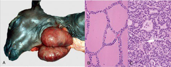
pathologic hyperplasia of the thyroid gland due to hyperthyroidism
atrophy
shrinkage of cells by loss of cell substance
causes of atrophy
atrophy of disuse
denervation atrophy
ischemia
inadequate nutrition
loss of endocrine stimulation
senile atrophy
tissue compression
protein degradation/decreased protein synthesis
atrophy is accompanied by…
increased autophagy, resulting in increased numbers of autophagic vacuoles (which are filled with dead cell parts)

normal brain vs atrophied brain
metaplasia is…
reversible
metaplasia
one cell type replaced with another cell type
metaplasia cause
cells sensitive to certain stressful stimuli will be replaced with a cell than can better withstand that stress. arises by altered differentiation pathway of stem cells. important protective features of normal epithelium are lost. may predispose malignant transformation is stimuli persist.
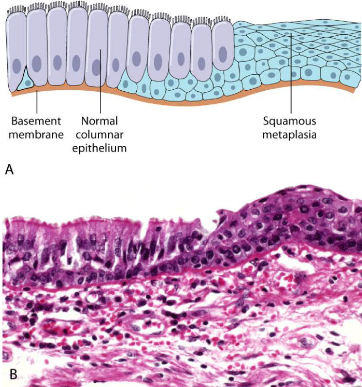
metaplasia
Question 1
Etiology is the term that refers to what element of pathology?
the cause of a disease of condition
Apoptosis differs from necrosis in that apoptosis is
Energy dependent, not associated with inflammation, and can occur under physiologic conditions
Question 3
Which of the following is NOT a feature of necrosis?
partitioning of the cytoplasm to form membrane-bound vesicles of cytosol and organelles
two phenomena that consistently characterize irreversibility
(1) Inability to reverse mitochondrial dysfunction
(2) profound disturbances in membrane function

changes in cell injury diagram
yes i understand this
hypoxia
oxygen deficiency
ischemia
loss of blood supply
the two main morphologic correlates of REVRESIBLE cell injury are
cellular swelling
fatty change

Which of the following BEST characterizes the lesion in the images provided?
Hyperplasia

Which of the following consistently characterize irreversible injury to cells?
a and b

Which of the following is true for the histology image below?
Squamous differentiation of respiratory epithelium in the bronchi of a habitual smoker
cellular swelling results from
inability to maintain ionic and fluid homeostasis
fatty change in cells occurs in
hypoxic, toxic, and metabolic injury to cells involved in fat metabolism
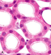
normal kidney tubules with viable epithelial cells

early (reversible) ischemic injury showing surface blebs, increased eosinophilia of cytoplasm, and swelling of occasional cells
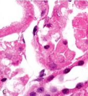
necrotic (irreversible) injury of epithelial cells, with loss of nuclei and fragmentation of cells and leakage of contents
necrosis
morphologic changes that accompany cell death: unable to maintain membrane integrity
features of necrosis
increased eosinophilia
nuclear changes
pyknosis
nuclear shrinkage with DNA condensation
karyorrhexis
nuclear fragmentation
karyolysis
nuclear dissolution

i looked at this diagram regarding necrosis and i understand what i am looking at

coagulative necrosis of the kidney (gross)

coagulative necrosis of the kidney (microscopic)
coagulative necrosis
tissue appears like solid mass in which proteins are denatured,
but the basic tissue architecture is preserved
liquefactive necrosis
dissolution of tissue, particularly common in hypoxic injury in
central nervous tissue

liquefactive necrosis of the brain
caseous necrosis
form of coagulative necrosis, tissue is yellow-white and crumbly, aka “cheesy”

caseous necrosis of the lung
fat necrosis
descriptive of focal areas of fat destruction, typically as result of lipolytic enzymes
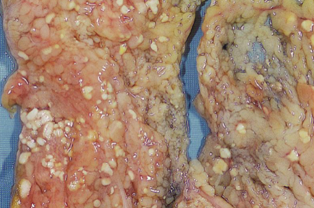
fat necrosis of the pancreas
fibrinoid necrosis
special form seen in blood vessels involving necrosis of the vessel walls together with deposition of amorphous eosinophilic protein; frequently associated with Ag-Ab complex deposition in walls of blood vessels

fibrinoid necrosis of an artery
reperfusion injury
Restoration of blood flow to ischemic but
otherwise viable tissues results in
exacerbated and accelerated injury
Increased production of reactive oxygen species leading to oxidative stress
Reperfusion delivers more leukocytes and cytokines
apoptosis
programmed, intentional cell death where the plasma membrane remains intact
apoptosis is…
…energy dependent
blebbing is…
partitioning (imagine squeezing a stress ball)

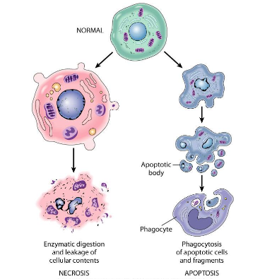
i looked at this diagram relating to necrosis vs apoptosis and i understand
deposition of lipids often appears as…
…clear vacuoles within cytoplasm and parenchymal walls
fatty change is also called…
…steatosis.
steatosis is…
the accumulation of free triglycerides
xanthomas
aggregates of cholesterol-containing “foamy” macrophages in skin and tendons
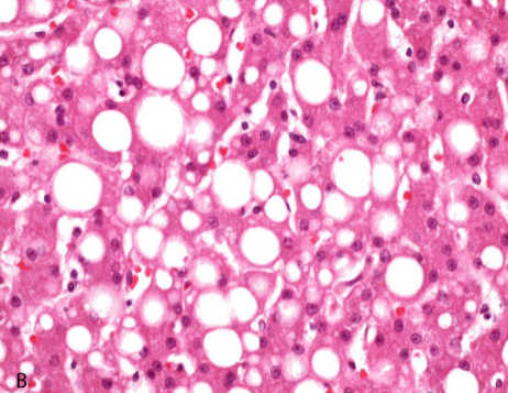
steatosis of the liver

protein reabsorption droplets in the renal tubules
protein accumulation occurs because…
…excess is presented to cells, or because cells synthesize excessive amounts
lipofuscin pigment
insoluble brown-yellow pigment that accumulates in tissues (especially heart, liver, brain) as a function of age or atrophy. Insoluble products of free radical catalyzed peroxidaton of lipid of organelle membranes

lipofuscin accumulation in cardiac muscle
hemosiderin pigment
hemoglobin-derived granular pigment that accumulates during
conditions of local or systemic iron excess. Initial
accumulations begin in mononuclear phagocytes in liver, bone marrow, spleen

hemosiderin accumulation in the liver under H&E

hemosiderin accumulation in the liver under Prussian Blue reaction
pathologic calcification
abnormal tissue deposition of calcium salts
dystrophic calcification
deposition of calcium at sites of cell injury and necrosis, occurs under normal serum calcium
metastatic calcification
deposition of calcium in normal tissues, reflecting a derangement in calcium metabolism
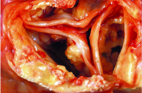
calcification of the aortic valve