Male GU Exam - Core Terminology & Definitions
 Call Kai
 Learn
Learn Practice Test
 Spaced Repetition
 Match
Match Flashcards
 Knowt Play
1/159

There's no tags or description
Looks like no tags are added yet.
Name | Mastery | Learn | Test | Matching | Spaced | Call with Kai | Chat |
|---|
No analytics yet
Send a link to your students to track their progress
160 Terms
Corpora cavernosa
Paired erectile tissue structures on the dorsal penis that engorge with blood during erection.
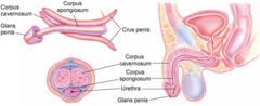
Corpus spongiosum
Column of erectile tissue surrounding the male urethra on the ventral side of the penis.
Prepuce
The foreskin covering the glans penis in uncircumcised males.
Smegma
Secretions of the glans penis that may collect under the foreskin of uncircumcised men.
Scrotal septum
Internal structure dividing the scrotum into two distinct compartments.
Normal post-pubertal testicle length
Normal adult testicular length measuring 4 to 5 cm.
Epididymis
Nodular, cord-like structure located on the posterior surface of the testicle.
Vas deferens
Stiff, tubular duct transporting sperm from the epididymis toward the ejaculatory duct.
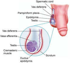
Cremasteric muscle
Muscle layer within the scrotum and spermatic cord that elevates the testicle.
Prostate gland location
Surrounds the urethra at the bladder neck and is roughly testicle-sized.
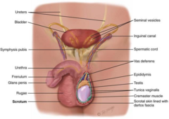
Prostate gland function
Contributes to the production of ejaculatory fluid.
Vascular mechanism of erection
Increased arterial dilation combined with decreased venous outflow within the corpora cavernosa.
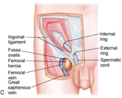
Inguinal canal
Tunnel medial and parallel to the inguinal ligament forming a pathway for the vas deferens.
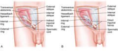
Internal inguinal ring
Deep entrance to the inguinal canal through abdominal wall musculature.
External inguinal ring
Superficial exit of the inguinal canal superior to the pubic tubercle.
Urethral meatus
Terminal external orifice of the urethra located on the glans penis.
Incarcerated hernia
Nonreducible hernia where tissue is trapped outside the abdominal cavity.
Strangulated hernia
Nonreducible hernia with compromised blood supply to protruded tissue requiring emergency surgery.
Cremasteric reflex
Elevation of the testicle and scrotum upon stroking the ipsilateral inner thigh.
Tanner staging system
Objective 5-stage classification tracking development sequence of secondary sexual characteristics.
Paraphimosis
Inability to return retracted foreskin back over the glans, leading to venous congestion and edema.
Phimosis
Inability to retract foreskin from its natural position over the glans penis.
Pathologic phimosis
Nonretractable foreskin resulting from scarring due to trauma, infection, or inflammation.
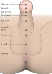
Hypospadias
Congenital urethral defect where the meatus opens on the ventral surface of the penis.
Peyronie disease
Fibrous band in the corpus cavernosum causing penile curvature or indentation during erection.
Hydrocele
Fluid accumulation between the parietal and visceral layers of the tunica vaginalis.
Spermatocele
Benign cystic accumulation located on the epididymis, often containing sperm.
Erectile mechanism in male genitalia
Engorgement of corpora cavernosa caused by increased arterial dilation and decreased venous outflow.
Inguinal canal anatomical path
Lies medial and parallel to inguinal ligament, forming a tunnel for vas deferens through abdominal wall.
Vas deferens physical palpation distinction
Palpates as a slightly stiff, tubular structure, distinguishing it from surrounding softer spermatic cord vessels.
Transillumination differential in scrotal masses
Fluid-filled masses (hydroceles, spermatoceles) transilluminate, whereas solid testicular masses or normal testes do not.
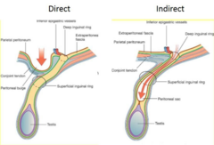
Indirect vs direct inguinal hernia origin
Indirect hernias are commonly congenital at internal ring; direct hernias are acquired from tissue weakness near external ring.
Inguinal hernia palpation finger tip vs side
An indirect hernia touches the tip of the examining finger; a direct hernia touches the side.
Incarcerated vs strangulated hernia relationship
An incarcerated hernia is nonreducible; it becomes strangulated when tissue blood supply is compromised.
Cremasteric reflex anatomical pathway
Stroking inner thigh triggers ipsilateral contraction of cremasteric muscle, elevating testicle and scrotum.
Phimosis vs paraphimosis definition difference
Phimosis is inability to retract foreskin; paraphimosis is retracted foreskin trapped behind glans causing constriction.
Physiologic vs pathologic phimosis distinction
Physiologic phimosis stems from congenital adhesions in newborns; pathologic phimosis results from scar tissue formation.
Paraphimosis ischemia risk
Persistent foreskin entrapment causes venous stasis and glans ischemia, turning glans blue or black.
Hypospadias anatomical defect
Congenital displacement where the urethral meatus opens on the ventral surface of the penis.
Primary syphilis vs genital herpes lesion presentation
Syphilitic chancres are typically painless single ulcers, whereas genital herpes causes painful fluid-filled vesicles or erosions.
Peyronie disease mechanism
Fibrous plaque development in corpus cavernosum causing penile curvature or indentation during erection.
Hydrocele anatomical fluid location
Accumulation of serous fluid between parietal and visceral layers of tunica vaginalis.
Spermatocele contents and location
Benign cystic accumulation containing dead sperm, located superiorly and separately on the epididymis.
Varicocele vascular origin
Abnormal tortuosity and dilatation of the pampiniform venous plexus within spermatic cord.
Primary vs secondary varicocele positional response
Primary varicoceles collapse when supine; secondary varicoceles show no positional change regardless of posture.
Varicocele fertility relationship
Varicocele represents the most common surgically correctable cause of male factor infertility.
Epididymitis vs orchitis anatomical sites
Epididymitis is inflammation of epididymis (often secondary to UTI); orchitis is acute inflammation of testicle.
Testicular torsion physical exam orientation
Testicle presents as high-riding with horizontal lie, with epididymis located anteriorly instead of posteriorly.
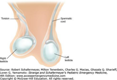
Testicular torsion diagnostic reflex sign
Absence of cremasteric reflex on affected side strongly indicates testicular torsion.
Klinefelter syndrome genetic and genital features
XXY genotype leading to small firm testes, gynecomastia, tall stature, poor muscle tone, and infertility.
Tanner staging clinical purpose
Objective 5-stage classification system tracking adolescent development of primary and secondary sexual characteristics.
Tanner Stage 2 vs Stage 3 genitalia expansion
Stage 2 involves initial scrotal/testicular enlargement; Stage 3 features significant penile length enlargement.
Tanner Stage 4 vs Stage 5 pubic hair distribution
Stage 4 hair is adult-textured but restricted to pubis; Stage 5 hair spreads to medial thighs.
Foreskin replacement procedural rule
Retracted foreskin must always be replaced over glans immediately after exam to prevent paraphimosis.
Vascular mechanism of penile erection
Increased arterial dilation increases blood flow into the corpora cavernosa while decreased venous outflow traps blood.
Sequence for foreskin inspection during male GU exam
Retract foreskin to inspect glans and meatus, then always replace foreskin immediately before proceeding.
Step-by-step technique for testicular palpation
Palpate each testicle between thumb and first two fingers, assessing firmness, symmetry, and presence of masses.
Palpation sequence from epididymis to spermatic cord
Begin at epididymis on posterior testicle, then palpate upward along spermatic cord to external inguinal ring.
Diagnostic mechanism of scrotal transillumination
Light passes through fluid-filled structures (hydrocele), while solid masses or normal testes block light transmission.
Procedure for inguinal hernia palpation
Insert gloved finger into inguinal canal through external ring and instruct patient to cough or perform Valsalva.
Tactile mechanism distinguishing indirect from direct hernias
Indirect hernias touch the tip of the examining finger; direct hernias touch the side of the finger.
Mechanism of hernia strangulation
Nonreducible tissue protrusion leads to compromised blood supply, causing ischemia and requiring prompt surgical intervention.
Procedure and physiological mechanism of cremasteric reflex
Stroke inner thigh with a blunt instrument; ipsilateral cremaster muscle contracts, elevating testicle and scrotum.
Anatomical path of the vas deferens through the groin
Travels from epididymis up spermatic cord, entering external inguinal ring and passing through internal ring via inguinal canal.
Step-by-step physiological progression of Tanner Stage 2
Testes/scrotum begin enlarging with scrotal skin reddening, minimal penile enlargement, and initial straight dark pubic hair at base.
Step-by-step physiological progression of Tanner Stage 3
Testes/scrotum enlarge further, penis lengthens significantly, and pubic hair becomes darker, coarser, and curly.
Step-by-step physiological progression of Tanner Stage 4
Penis grows in length and diameter with glans development, testes reach near-adult size, and pubic hair darkens without thigh spread.
Step-by-step physiological progression of Tanner Stage 5
Penis, testes, and scrotum reach full adult size and shape, with pubic hair spreading to medial thighs.
Pathophysiologic mechanism of Paraphimosis progression
Retracted foreskin behind glans creates constrictive band, causing venous congestion, edema, glans ischemia, and blue discoloration.
Mechanism differentiating physiologic from pathologic phimosis
Physiologic phimosis stems from congenital adhesions that resolve over time; pathologic phimosis results from fibrotic tissue scarring.
Developmental mechanism of Hypospadias
Congenital failure of urethral fold closure, causing urethral meatus to open on ventral surface of penis.
Pathophysiologic mechanism of Peyronie disease
Fibrous plaque develops within corpus cavernosum, causing penile curvature or indentation during erection.
Fluid accumulation mechanism in Hydrocele
Serous fluid collects between parietal and visceral layers of tunica vaginalis surrounding testicle.
Pathophysiologic mechanism of Varicocele formation
Incompetent valves cause tortuosity and dilation of pampiniform plexus veins within spermatic cord.
Positional diagnostic mechanism for primary vs secondary varicocele
Primary varicocele decompresses and disappears when supine; secondary varicocele remains unchanged in supine position.
Pathophysiologic mechanism of Testicular Torsion
Testis twists on spermatic cord, occluding venous and arterial blood supply and causing rapid testicular ischemia.
Mechanism of Cremasteric reflex loss in testicular torsion
Ischemia and mechanical twisting of spermatic cord disrupt reflex arc, causing absence of cremasteric response.
Anatomical displacement mechanism in testicular torsion
Spermatic cord shortening causes testicle to become high-riding with horizontal lie and anteriorly rotated epididymis.
Physical exam procedure for femoral hernia detection
Inspect and palpate anterior thigh region over femoral canal while patient stands and performs Valsalva maneuver.
Pathophysiologic mechanism of direct inguinal hernia
Acquired weakness in posterior wall of inguinal canal causes tissue to bulge directly forward through Hesselbach triangle.
Pathophysiologic mechanism of indirect inguinal hernia
Persistent patent processus vaginalis allows bowel or tissue to pass through internal inguinal ring into inguinal canal.
Clinical Maneuver: Cremasteric Reflex Test
Stroke inner thigh with a blunt instrument; normal result is elevation of testicle and scrotum on stroked side.
Clinical Case: Scrotal "Bag of Worms"
Soft mass superior to testicle feeling like a "bag of worms" that worsens standing indicates a varicocele.
Physical Exam: Primary vs. Secondary Varicocele
Primary varicocele decompresses/disappears when supine; secondary varicocele shows no change with position changes.
Physical Exam: Inguinal Hernia Inspection & Palpation
Inspect groin standing; palpate inguinal canal while patient performs a Valsalva maneuver or coughs.
Hernia Exam: Tip of Finger Touch
A herniating bulge touching the tip of the examining finger in the canal indicates an indirect inguinal hernia.
Hernia Exam: Side of Finger Touch
A herniating bulge touching the side of the examining finger in the canal indicates a direct inguinal hernia.
Special Test: Transillumination of Scrotal Mass
Fluid-filled hydrocele lights up (transilluminates); solid testicular mass or large testicle does not transilluminate.
Clinical Emergency: Testicular Torsion Findings
High-riding testicle with horizontal lie, anterior epididymis, severe acute pain, and absent cremasteric reflex.
Clinical Rule: Prepuce Retraction Step
Always replace (reduce) retracted foreskin over glans immediately after exam to prevent paraphimosis.
Clinical Manifestation: Paraphimosis Presentation
Painful swelling, glans edema, constrictive preputial band, and potential blue/black ischemic glans discoloration.
Clinical Manifestation: Pathologic Phimosis Cause
Inability to retract foreskin due to preputial scarring from prior trauma, inflammation, or recurrent infection.
Clinical Presentation: Syphilitic Chancre
Presents as a single, firm, typically painless ulceration on penile skin during primary syphilis.
Clinical Differentiation: Genital Herpes vs. Chancre
Genital herpes causes painful fluid-filled vesicles/ulcers (HSV-2); syphilitic chancres are characteristically painless.
Clinical Finding: Condyloma Acuminata Inspection
Presents as verrucous, genital wart-like lesions on penile mucosa or skin caused by HPV.
Clinical Finding: Peyronie Disease Manifestation
Fibrous plaque in corpus cavernosum causing pain, curvature, or indentation of penis during erection.
Clinical Findings: Klinefelter Syndrome Physical Phenotype
Tall stature, gynecomastia, decreased muscle tone, small testes, and lower IQ associated with XXY genotype.
Palpation Finding: Spermatocele Location
Painless, mobile, transilluminating cystic mass located on the epididymis, separate from main testicle.
Clinical Significance: Varicocele and Fertility
Varicocele is recognized as the most common surgically correctable cause of male infertility.
Clinical Context: Newborn Hydrocele Course
Common cause of painless scrotal swelling in newborns; majority resolve spontaneously by age 2.