Swine Infectious Diseases: Circulatory, skin, reproductive, vesicular, emerging
1/74
There's no tags or description
Looks like no tags are added yet.
Name | Mastery | Learn | Test | Matching | Spaced |
|---|
No study sessions yet.
75 Terms
What was mycoplasma suis reclassified as?
M. suis has been reclassified from Eperythrozoon suis
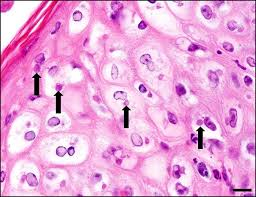
Etiology of Eperythrozoon suis?
These are epicellular and membrane-bound
intracellular, round to oval organisms that are found
within or attached to the outer surface of erythrocytes
and free within the plasma (Groebel et al., 2009). They
change size and shape as they mature, which gives the
microscopic appearance of infection by two separate
organisms. They stain well with Giemsa but not with
Gram stain.
What is the reservoir for Eperythrozoo suis?
The reservoir for M. suis is domestic swine
What vector and non vector transmission dynamics transmit Eperythrozoon suis?
Transmission is mechanical by blood-sucking arthropods,
primarily lice, or reuse of blood-contaminated
needles, snares, and surgical or tattoo instruments.
Most common clinical sign of Eperythrozoo suis?
Acute disease is characterized by
anemia
Clinical signs and what age is affected for Eperythrozoo suis?
Acute disease is usually
seen in suckling or newly weaned piglets or other pigs
that have been stressed, and consists of fever of 40–42°C,
anemia, jaundice, pale mucous membranes, cyanosis of
the ears, weakness, and poor weight gain.
Eitiology of Greasy Skin Disease / Exudative dermatitis in pigs?
Staphylococcus hyicus is a gram-positive coccus considered to be normal flora on the skin of pigs. This bacterium is very persistent in the environment.
Pathology of Staph hyicus?
At least six antigenically distinct exfoliative toxins (ExhA, ExhB, ExhC ExhD, ShetA, ShetB). These toxins target the stratum granulosum in the epidermis and are similar to S. aureus toxins.

Classic picture of what skin disease?
Greasy skin disease (Staph Hyicus)
Clinical signs and what age groups for greasy pig disease?
Aged 5 days to 2 months, Lesions progress to an exudative dermatitis characterized by exfoliation and crusting, which
begins in the groin, axillae, behind the ears, and on areas
of damaged skin. Erosions at the coronary band of hooves
and vesicles or ulcers in the mouth and on the tongue
and snout are common findings
Is greasy skin disease itchy?
NO!
DDX for greasy skin disease?
Differential diagnoses should include swine
pox, mange, ringworm, and pityriasis rosea.
Is greasy skin disease treatable?
Treatment with antibiotics is challenging
due to resistance to beta lactams, erythromycin,
streptomycin, tetracycline, and sulfonamide. The choice,
therefore, should be based on sensitivity testing. Topical
Etiology of swine pox
Swine pox virus is the only member of the
genus Suipoxvirus, family Poxviridae. DNA enveloped
Transmission dynamics swine pox
The pig is the only known host of this virus, and although worldwide in distribution, it exists primarily in herds where poor
sanitation is practiced. The reservoir is infected swine, as
the virus is host-restricted. The virus may persist in an
active form in dry skin scabs for up to 1 year. Although
horizontal transmission may occur via nasal and oral
secretions coming in contact with abraded skin, the
primary method of transmission is mechanical via the
pig louse. Flies and mosquitoes can also carry the viral
particles. Once the virus is established within a herd, it
usually persists.
What area of the skin does swine pox replicate in?
The virus replicates in the cells of the stratum spinosum
Clinical progression of swine pox
Maccular form (reddening), followed by 1- to 6-mm-diameter papules (reddening with edema); transient vesicles (flui
within the lesion), then pustules (umbilicated, ischemic),
and finally, crusts (brown to black in color).
How can location of pox on the pig help lead to the transmission cause?
location of the lesions follows the vector
preferences, that is, the pig louse attacks the lower parts
of the body, while flies feed predominantly over the top
of the body.
What is the hallmark pathologic finding of swine pox?
The presence of intracytoplasmic
inclusion bodies along with central nuclear clearing
in affected epithelial cells is a hallmark sign of this disease.
What is the most significant ectoparasite of swine?
Sarcoptes mange, Sarcoptes scabiei var. suis
Common transmission of mange to piglets?
Nursing piglets obtain the mites from
an infected sow through direct contact
What group of pigs are the primary reservoirs for mange?
Breeding sows with hyperkeratotic encrustations in their ears are the primary reservoirs of mites.
Is mange itchy?
YES!!!
Clinical signs of mange?
acute pruritic or allergic hypersensitive form affects younger, growing pigs. This is characterized by an intensely pruritic, erythematous papular dermatitis on the ventral abdomen, flank, and rump.
chronic or hyperkeratotic form is typically found
in mature sows and boars. Thick, crusty scabs begin on
the pinnae and spread to the neck and head. The papules are manifestations of the hypersensitivity reaction, contain eosinophils, mast cells, and lymphocytes, and have an associated eosinophilic perivasculitis.
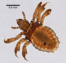
What is the only species of louse that affects pigs?
Haematopinus suis, sucking lice and are the only species of louse that affects swine
Transmission of Haematopinus suis?
this louse is host-specific and will not survive very long (less than 2–3 days) off the host.
What is Haematopinus suis a vector for in swine?
It is considered a vector for swine pox and M. suis
Clinical signs of Haematopinus suis in young pigs?
Young pigs may show pruritus, poor growth, and anemia.
What areas do Haematopinus suis preference in swine?
have a predilection for the skin on the flank area, neck, axilla, groin, and the inner ears.
Brucellosis etiology in pigs, which serovar is most common?
Brucella suis, particularly biovars 1, 2, and
3, is the only species of Brucella that causes systemic
infection and clinical disease, including infertility, in
swine. Biovar 3 is currently the most common cause
of this disease in swine. Morphologically
What are potential reservoirs for B. suis and what serovars have been eradicated?
Domestic swine populations are the primary sources for B. suis.
Feral pigs are also reservoirs in areas where
contact with domestic swine can occur. In the US, B. suis biovars 1 and 3 have been eradicated.
B. suis is present in semen of infected boars and can be spread by
natural breeding or artificial insemination.
Pathogenesis of Brucella suis?
After mucosal exposure to organisms,
they enter through follicle-associated epithelial cells (M
cells) or by phagocytosis, travel to the local lymph nodes,
gain entrance to macrophages and neutrophils, and multiply.
Clinical signs of brucella suis?
The clinical signs of B. suis
infection vary with the herd and range from no obvious
disease to the classical signs, which include abortion, infertility,
metritis, orchitis, lameness, spondylitis, and posterior
paralysis. Clinical disease in piglets of weaning age usually
consists of spondylitis and posterior paralysis
Microscopic and histopathologic lesions of brucella suis?
Microscopic lesions consist of granulomatous inflammation in the endometrium, uterine glands, and placenta. Abscesses in the kidneys, spleen, ovaries, lungs, brain, and other tissues may be seen
What BSL should brucella be?
BSL 3. No longer reportable as of NOV 2024
What species of leptospirosis affect pigs?
Leptospira interrogans and L. borgpetersenii
Etiology of leptospirosis and what serovar is most commonly clinical in pigs?
gram negative, motile aerobic spirochetes. The serovar Pomona
is the most common cause of clinical leptospirosis in
swine,
Transmission of leptospirosis in pigs?
Leptospires are shed from carrier animals in urine and genital fluids into the environment.
Venereal transmission is thought to be the mode of
spread for serovar Bratislava because sows and boars
harbor it in the reproductive tract
Clinical signs of leptospirosis in pigs
The acute form is characterized
by a mild transient anorexia, listlessness,
diarrhea, and pyrexia that resolves within a week and
usually goes unrecognized.
The acute form is characterized by a mild transient anorexia, listlessness, diarrhea, and pyrexia that resolves within a week and
usually goes unrecognized.
Infertility of the sow is seen following infections due to serovar
Bratislava;
Necropsy findings of leptospirosis in chronic forms
In chronic disease,
lesions are confined to the kidneys and consist of small
gray lesions on the renal cortex.
Etiology of porcine parvovirus?
PPV is a disease of swine characterized by embryonic
and fetal infection which is manifest as stillbirths, mummification,
embryonic death, and infertility (SMEDI)
when susceptible sows and gilts are exposed to the
virus between 6 and 70 days of gestation. DNA Nonenveloped
Porcine parvovirus is one of the major infectious causes
of embryonic and fetal death
Transmission of parvovirus in pigs
It is ubiquitous
Gilts are most commonly infected oronasally and prenatal pigs are infected transplacentally.
Nursing pigs absorb protective PPV antibody from
colostrum.
The major reservoir for PPV is environmental. The
virus is thermostable and resistant to many disinfec
tants.
Virus replicated first in the tonsils
What is the crown rump size of the mumified fetuses infected with parvovirus?
Most of the infected fetuses have a crown–rump length of 17 cm
or less because those infected after day 70 are able to
respond to the viral assault and survive
Control of parvovirus in the USA
Vaccines are used extensively in the US. They are administered
several weeks before conception
necropsy findings of pig parvovirus
Gross lesions are confined to the placenta,
which may be edematous and have white, mineralized
deposits and stunted fetuses with prominent blood vessels
on their surfaces, petechial hemorrhages, edema,
enlarged dark liver and kidneys, serosanguinous flui
in body cavities, and mummificatio
Etiology of porcine reproductive and respiratory syndrome?
The causative agent is a single-stranded
RNA virus classified in the order Nidovirales, family
Arteriviridae, and genus Arterivirus. This agent shares
structural and functional organization with others in the
genus, including lactate dehydrogenase-elevating virus,
equine arteritis virus, and simian hemorrhagic fever
virus. These viruses in general are known to have high
rates of mutation.
Hallmark signs include reproductive disorders,
high piglet mortality, and respiratory disease seen in a
wide age range of animals.
What are the two important serotypes of porcine reproductive and respiratory syndrome?
1 (Lelystad) found primarily in the EU and Type 2 (VR-
2332), found primarily in the US
Transmission dynamics porcine reproductive and respiratory syndrome?
This virus is spread predominantly through direct contact between
infected and naive pigs, although the route of fetal PRRSV
infection has not been identified. Once infected, pigs
become persistently infected.
The virus establishes a foothold by infecting macrophages
located within mucosal surfaces. The virus is believed to
be limited to domestic swine. The disease does persist
in infected swine in a transmissible, viable state, often
without stimulating antibody production, thereby making
serologic screening for the disease inaccurate.
The virus has been found in serum, oropharyngeal fluids, semen, feces, and urine, and animals are susceptible via intranasal, intramuscular, oral, intrauterine and vaginal exposure
Pathogenesis of Porcine repro and resp syndrome?
The virus has been shown to enter via
the nasal epithelium, bronchial epithelium, and tonsilar
and pulmonary macrophages, followed by replication in
alveolar macrophages, with a subsequent viremia and
spread to lymphoid organs and lungs.
Late gestational abortions typically occur when animals are infected during the third trimester and can occur sporadically or sweep throughout the population of animals
Clinical signs of PRRS?
Clinical signs
in infected females vary from none to anorexia, fever,
pneumonia, agalactia, red/blue discoloration of ears
and vulva, subcutaneous edema, and a delayed return
to estrus.
Clinical signs in PRRSV-infected newborn pigs also
vary in frequency and severity. Dyspnea and tachypnea
are the most characteristic clinical signs, with other signs
including periocular and eyelid edema, conjunctivitis,
blue discoloration of the ears, diarrhea, and CNS signs.
Mortality can reach 100%.
As the pigs reach postweaning
age, the clinical signs shift to include fever, pneumonia,
failure to thrive, and significant mortality caused
by otherwise non-life-threatening concurrent bacterial
infections
Necropsy findings of PRRS?
Gross lesions in young piglets include mottled lungs with tan foci of consolidation; lymphadenopathy of the mesenteric and middle
iliac nodes, which are tan and may contain cysts, moderately
enlarged and rounded hearts, and clear fluid in
the pericardial space and abdominal cavity.
Etiology foot and mouth disease?
Foot-and-mouth disease virus (FMDV) is
in the family Picornaviridae, genus Aphthovirus. RNA Nonenveloped
Transmission dynamics of Food and mouth disease? What family of animals does it most commonly affect?
FMDV affects members of the order
Arteriodactyla. All secretions
and excretions from infected animals contain infectious
virus. FMDV can remain infectious within the
environment for extended periods.
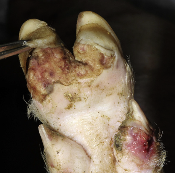
Clinical signs foot and mouth disease pigs?
Pigs display fever and
formation of vesicles in and around the mouth and feet
(Fig. 16.26). Lesions on the feet are often interdigital,
with the coronary band being especially predisposed
due to the vascularity. Lesions on the feet result in the
animal being lame and often ‘dog-sitting’.
Oral lesions most commonly affect the tongue, and
foot lesions are most often interdigital, at the heel bulb,
and coronary bands.
Etiology of swine vesicular disease?
Swine vesicular disease virus is an
Enterovirus in the family Picornaviridae. RNA nonenveloped
Clinical signs of Swine vesicular disease?
Clinically, pigs have mild
fever with rare lameness.
Why is swine vesicular disease reportable?
Indistinguishable from Foot and Mouth disease
Etiology vesicular xanthema
Vesicular exanthema of swine (VES) is caused by the
vesicular exanthema of swine virus, genus Vesivirus in
the Caliciviridae family. RNA nonenveloped. Indistinguishable from foot and mouth disease
Eitiology of vesicular stomatitis virus?
Vesicular stomatitis (VS) infection in pigs is indistinguishable
from FMD, and therefore is classified a notifiable
disease (Health, 2013a). Vesicular stomatitis virus
is in the genus Vesiculovirus and family Rhabdoviridae. RNA Envelped. Also a zoonotic disease.
Eitiology of classical swine fever?
Etiology Classical swine fever (CSF) virus is in the
family Flavivirus, genus Pestivirus. It is also known as
hog cholera in the literature. RNA enveloped.
Clinical signs of classical swine fever
Animals with CSF have
pyrexia, anorexia, lethargy, conjunctivitis, respiratory
signs, and constipation followed by diarrhea
Necropsy findings classical swine fever
hemorrhage of the peripheral lymph
nodes and renal petechiae and ecchymoses are present,
Etiology nipah virus?
Nipah virus is a single-stranded negative
sense enveloped RNA virus in the family Paramyxovirus, genus
Henipavirus. There are strain differences between isolates
from different geographic regions
Transmission of nipah virus?
Initially identified in Malaysia in 1999, the virus has since emerged in Bangladesh and India. Pigs are an amplifying host; however,
bats are the reservoir host and secrete virus in urine
where pigs may ingest items containing infectious viral
particles and become infected (Williamson and Torres-
Velez, 2010). Close contact is required for transmission
Clinical signs of nipah virus?
Pigs are asymptomatic or have acute
febrile disease with respiratory/CNS signs
What is the BSL for nipah and is it zoonotic?
Highly zoonotic and BSL 4
Etiology of Porcine lymphotrophic herpesvirus (PLHV)?
DNA Enveloped, porcine lymphotropic disease with high mortality, similar to that of human post-transplantation lymphoproliferative disease,
What herpesvirus is a risk to humans from xenotransplantation?
There is also concern that pig–human xenotransplantation may result in human disease from this virus
PLHV infects which cells?
B cells
Clinical signs of PLHV?
Clinical signs included lethargy, fever, anorexia, and enlarged lymph nodes
Ovine herpesvirus 2 etiology?
A naturally occurring disease similar to malignant
catarrhal fever (MCF) has been reported in pigs, although
rare and poorly documented. Pigs becoming ill
after having contact with sheep.
Clinical signs for ovine herpes virus in pigs?
Pigs with MCF display high persistent fever, anorexia, depression, recumbency, foul-smelling nasal discharge, ocular discharge, bilateral corneal edema, keratoconjunctivitis, ataxia, tremors, and possible convulsions
Hepatitis E etiology and clinical signs in pigs?
Infection in pigs is primarily without clinical signs.
Can pigs get ebola?
Yes
Japanese encephalitis etiology and clinical signs in pigs?
Japanese encephalitis virus (JEV) is a member of the
family Flaviviridae, genus Flavivirus. While mosquitos
transmit the agent, the pig is a natural reservoir of JEV
(Grand, 2012). Clinical signs in pigs include testicular
degeneration, infertility, mummified fetuses, and piglets
with birth defects, reproductive failure