Human Anatomy Module 1
 Call Kai
 Learn
Learn Practice Test
 Spaced Repetition
 Match
Match Flashcards
 Knowt Play
1/46

Earn XP
Description and Tags
The Skeletal System
Name | Mastery | Learn | Test | Matching | Spaced | Call with Kai | Chat |
|---|
No analytics yet
Send a link to your students to track their progress
47 Terms
Regional Approach
studying one region or part at a time
Systemic Approach
studying one system at a time
Anatomical Position
Standing upright with feet together
Hands by the side
Face looking forward.
The mouth is closed and the facial expression is neutral.
The rim of bone under the eyes is in the same horizontal plane as the top of the opening to the ear.
The eyes are open and focused on something in the distance.
The palms of the hands face forward with the fingers straight
The toes point forward.

Coronal planes
vertically divides the body into anterior and posterior parts

Sagittal planes
vertically divides the body into right and left parts

Median sagittal plane
divides the body into equal right and left parts
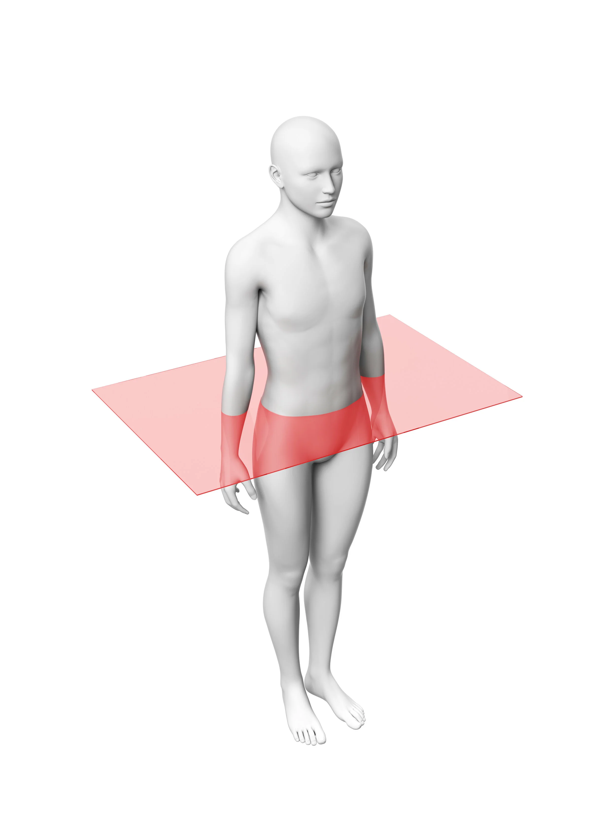
Transverse, horizontal, or axial plane
horizontally divides the body into superior and inferior parts
Anterior or Ventral
to the front
Posterior or Dorsal
to the back
Medial
closer to the midline
Lateral
further from the midline
Superior
closer to the top
Inferior
further from the top
Proximal
closer to attachment
Distal
further from the top
Cranial
toward the head
Caudal
toward the tail
Superficial
closer to the surface
Deep
further from the surface
Right hypochondriac region
Top left square in the 3×3 grid of the abdomin
Epigastric region
Center top square in the 3×3 grid of the abdomin
Left Hypochondriac region
Top right square in the 3×3 grid of the abdomin
Right Lateral Region
Left square of the middle row in the 3×3 grid of the abdomin
Umbilical Region
Center square in the middle row in the 3×3 grid of the abdomin
Left Lateral Region
Right square in the middle row in the 3×3 grid of the abdomin
Right inguinal region
Bottom left square in the 3×3 grid of the abdomin
Pubic region
Bottom center square in the 3×3 grid of the abdomin
Left Inguinal Region
Bottom right square in the 3×3 grid of the abdomin
Right Upper Quadrant (RUQ)
Top left square in the 2×2 grid of the abdomin
Left Upper Quadrant (LUQ)
Top right square in the 2×2 grid of the abdomin
Right lower quadrant (RLQ)
Bottom Left square in the 2×2 grid of the abdomin
Left Lower Quadrant (LLQ)
Bottom Right square in the 2×2 grid of the abdomin
X-ray (Plain Radiography)
X-rays pass through the body, and are attenuated (reduced in energy) by tissue
Air attenuates x-rays a little
Fat attenuates more than air
Bone attenuates the most
X-ray with Contrast
IV iodine is given to the patient before x-rays are taken in order to see parts of the body more clearly
Ultrasound (US)
a very-high-frequency sound wave interpreted by a powerful computer, and a real-time image is produced on the display panel
Doppler ultrasound
uses the doppler effect to determine flow and direction (usually used on blood vessels)
Computed Tomography (CT)
obtains a series of images of the body (slices) in the transverse plane
Magnetic Resonance Imaging (MRI)
uses a strong magnetic field that is reflected by the hydrogen ions found in the water of our cells
Axial portion
The head, neck, and trunk
Appendicular Portion
The upper and lower limbs
Cranial cavity
Houses the brain
Vertebral canal (Spinal Cavity)
Contains the spinal cord
Thoracic cavity
houses the lungs and thoracic viscera
Abdominopelvic cavity
contains abdominal and pelvic viscera
Serous membrane
a double layered membrane that secretes serous fluid (slippery fluid that prevents friction between layers) found in the tharacic and abdominopelvic cavities
Visceral layer
the inner layer of the serous membrane, which covers an organ
Parietal layer
the outer layer of the serous membrane, which lines the wall of the the cavity