Concepts of Care for Patients with Cardiac Conditions (NUR 411)
 Call Kai
 Learn
Learn Practice Test
 Spaced Repetition
 Match
Match Flashcards
 Knowt Play
1/29

There's no tags or description
Looks like no tags are added yet.
Name | Mastery | Learn | Test | Matching | Spaced | Call with Kai | Chat |
|---|
No analytics yet
Send a link to your students to track their progress
30 Terms
Valvular Heart Disease
Acquired valvular dysfunctions that prevent the heart valves from functioning properly…
i.) Mitral stenosis → valve leaflets fuse & become stiff & chordae tendinae contract/shorten
ii.) Mitral regurgitation → incomplete closure (during systole) allows backflow of blood into the left atrium (when left ventricle contracts)
iii.) Mitral valve prolapse (MVP) → valvular leaflets enlarge & prolapse in the L atrium during systole
iv.) Aortic stenosis → aortic valve orifice narrows & obstructs L ventricular flow during systole
v.) Aortic regurgitation → aortic valve leaflets do not close properly (during diastole), allowing blood to flow from aorta into L ventricle
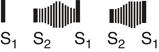
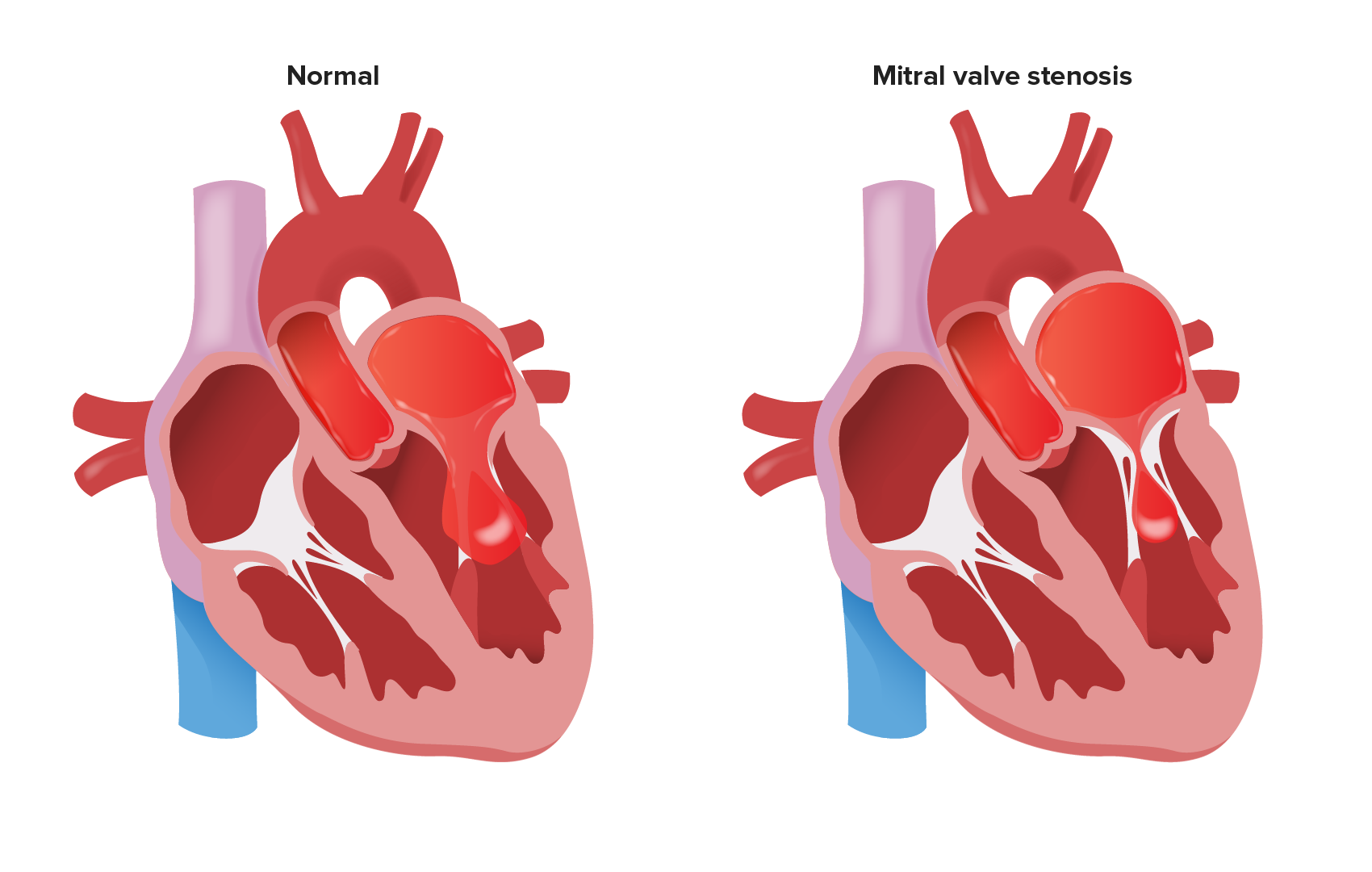
Mitral Stenosis
Mitral valve leaflets fuse/stiffen & the chordae tendineae contract/shorten…
i.) Most commonly caused by rheumatic fever/carditis
ii.) L atrial pressure rises → pulmonary artery pressure elevates → R ventricle hypertrophies
iii.) Pulmonary congestion & R-sided HF occur first → preload ↓ & CO falls
Clinical Manifestations of Mitral Stenosis
Mild Stenosis
i.) Usually asymptomatic
Progressive Stenosis
i.) Dyspnea on exertion
ii.) Orthopnea
iii.) Paroxysmal nocturnal dyspnea
iv.) Palpitations
v.) Dry cough
vi.) Fatigue
Advanced Stenosis
i.) Hemoptysis (coughing up blood)
ii.) Pulmonary edema
iii.) R-sided HF (signs & symptoms)
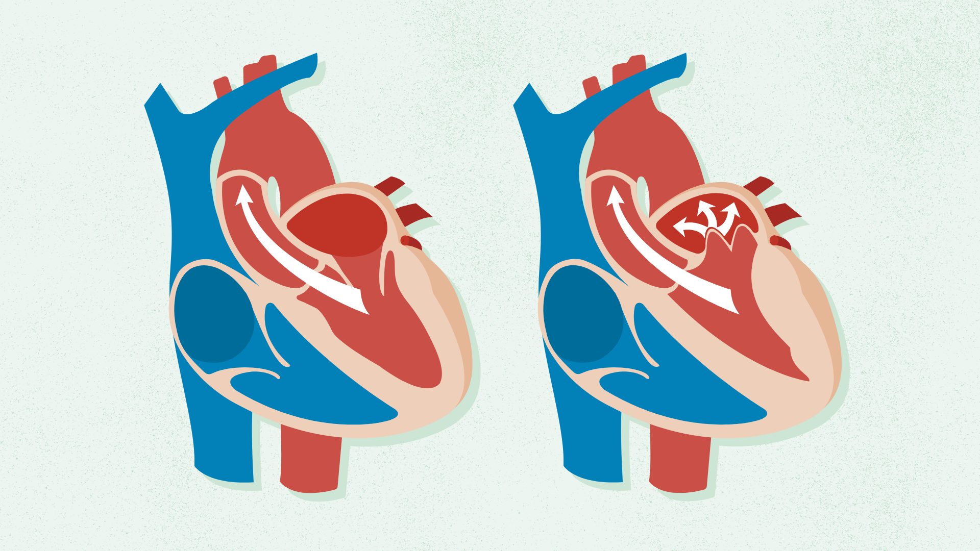
Mitral Valve Prolapse (MVP)
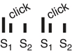
Valvular leaflets enlarge & prolapse into the L atrium during systole (usually benign)…
i.) Associated w/ Marfan syndrome & familial tendency
ii.) Normal HR & BP are expected findings → mid-systolic click & late systolic murmur (at apex) possible
iii.) May progress to mitral regurgitation
Clinical Manifestations of Mitral Valve Prolapse (MVP)
Most patients are asymptomatic…
i.) Atypical chest pain (sharp, localized to L side of chest)
ii.) Palpitations
iii.) Exercise intolerance
iv.) Dizziness
v.) Syncope
Mitral Regurgitation
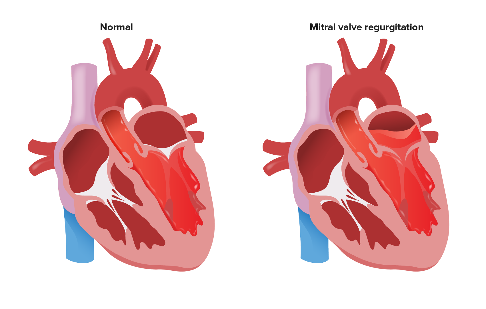
Fibrotic & calcific changes prevent the mitral valve from closing completely during systole…
i.) Blood flows backward from L ventricle to L atrium (systole)
ii.) Regurgitant blood flows back to L ventricle along w/ normal flow (diastole) → higher volume must ejected during next systole
iii.) L atrium & L ventricle dilate/hypertrophy to compensate for ↑ volume/pressure
iv.) Commonly caused by mitral valve prolapse, rheumatic heart disease, & MI
Clinical Manifestations of Mitral Regurgitation
Disease progression is slow, w/ onset of symptoms occurring when L ventricle fails…
i.) Fatigue & chronic weakness (from ↓ CO)
ii.) Dyspnea on exertion
iii.) Orthopnea
iv.) Atypical chest pain
v.) Palpitations
vi.) Atrial fibrillation (AF)
vii.) R-sided HF (signs & symptoms)
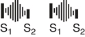
viii.) High-pitched systolic murmur (at the apex)
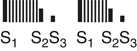
ix.) S3 (severe regurgitation)
Aortic Stenosis
Aortic valve orifice narrows, obstructing L ventricular outflow during systole…
i.) Most common cardiac valve dysfunction "(‘wear & tear” disease);
ii.) Ventricular hypertrophy caused by ↑ afterload
iii.) Surgery indicated when surface area is ≤ 1 cm
iv.) L ventricle fails → blood pools in L atrium → pulmonary congestion
v.) CO becomes fixed as stenosis worsens; R-sided HF occurs late in disease

Common Causes of Aortic Stenosis
Younger/Middle-Aged Adults
i.) Congenital bicuspid or unicuspid aortic valves
ii.) Rheumatic aortic stenosis (occurs w/ rheumatic mitral valve disease)
Older Adults
i.) Atherosclerosis
ii.) Degenerative calcification
Clinical Manifestations of Aortic Stenosis
Classic Triad
i.) Dyspnea on exertion
ii.) Angina on exertion
iii.) Syncope on exertion
Additional Manifestations
i.) Fatigue
ii.) Orthopnea
iii.) Paroxysmal nocturnal dyspnea
iv.) Palpitations
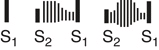
v.) Systolic crescendo-decrescendo murmur
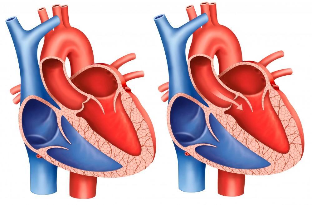
Aortic Regurgitation
Aortic valve leaflets do not close properly during diastole…
i.) The annulus (valve ring) may be dilated, loose, or deformed
ii.) Blood flows backward from aorta into l ventricle during diastole
iii.) L ventricle dilates to accommodate greater blood volume → eventually hypertrophies
Common Causes of Aortic Regurgitation
Usually results from nonrheumatic conditions…
i.) Infective endocarditis
ii.) Congenital anatomic aortic valvular abnormalities
iii.) Hypertension
iv.) Marfan syndrome
Clinical Manifestations of Aortic Regurgitation
Patients remain asymptomatic for years due to L ventricular compensation…
i.) Dyspnea on exertion\
ii.) Orthopnea
iii.) Paroxysmal nocturnal dyspnea
iv.) Palpitations
v.) Nocturnal angina w/ diaphoresis
vi.) Fatigue
vii.) Sinus tachycardia
viii.) Bounding arterial pulse
ix.) Widened pulse pressure (↑ SBP w/ ↓ DBP)
x.) Decrescendo diastolic murmur
Interventions/treatment for Valvular Disease
Drug Therapy
i.) Diuretics → reduce fluid overload & pulmonary/systemic congestion (i.e. Furosemide, Hydrochlorothiazide, Spironolactone)
ii.) Beta Blockers → control HR to improve cardiac function; manage AF & ventricular response (i.e. Metropolol)
iii.) ACE Inhibitors → reduce afterload & improve CO (i.e. Lisinopril)
iv.) Digoxin → improve contractility to aid flow of blood
v.) Vasodilators (Calcium Channel Blockers) → reduce regurgitant flow in stenosis (i.e. Losartan)
Surgical Interventions
i.) Valve replacement (require Warfarin & INR goal of 3.0-4.0)
ii.) Balloon valvuloplasty
Diagnostic Imaging for Valvular Disease
Echocardiography
i.) Primary diagnostic test
ii.) Measures chamber size, EF, & flow gradient across valves
Chest X-Ray
i.) Mitral Stenosis → L atrial enlargement, prominent pulmonary arteries, R ventricular enlargement
ii.) Mitral Regurgitation → cardiac shadow increase
iii.) Aortic Stenosis → L ventricular enlargement, pulmonary congestion
iv.) Aortic Regurgitation → L atrial/ventricular dilation, pulmonary congestion (if HF present)
Electrocardiogram (ECG)
i.) Evaluate heart rhythm
ii.) Atrial fibrillation commonly found in mitral stenosis/regurgitation & aortic stenosis
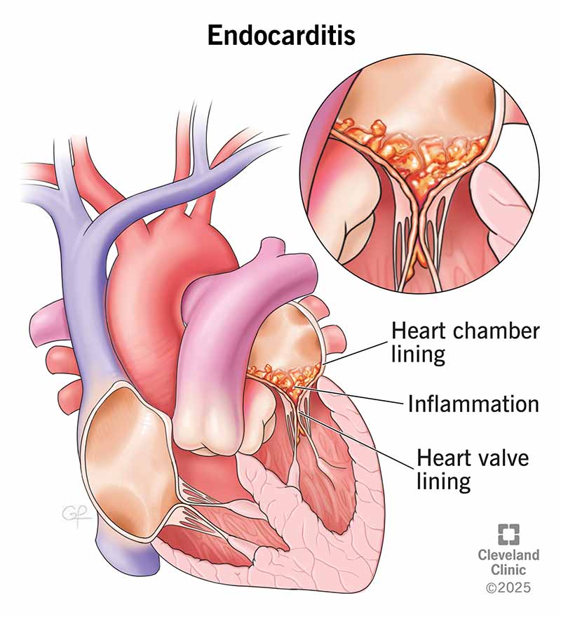
Infective Endocarditis
Infection of endocardium & heart valves ; often occurs w/ those who use IV drugs…
i.) Blood flows rapidly from high-pressure to low-pressure zone → erodes endocardium
ii.) Platelets and fibrin adhere → form vegetative lesion
iii.) Bacteria trapped during bacteremia → deposited in vegetation
iv.) Vegetation grows → destroys endocardium & valve
v.) Valvular insufficiency or stenosis results
vi.) Vegetations may embolize into systemic circulation
Clinical Manifestations of Infective Endocarditis
Cardiovascular
i.) Murmurs
ii.) S3 or S4 heart sounds
iii.) R-side or L-sided HF
Embolic
i.) L-sided involvement → spleen, kidneys, GI tract, brain, extremities
ii.) R-sided involvement → pulmonary emboli
iii.) Splenic infarction (sudden ABD pain radiating to L shoulder, rebound tenderness)
iv.) Renal infarction (flank pain to groin w/ hematuria or pyuria)
v.) Neurologic changes
vi.) Pleuritic chest pain, dyspnea, cough
Integumentary
i.) Petechiae (mucous membranes, palate, conjunctivae, skin above clavicles)
ii.) Splinter hemorrhages
iii.) Osler nodes (palms/soles)
iv.) Janeway lesion (flat, reddened maculae on hands/feet)
v.) Roth spots (hemorrhagic lesions that appear as round or oval spots on retina)
Interventions/Treatment for Infective Endocarditis
Antibiotic Therapy
i.) Long-term (weeks to months) IV therapy
Oral Hygiene
i.) Proper oral hygiene is the BEST intervention for prevention
ii.) Brush ≥2×/day w/ a soft toothbrush
iii.) Avoid irrigation devices & flossing
Surgical Intervention
i.) Valve repair or replacement if indicated
ii.) Vegetation excision
Pericarditis
Inflammation/alteration of the pericardium…
i.) Acute pericarditis r/t infection, autoimmune disorders, MI, or trauma
ii.) Chronic (constrictive) pericarditis r/t TB, radiation therapy, trauma, renal failure, or metastatic cancer
iii.) Pericardium becomes rigid → prevents adequate ventricular filling → cardiac failure

Clinical Manifestations of Pericarditis
Acute Pericarditis
i.) Substernal precordial pain (radiating to left neck, shoulder, or back)
ii.) Grating, oppressive pain aggravated by breathing (especially inspiration), coughing, swallowing
iii.) Worse when supine; relieved by sitting up and leaning forward
iv.) Pericardial friction rub (scratchy, high-pitched sound at left lower sternal border)
v.) Elevated WBC count & fever
vi.) ST elevation in all leads
vii.) Atrial fibrillation (AF) common
viii.) Echocardiogram shows pericardial effusion
Chronic Pericarditis
i.) Signs of R-sided HF
Interventions/Treatment for Pericarditis
Drug Therapy
i.) NSAIDs (first-line for pain)
ii.) Corticosteroids
iii.) Colchine (NSAID adjunct to reduce symptoms/recurrence)
Positioning
i.) Sitting upright & leaning slightly forward
Pericardiocentesis
i.) Performed for fluid accumulation around heart
Acute Cardiac Tamponade
Emergency condition characterized by rapid fluid accumulation in the pericardium that compresses the heart & impairs CO…
i.) Compression of atria & ventricles leads to ↓ CO
ii.) Small volumes (20-50 mL) cause tamponade if accumulated rapidly

Common Causes of Acute Cardiac Tamponade
i.) Pericarditis
ii.) Ventricular wall rupture (from acute MI)
iii.) Cancer
iv.) Aortic dissection
v.) Complications from invasive procedures
vi.) Post-CABG (blood clot in pericardium)
Clinical Manifestations of Acute Cardiac Tamponade
i.) Muffled/distant heart sounds
ii.) JVD
iii.) Paradoxical drop in SBP (during inspiration)
iv.) Hypotension
v.) Tachycardia
vi.) Narrowed pulse pressure
vii.) Fluid accumulation (confirmed via ECG or chest X-ray)
Interventions/Treatment for Acute Cardiac Tamponade
Pericardiocentesis
i.) 8 in, 16- or 18 G needle inserted into pericardial space
ii.) Performed under echocardiographic or fluoroscopic guidance
iii.) Catheter inserted to withdraw all available fluid; temporary pericardial drain may be placed
iv.) Monitor pulmonary artery & R atrial pressures during procedure (should normalize as fluid removed)
Post-CABG
i.) Volume expansion + emergency sternotomy w/ drainage
Recurrent Cases
i.) Pericardial window (removes portion of pericardium to drain into pleural space)
ii.) Pericardiectomy (complete removal of pericardium)
Rheumatic Carditis
Sensitivity response after upper respiratory infection w/ group A beta-hemolytic streptococci…
i.) Inflammation occurs in all three layers of the heart
ii.) Aschoff bodies (small nodules in myocardium) form & are replaced by scar tissue
iii.) Pericardium thickens w/ exudate; serosanguineous pleural effusion may develop
iv.) Hemorrhagic & fibrous lesion form on valve surfaces → mitral/aortic stenosis or regurgitation

Clinical Manifestations of Rheumatic Carditis
i.) Inflammation of heart layers
ii.) Impaired myocardial contractile function
iii.) Thickened pericardium
iv.) Valvular damage
v.) S/S of HF
Interventions/Treatment for Rheumatic Carditis
Antibiotic Therapy
i.) Penicillin (drug of choice)
ii.) Erythromycin (penicillin-sensitive patients)
ii.) Begin course immediately & continue for a full 10 days
Symptom Management
i.) Maintain hydration
ii.) Antipyretics
iii.) Encourage adequate rest
Patient Education
i.) Recurrence most likely due to Streptococcus reinfection
ii.) Lifelong antibiotic prophylaxis required
Cardiomyopathy
Subacute or chronic disease of cardiac muscle classified based on structural & functional abnormalities…
i.) Dilated (DCM) → extensive myofibril damage that causes ventricular dilation & impaired systolic function
ii.) Hypertrophic (HCM) → asymmetric ventricular hypertrophy that causes diastolic filling problems & outflow obstruction; mitral valve abnormalities contribute
iii.) Restrictive → sarcoidosis/amyloidosis cause stiff ventricles that restrict diastolic filling; poor prognosis
iv.) Arrhythmogenic R Ventricular → myocardial tissue replaced w/ fibrous/fatty tissue; familial cause

Interventions/Treatment for Cardiomyopathy
Dilated & Restrictive Cardiomyopathy
i.) Diuretics → manage fluid overload
ii.) Vasodilators → improve flow
iii.) Cardiac glycosides → improve CO (i.e. Digoxin)
iv.) Beta-Blockers → block sympathetic stimulation & tachycardia (i.e. Metoprolol)
v.) Antidysrhythmics
vi.) Avoid causative toxins (i.e. alcohol)
Hypertrophic Cardiomyopathy
i) Negative inotropic agents → reduce outflow obstruction & HR to improve angina, dyspnea, syncope (i.e. Carvedilol, Verapamil)
ii.) Prohibit strenuous exercise
iii.) Avoid excess alcohol & dehydration
iv.) Implantable cardioverter/defibrillator (ICD)
v.) Ventricular septal myectomy → excise septum to widen outflow tract
vi.) Percutaneous alcohol septal ablation → alcohol injected into septal artery to cause small infarction & remodeling to reduce obstruction
Arrhythmogenic R Ventricular Cardiomyopathy
i.) Radiofrequency catheter ablation
ii.) Implantable defibrillator (if unresponsive to drugs)