14 Topography of the abdomen of the horse
 Call Kai
 Learn
Learn Practice Test
 Spaced Repetition
 Match
Match Flashcards
 Knowt Play
1/54

Earn XP
Description and Tags
W4 Elnady
Name | Mastery | Learn | Test | Matching | Spaced | Call with Kai | Chat |
|---|
No analytics yet
Send a link to your students to track their progress
55 Terms
What is the difference between the abdomen and the abdominal cavity?
The abdomen is the external part of the body that can be seen and touched, while the Abdominal cavity is the internal space within the body that holds the abdominal organs and is not visible or palpable.
Why is the paralumbar fossa smaller in the horse than the cow?
the horse has 18 pairs of ribs and the cow inly has 13 pairs of ribs

What are the layers of the abdominal wall?
EAO, IAO, TA
they have a well-developed cutaneous trunci and tunica flava abdominis. Has subperitoneal fat

What is important about the prepubic tendon in the horse?
it supports the majority of the weight of the abdominal viscera and is susceptible to rupture in the pregnant mare

What are the effects of heaves in the horse?
it causes hypertrophy of the external abdominal oblique, however, the aponeurosis does not increase in size so it creates a margin.
it creates a heaves line

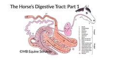
What is the difference between horses and bovine on where fermentation happens?
Horses are POST-gastric fermenters (hindgut fermenters) and bovine are PRE-gastric fermenters (foregut fermenters)

What are the parts of the equine stomach?
1. cardiac
2. fundus
3. saccus cecus (bind pouch at esophageal opening)
4. body
5. pyloric = antrum, canal, and pylorus

what type of stomach do horses have?
simple, composite (contains glandular and nonglandular parts)

why can't the horse's stomach be palpated externally?
it is high and you cannot reach it from the ventral part
Why can't horses vomit?
the cardiac sphincter is very strong so horses rarely vomit AND the stomach does not rest on the floor of the abdomen (in humans, when they vomit, their abdominal muscles contract)
What is the greater omentum of the horse attached to?
the greater omentum (o) is attached to the ventral surface of the transverse colon (f); it is a landmark during exploratory surgery
The greater omentum is very small in the horse
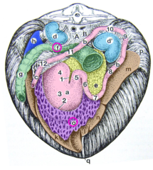
What ligament is unique to the horse?
the renosplenic (nephrosplenic) ligament which is part of the greater omentum.
The fixed viscera includes ______________
1. descending duodenum
2. transverse colon
3. descending colon
Where is the descending duodenum located in all domestic species?
dorsal in the right side of the abdomen
it has a short mesoduodenum which contains the right lobe of the pancreas
Where is the transverse colon located in all domestic species?
it runs from RIGHT TO LEFT and is cranial to the cranial mesenteric artery
Where is the descending colon located in all domestic species?
dorsal in the left side of the abdomen
Where is the jejunum located?
in the left dorsal part of the abdomen
Where can the jejunum get entrapped?
in the epiploic foramen and the tunica vaginalis
How can you tell the ileum from the jejunum?
it is firmer than the jejunum (has thicker walls) and has the ileocecal fold

What are the parts of the cecum?
base, body and apex (most cranial part)

What are some defining characteristics of the cecum?
it has 4 teniae and has haustra (sacculations)

Where is the base of the cecum located?
partly in the right paralumbar fossa and partially covered by the ribs
the ileum enters and the right ventral colon leaves here
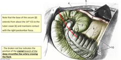
Where is the body of the cecum located?
it sinks within the abdomen to lie on the floor between the ventral parts of the ascending colon

Where would you perform auscultation and trocarization of the cecum?
in the right paralumbar fossa

Where is the apex of the cecum located?
caudal to the xiphoid process between the right and left part of the ascending colon

What are the two cecal openings?
ileocecal opening and cecocolic opening

What are the three cecum mesenteric attachments?
1. ileocecal fold
2. cecocolic fold (landmark for surgery)
3. cecoduodenal ligament

Where does the ascending colon start and end?
it starts at the cecocolic orifice and ends at the transverse colon

How can you differentiate between the dorsal and ventral colons?
the right and left ventral colons have haustra, whereas the left and right dorsal colons do not
True/false- the ascending colon is fixed
FALSE- the ascending colon is not fixed except at the beginning (at the base of the cecum) and its end (transverse colon) but the ventral and dorsal colons are connected by short intercolic ligament
it moves like a piston!

What are the characteristics of the descending/small colon?
sacculations and a long des. mesocolon
it has two tenia
fecal ball appearance

Where are the potential sites of obstruction?
ileocecal opening, cecocolic opening, pelvic flexure, diaphragmatic flexure, when RDC becomes the transverse colon
AKA where sudden diameter changes occur and where flexures occur
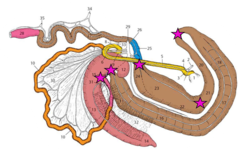
Where is the most common site of obstruction in horses?
pelvic flexure

during an exploratory celiotomy, where is the most common site for performing an enterotomy to empty the colon?
the pelvic flexure because it is easily exteriorized and can be placed on an operating tray to minimize contamination of the abdomen during surgery
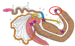
Where is the liver of the horse located?
entirely under the ribs. Oriented obliquely

true/false- horses have a gallbladder
FALSE- there is no gallbladder in equine
normally, the gallbladder stores bile and secretes it when needed. However, in the horse it is just secreted when they need it. No storage
why is it difficult to obtain a liver biopsy in the horse?
because the liver is completely overlapped by the lung
Where is the epiploic foramen located?
The epiploic foramen is located in the right dorsal abdomen ventral to the caudate lobe of the liver
it is between the caudal vena cava and caudate lobe of the liver dorsally and the portal vein and gastropancreatic fold ventrally

What are the parts of the pancreas?
it is triangular shaped with a body and 2 lobes (R and L)
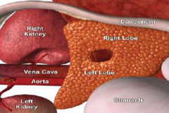
what is unique about the pancreas in the horse?
the body is perforated by a portal ring where the pancreatic tissue surrounds the portal vein

Where is the pancreas of the horse loacted?
primarily positioned to the right sublumbar, caudal to the stomach and liver
it extends between the right and left kidneys

What are the surfaces of the spleen?
parietal and visceral
Where is the spleen located?
the base is ventral to the last three ribs. The caudal margin is convex and initially parallel to the costal arch and can be palpated per rectum

What are the ligaments of the spleen?
1. gastrosplenic ligament
2. phrenicosplenic ligament (diaphragm and spleen)
3. nephrosplenic (renosplenic) ligament

What is the clinical significance of the renosplenic ligament?
it forms a shelf upon which the left colon can become lodged when displaced

How does the shape of the right and left kidneys differ?
The right kidney is heart shaped and the left kidney is bean shaped

Where is the right kidney located?
it contacts the caudate lobe of the liver and is ventral to the last 3 ribs and transverse process of L1

where is the left kidney located?
medial to the spleen ventral to the last rib and transverse process of L1-3
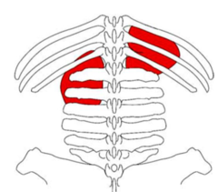
What are the visceral projections on the RIGHT side?

Where is the descending duodenum located and why is this significant?
it lies lateral to the base of the cecum and should be avoided during trocarization

What viscera is projected on the LEFT side?

what organs are projected ventrally?
the colon (sacculated)
ventral colon (right and left)
cecum (body and apex)
jejunum and descending colon
gravid uterus

If the large colons appear smooth upon linea alba exposure, one should immediately suspect what????
torsion of the large colons

the ventral colon is _______________
sacculated
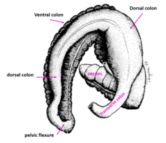
The dorsal colon is _____________
smooth
