Comprehensive BIOS 1710 Homeostasis, Nervous, Endocrine, and Immune Systems
1/223
There's no tags or description
Looks like no tags are added yet.
Name | Mastery | Learn | Test | Matching | Spaced |
|---|
No study sessions yet.
224 Terms
Homeostasis
Active maintenance of stable conditions inside of cells and organisms, environment outside of cells may change, but environment inside of cells remain relatively constant.
Examples of conditions maintained by homeostasis
Heart rate, blood pressure, blood sugar, blood pH, and other concentrations.
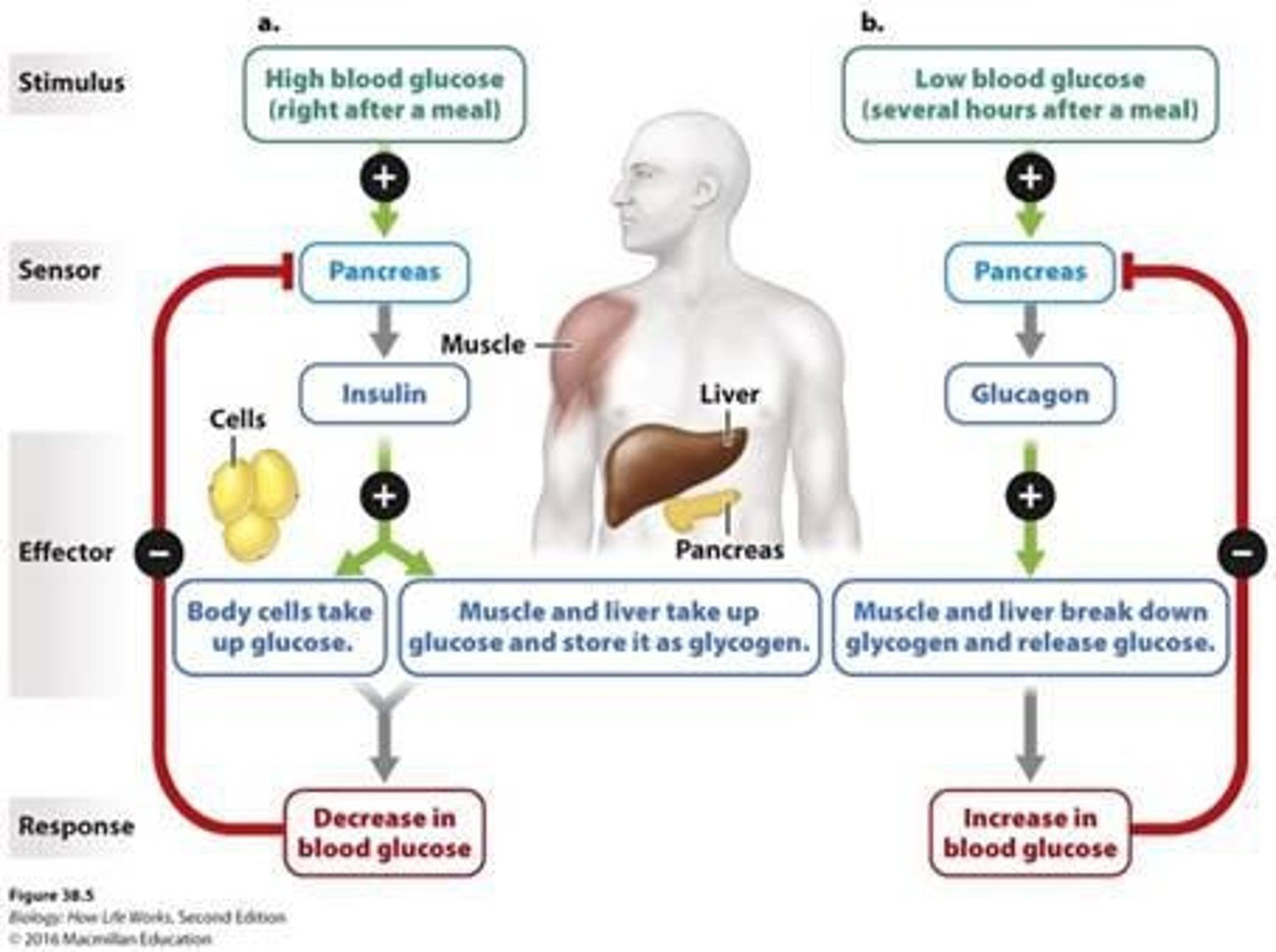
Blood glucose levels
Maintained by insulin and glucagon (pancreas), insulin lowers blood sugar and glucagon raises it.
Calcium levels in blood
Maintained by parathyroid hormone (PTH) and calcitonin; low blood calcium = PTH stimulates bone breakdown and kidney reabsorption.
Factors affecting homeostasis - External
Temperature (heatstroke/hypothermia), oxygen availability (changes in breathing/heartrate), toxins/pathogen (viruses, bacteria).
Factors affecting homeostasis - Internal
Metabolic activity (exercise increasing CO2, heat, waste), hormonal changes (puberty, pregnancy, aging), genetics (inherited disorders impairing regulation - diabetes, cystic fibrosis).
Factors affecting homeostasis - Lifestyle/Behavior
Diet and hydration (glucose, electrolytes), physical activity (circulation, metabolism), stress (psychological) - activates HPA axis (altering cortisol and other hormones), substance use.
Negative feedback failure
Diabetes (insulin feedback doesn't control blood glucose).
Positive feedback out of control
Blood clotting disorders or cytokine storm in severe infection.
Negative feedback mechanism
A stimulus acts on a sensor that communicates with an effector, which produces a response that opposes the initial stimulus/sensor detects stimulus and signals effector - keeps responding until reaching set point.
Positive feedback mechanism
Control mechanism where a change triggers responses that amplify the change, moving further from the starting state (childbirth) oxytocin increases contractions, which causes more oxytocin release until delivery.
Endothermic animals
Mammals, birds - generate and maintain body heat through internal metabolic processes.
Ectothermic animals
Reptiles, amphibians, fish, invertebrates - rely on external environment for body heat.
Nervous system evolution
No nervous system → simple nerve nets (sponges → cnidarians).
Bilateral symmetry in nervous systems
Cephalization (head concentration of sensory organs + processing centers).
Nerve cords and ganglia
Found in flatworms, annelids, arthropods.
Centralization and specialization
True brain and spinal cord (vertebrates).
Complex brains
Evolve in higher vertebrates, especially mammals, supporting memory, learning, reasoning, and social behavior.
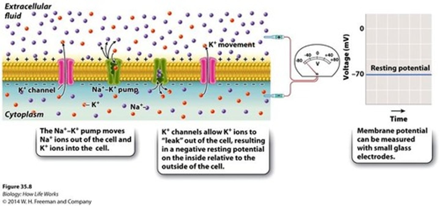
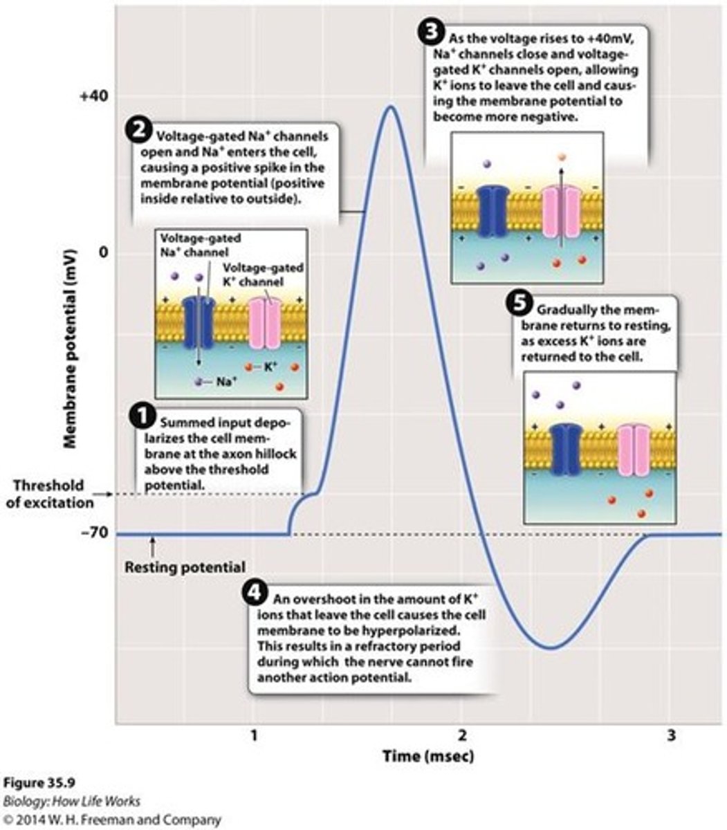
Resting membrane potential
The steady electrical charge difference across a neuron's plasma membrane when the cell is not transmitting an action potential. It's usually about -70 mV, with the inside more negative than the outside.
Ion concentration gradients
Na⁺ (sodium): higher outside the cell. K⁺ (potassium): higher inside the cell.
Na⁺/K⁺ pump
Uses ATP to pump 3 Na⁺ out and 2 K⁺ in. Makes the inside more negative and maintains ion gradients.
Potassium leak channels
Some K⁺ leaks back out, carrying positive charge with it. This movement leaves behind more negative charges inside.
Stable electrochemical balance
A stable electrochemical balance is reached, where the outward K⁺ movement is balanced by the inward pull of negative charges inside the cell.
Voltage of neurons
When no signal is present, it results in more negative ions inside the cell, some of the ions cannot cross the cell's membrane, making the inside negatively charged relative to the outside.
Membrane polarization
The membrane can be described as polarized due to the difference between charges of the inside and outside of the cell.
Action Potential
A signal that carries information from the cell body to the ends of the axon when depolarization at the axon hillock is strong enough.
Neurons
Cells that transmit information via action potentials and synapses, consisting of dendrites, cell body, and axon.
Glial Cells
Cells that provide structural support, insulate axons with myelin, regulate the extracellular environment, and help repair neurons.
Myelin Sheaths
Insulating layers formed by glial cells that enable faster signal conduction through saltatory conduction.
Resting Potential
The electrical potential of a neuron at rest, typically around -70 mV, maintained by the Na⁺/K⁺ pump and leaky K⁺ channels.
Depolarization
The phase where the threshold of approximately -55 mV is reached, causing voltage-gated Na⁺ channels to open and Na⁺ to rush in.
Rising Phase
The phase during which more Na⁺ channels open due to positive feedback, peaking at about +40 mV.
Falling Phase (Repolarization)
The phase where Na⁺ channels inactivate and K⁺ channels open, allowing K⁺ to leave the cell and the voltage to drop.
Hyperpolarization
The phase where K⁺ efflux overshoots, making the membrane potential more negative than the resting potential.
Refractory Period
The time after an action potential during which Na⁺ channels are inactivated, preventing immediate re-firing and ensuring unidirectional signal travel.
Propagation Velocity
The speed at which action potentials travel, influenced by axon diameter, myelination, and membrane properties.
EPSP (Excitatory Postsynaptic Potential)
A depolarization caused by Na⁺ influx that makes a neuron more likely to fire.
IPSP (Inhibitory Postsynaptic Potential)
A hyperpolarization caused by Cl⁻ influx or K⁺ efflux that makes firing less likely.
Temporal Summation
The process where repeated inputs at one synapse over time add together.
Spatial Summation
The process where inputs from multiple synapses on dendrites add together.
CNS (Central Nervous System)
The part of the nervous system that includes the brain and spinal cord, responsible for processing information and issuing commands.
PNS (Peripheral Nervous System)
The part of the nervous system that includes nerves carrying signals to and from the CNS.
Afferent Neurons
Neurons that carry sensory input to the CNS.
Efferent Neurons
Neurons that carry commands from the CNS to muscles and glands.
Somatic Nervous System
The part of the nervous system responsible for voluntary control of skeletal muscles.
Autonomic Nervous System
The part of the nervous system responsible for involuntary control of smooth muscle, heart, and glands.
Sympathetic Nervous System
The division of the autonomic nervous system responsible for the 'fight-or-flight' response.
Parasympathetic Nervous System
The division of the autonomic nervous system responsible for the 'rest-and-digest' response.
Sensory Receptors
Cells that convert physical or chemical stimuli into electrical signals (nerve impulses).
Chemoreceptors
Sensory receptors that respond to taste (gustation) and smell (olfaction).
Mechanoreceptors
Sensory receptors that respond to touch, pressure, vibration, hearing, and balance.
Electromagnetic Receptors
Sensory receptors that respond to light and vision.
Modality Requirements
Specific conditions necessary for effective sensory transduction for each modality, such as binding of molecules for olfaction and taste.
Vision
light activates opsin proteins in photoreceptor cells
Three types of eyes
eyecups (flatworms), compound eyes (arthropods), single-lens eyes (vertebrates, cephalopods)
Retina
photoreceptors = rods (light intensity) and cones (color, wavelengths)
Color vision
different cone types sensitive to short, medium, or long wavelengths
Divisions of the Brain
hindbrain, midbrain, forebrain. Mammals have expanded forebrain (cerebral cortex)
Cerebrum lobes
Frontal - planning, decision-making, motor control. Parietal - somatosensory processing. Temporal - auditory, memory, language. Occipital - vision.
White matter
myelinated axons (communication)
Gray matter
neuron cell bodies (processing)
Localization
functions mapped to specific cortical areas
Topographical maps
sensory and motor cortex represent body regions proportionally to sensitivity/motor control
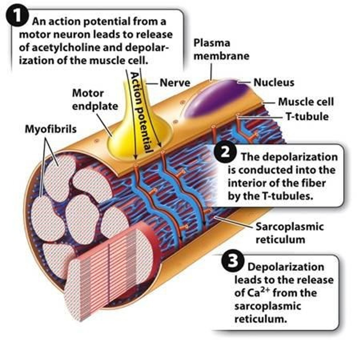
Synaptic transmission steps
Action potential reaches axon terminal.
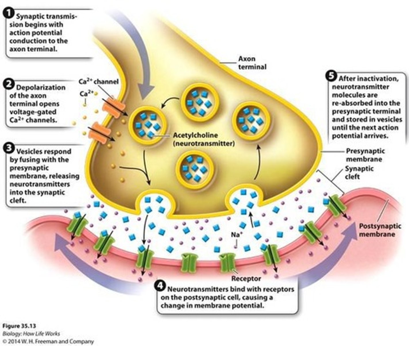
Calcium channels in synaptic transmission
Voltage-gated Ca²⁺ channels open; Ca²⁺ flows into presynaptic terminal.
Neurotransmitter release
Ca²⁺ triggers synaptic vesicles to fuse with membrane → neurotransmitter (ACh) released into synaptic cleft.
ACh function
ACh binds receptors on muscle fiber's sarcolemma → opens ion channels → depolarization.
Resetting the synapse
ACh is broken down by acetylcholinesterase, resetting the synapse.
Communication at the synapse
Ca²⁺ influx into the presynaptic neuron is the signal to release neurotransmitters.
Hormone definition
Chemical messengers produced and secreted by neurosecretory cells in the endocrine glands into the bloodstream.
Hormone vs. neurotransmitter
Hormone = released by endocrine glands into the bloodstream, travels long distances to reach target cells, effects - generally slow to start but last longer. Neurotransmitter = released by neurons into the synaptic cleft, acts on nearby cells (very short distance), effects - immediate but short lived.
Negative feedback example
Insulin and blood glucose - when blood sugar rises after eating, the pancreas releases insulin which helps absorb glucose, this lowers blood sugar, once blood sugar levels return to normal, insulin secretion decreases/ this maintains stable glucose levels.
Positive feedback example
Oxytocin during childbirth - baby's head pushes on the cervix, this signals the brain, then oxytocin is released, causing stronger uterine contractions, this pushes the baby further down leading to more oxytocin/ cycle continues until delivery.
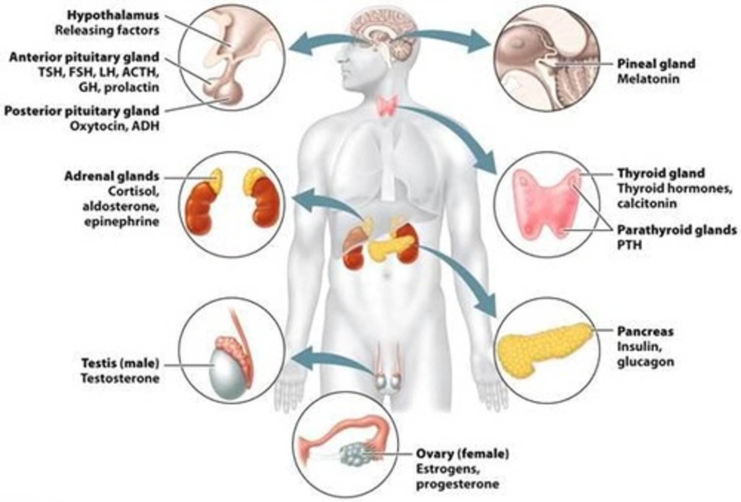
Major endocrine glands
Hypothalamus, pituitary gland (anterior and posterior), pineal gland, thyroid gland, parathyroid glands, adrenal glands (cortex and medulla), pancreas (islets of Langerhans), gonads (ovaries, testes).
Endocrine function tissues
Other tissues with endocrine function - kidneys, heart, stomach, small intestine, adipose tissue, placenta.
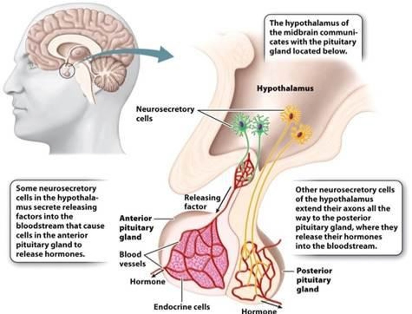
Hypothalamus hormones
Releasing hormones (stimulates pituitary) - TRH (thyrotropin-releasing hormone), CRH (corticotropin), GnRH (gonadotropin), GHRH (growth hormone). Inhibiting hormones - somatostatin (inhibits GH, TSH), dopamine (inhibits prolactin). Also produces - oxytocin and ADH(antidiuretic hormone) - stored and released by posterior pituitary.
Anterior pituitary hormones
growth hormone (GH), prolactin (PRL), thyroid-stimulating (TSH), adrenocorticotropic (ACTH), follicle-stimulating (FSH), luteinizing hormone (LH).
Posterior pituitary hormones
releases oxytocin and antidiuretic hormone (ADH/ made in hypothalamus).
Hypothyroidism
Too little thyroid hormone leading to low metabolism, fatigue, weight gain, slowed heart rate, depression, and can cause growth and developmental delays in children/myxedema coma (life-threatening).
Hyperthyroidism
Too much thyroid hormone resulting in high metabolism, weight loss, heart palpitations, anxiety, tremors, bulging eyes/throid storm (medical emergency).
Nervous System
Detects stress.
Hypothalamus
Part of the brain that receives neural information, produces releasing hormones that control the pituitary gland/signals pituitary.
Pituitary Gland
Releases hormones that regulate other endocrine glands.
Nervous System and Endocrine System Interaction
The nervous system provides rapid, precise signaling, while the endocrine system coordinates longer-lasting, widespread changes. They often work together.
Fight-or-Flight Response
Example of interaction: the sympathetic nervous system activates the adrenal medulla to release epinephrine and norepinephrine, producing faster heart rate, increased breathing, and metabolic changes that prepare an animal to respond to threats.
Hormone Release Situations
Three situations in which hormones are released: Nervous system signals, physiological changes (internal cues), and environmental cues.
Epinephrine/Norepinephrine
Released from adrenal medulla during stress (fight-or-flight).
Parathyroid Hormone (PTH)
Secreted when blood calcium is low.
Calcitonin
Secreted when calcium is high.
PTTH (Prothoracicotropic Hormone)
Released from the insect brain after feeding; stimulates the prothoracic gland.
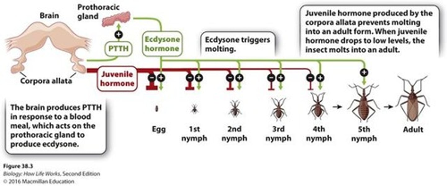
Ecdysone
Steroid hormone released by the prothoracic gland; triggers molting and tissue reorganization.
Juvenile Hormone (JH)
Secreted by the corpora allata; keeps the insect in a juvenile state.
Hormone Interaction for Molting
High JH + ecdysone → molting into another juvenile stage. Low JH + ecdysone → metamorphosis into the adult stage.
Juvenile Hormone as Insecticide Target
Juvenile hormone (JH) is a prime target; artificially raising JH levels prevents larvae from completing metamorphosis into fertile adults.
Anterior Pituitary
Arises from epithelial cells of the mouth; communicates with the hypothalamus indirectly by releasing hormones into a portal blood system.
Posterior Pituitary
Originates from neural tissue; neurosecretory cells in the hypothalamus extend axons directly into the posterior pituitary, releasing hormones straight into the bloodstream.
Tropic Hormones
Hormones that act on other endocrine glands to stimulate them to release their own hormones.
TSH (Thyroid-Stimulating Hormone)
Acts on the thyroid gland to stimulate the release of thyroid hormones (T3, T4).
FSH & LH (Gonadotropins)
Act on ovaries/testes to stimulate the release of estrogen, progesterone, testosterone.
ACTH (Adrenocorticotropic Hormone)
Acts on the adrenal cortex to stimulate the release of cortisol.