NEUROSCI EXAM 2- GONNA SLAY
1/100
There's no tags or description
Looks like no tags are added yet.
Name | Mastery | Learn | Test | Matching | Spaced | Call with Kai |
|---|
No analytics yet
Send a link to your students to track their progress
101 Terms
change blindness
A visual perception phenomenon that occurs when a person fails to notice a significant change to a visual scene, even when they are paying attention. It’s thought to happen when visual attention is diverted away from the change
What is Attention? William James (1890)
“Everybody knows what attention is. It is the taking possession by the mind, in clear and vivid form, of one out of what seem several simultaneously possible objects or trains of thought. Focalizations, concentrations of consciousness are of its essence. It implies withdrawal from some things in order to deal effectively with others…”
Attention
System that helps to select relevant information for processing, while filtering out irrelevant information
Why do we need attention?
Information overload:
We have to deal with an incredible amount of sensory information
“…amount of information coming down the optic nerve - estimated to be in the range of 108 – 109 bits/sec – far exceeds what the brain is capable of fully processing & assimilating into conscious experience.” C. Koch (2000)
Limited neural capacity:
Energy metabolism can support only about 1 spike in 10 seconds per neuron on average
Attention simplifies information processing!
By boosting relevant stimuli
By eliminating irrelevant stimuli from consideration
Type: Overt Attention
Eyes orient to source
Examples:
Looking at someone when they speak
Looking toward a loud noise
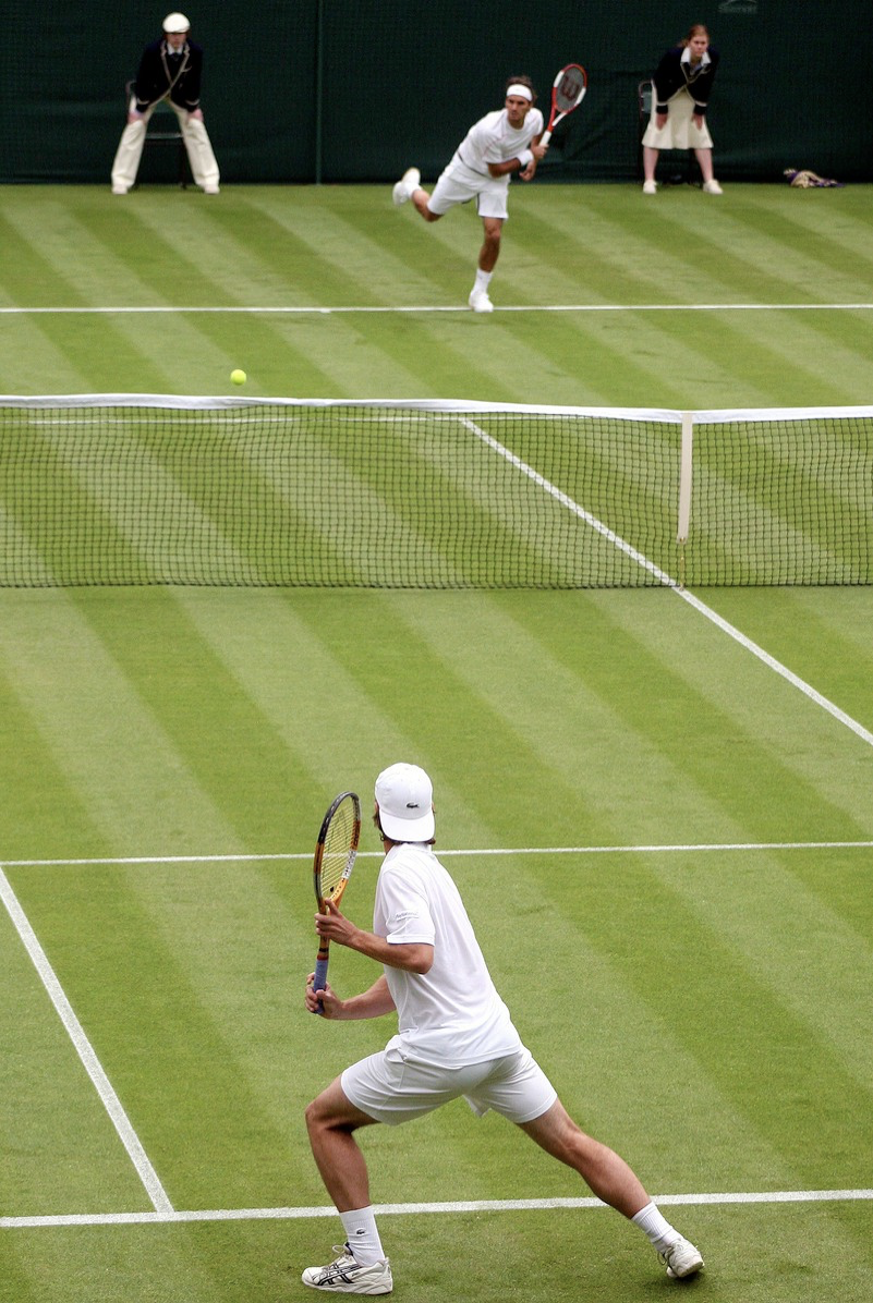
Moving your eyes to follow the passes in the first video
Moving our eyes allows us to select & process relevant parts of the display
Most direct way to deploy attention
Type: Covert Attention
Eyes do NOT orient to source
Examples:
Looking at someone while you type
Staring at your phone while you eavesdrop on a conversation at Starbucks
We don’t have to move our eyes to attend to different parts of our environment
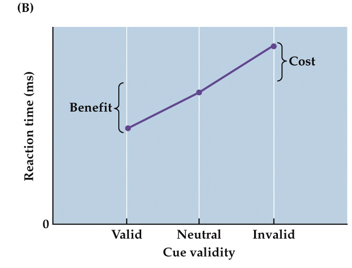
Type: Endogenously Cued Attention
Directed, volitional selection of information that is goal-driven and internally cued (WW); benefit for validly-cued targets / cost for invalidly-cued targets
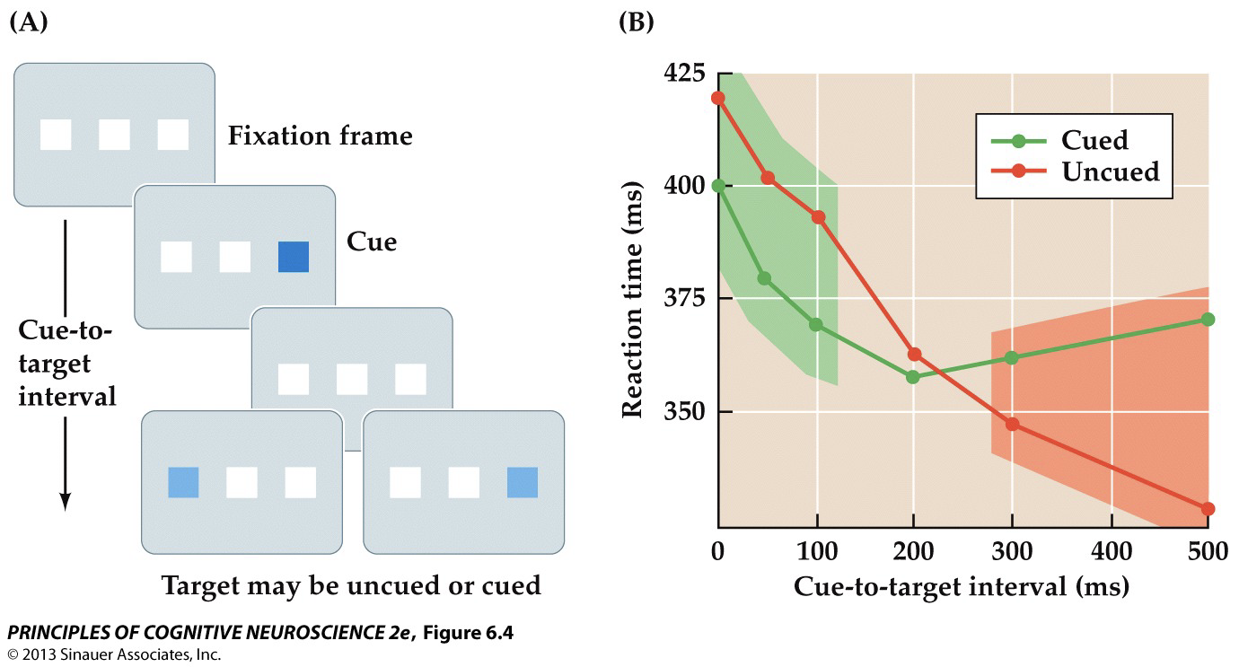
Type: Exogenously Cued Attention
Automatic, reflexive selection of information that is stimulus-driven and externally cued; benefit for cued targets w/ short cue-to-target intervals / cost for cued targets w/ long intervals… If the target shows up after a longer delay ( < 200 ms), your brain may actually inhibit returning to that cued spot— this is called Inhibition of Return (IOR), specific to exogenous cuing — it helps you avoid getting "stuck" looking at the same place over and over… so, you're slower to respond to targets at that location
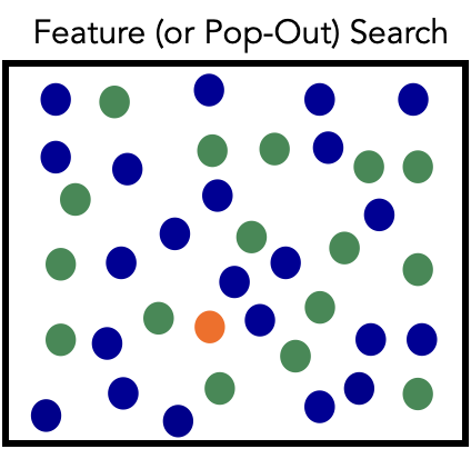
Feature (pop-out) search
Parallel selection- your brain can process multiple items at the same time, across the visual field — you don’t need to scan each one individually
Target is defined by a single feature that immediately stands out (color, shape, size); search is fast & independent of distractor number (as # goes up, time it takes to find target remains constant); RTs are (essentially) flat
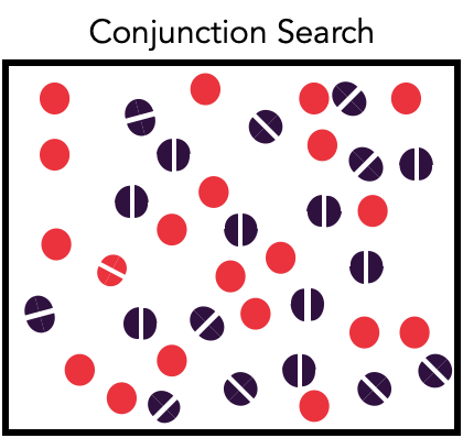
Conjunction search
Serial selection- checks one item at a time (focused attention)
Target requires a combination of features, making it harder to find; search time (RT) increases as the # of distractors (set size) increases; steeper slope for absent > present
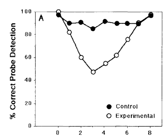
Attentional blink
A phenomenon that occurs when exposed to a rapid stream of visual presentation/ RSVP, & you fail to notice a 2nd target (T2) if it appears shortly after the 1st (T1); typically, the "blink" happens if T2 appears within 200-500 ms after T1; blink→ 1st attentional window closed, 2nd window not yet open
Task: ID white letter (T1) & detect X (T2) if present (control→ 1 task, exp→ 2)
1 location, everything is relevant + attended to (but still limited capacity to process)
Classic approach
Develop theories based on behavior
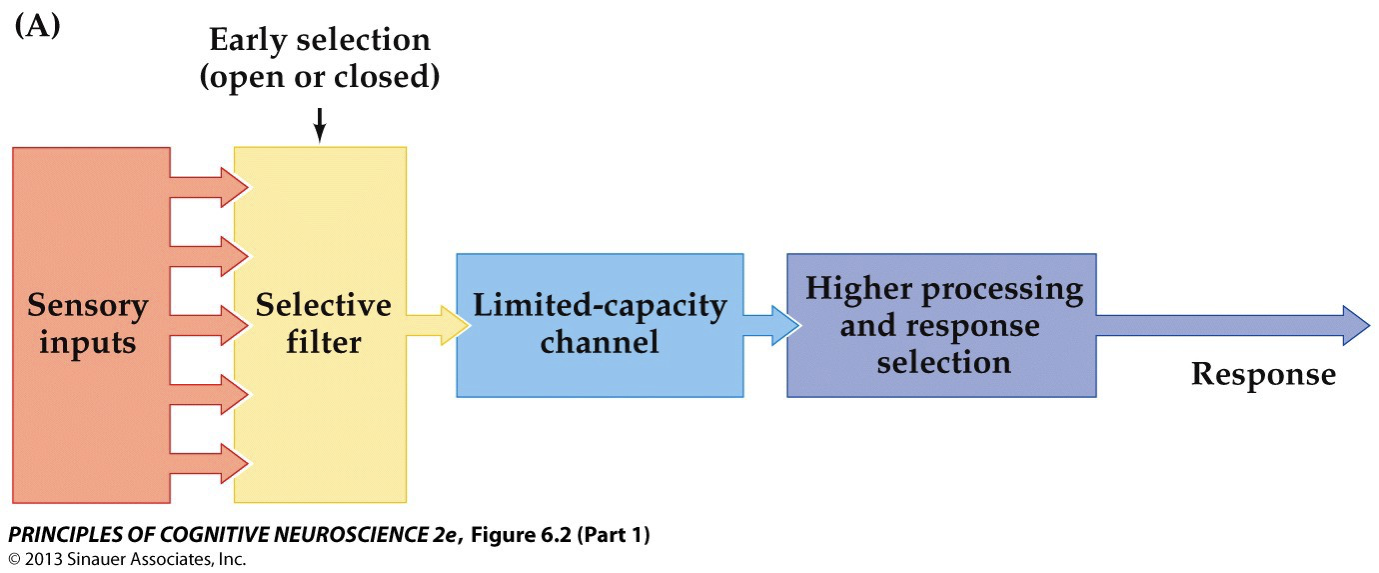
Boradbent’s Early Selection Model (1958)
Only the selected information is passed through; everything else is lost
Sensory inputs, selective filter (early selection, open to relevant info, closed to irrelevant info), limited-capacity channel, higher processing & response selection, response
Auditory Attention: Shadowing
Stimuli: 2 ears receive different information
Task: Participant repeats one stream aloud
Cherry (1953)
Attended stream: content reported accurately
Unattended stream: could only report pitch (M/F)
Moray (1959)
Don’t hear irrelevant words in unattended stream, even repeated 35 times
33% hear own name in unattended stream (“Cocktail party effect”)
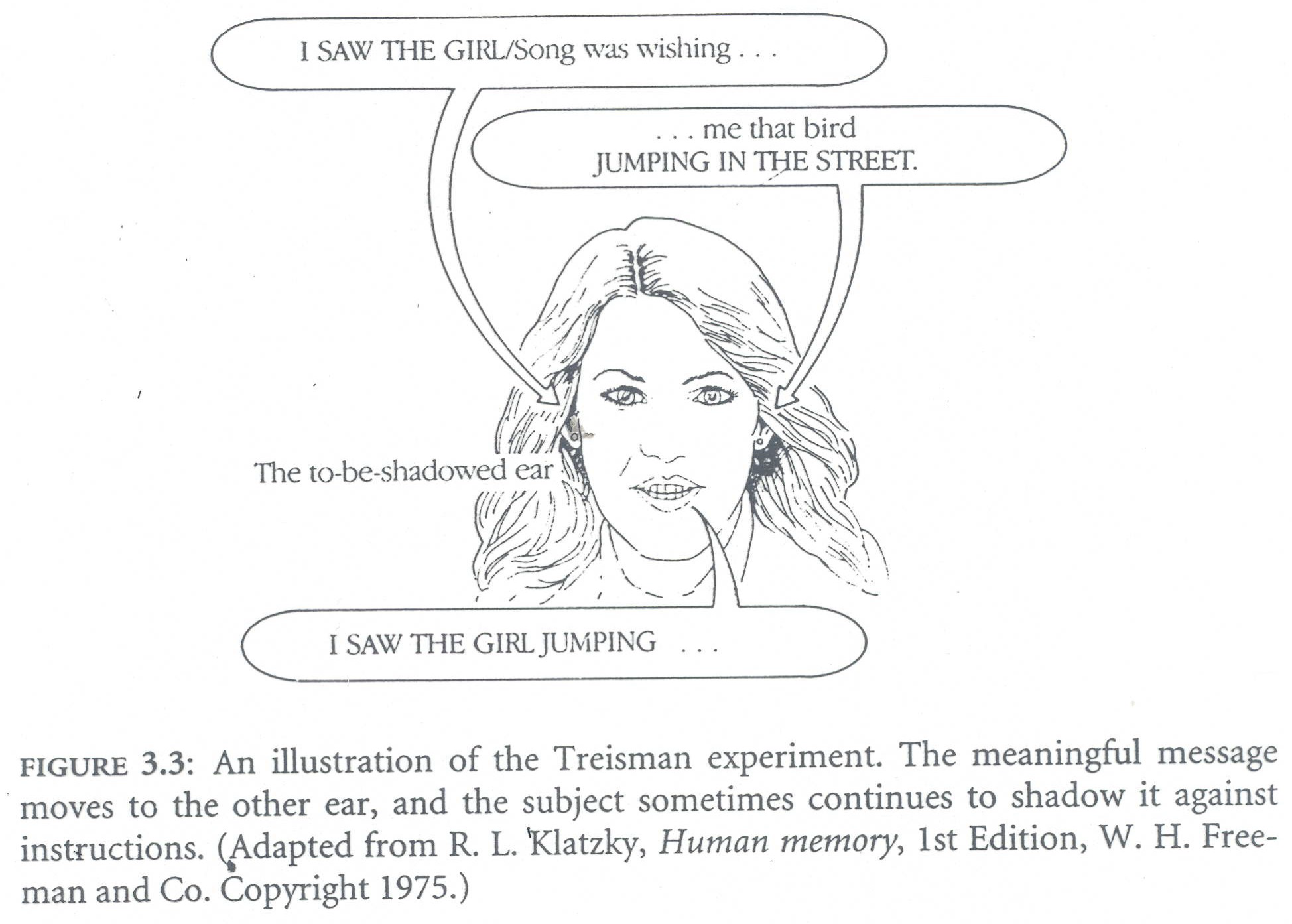
Treisman (1960)
Participants continued to shadow meaningful content even from unattended stream
Implies processing in US up to at least semantic level
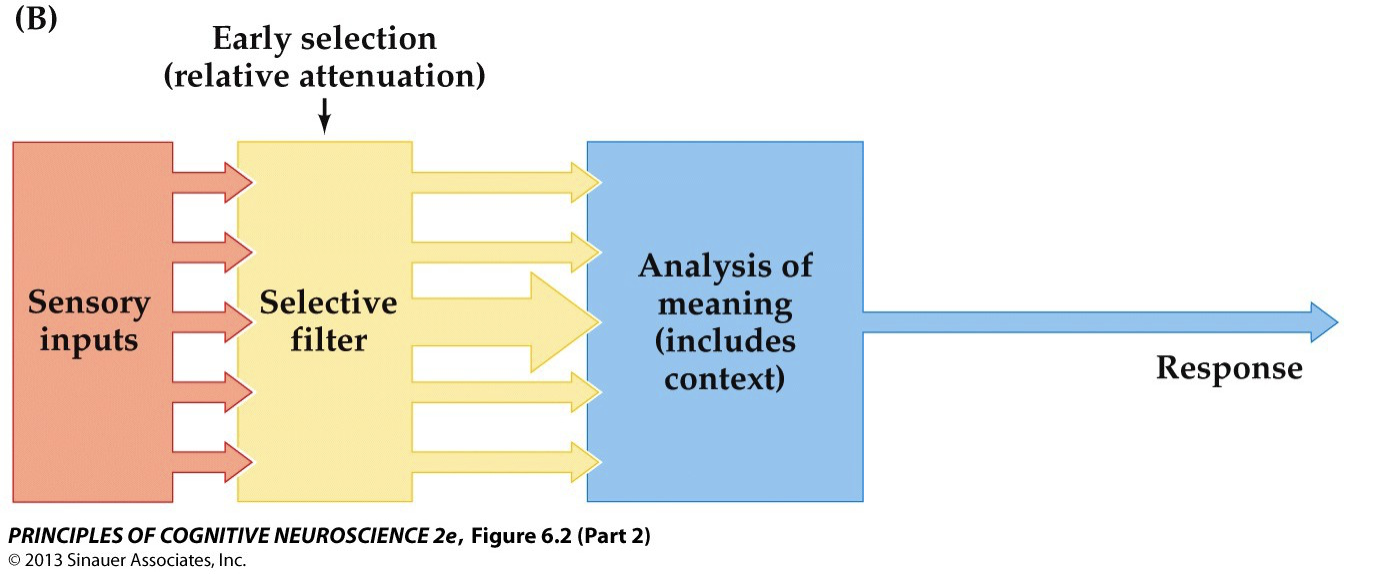
Treisman’s Attenuation Model
Attended information passed through; unattended information attenuated but salient information can still get through
Sensory inputs, selective filter (early selection→ relative attenuation), analysis of meaning (includes context), response
Attention in the Brain
Greater # of spikes (APs) when attention is engaged (seen w/ EEG, PET, fMRI)
Influence of attention observed at multiple levels of processing
Early Selection
happens before conscious awareness (IOW, APs before time of CA); for both endogenously & exogenously cued spatial attention, the effects on the sensory pathways appear to entail a modulation of early sensory processing activity in the visual sensory cortex
Late Selection
happens at (or after) the stage of conscious awareness
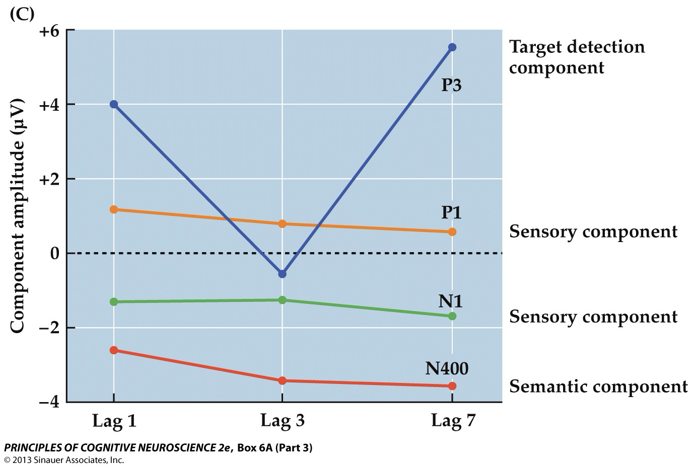
Attentional Blink Affects Neural Processing at
… Level of Conscious Perception; ERP Components→ only P3 (conscious detection) was affected (Lag 3 = T2 is the 3rd item after T1 (T1, distractor, distractor, T2); P1 & N1 components did not differ as a function of whether the target occurred during the blink period→ sharp contrast to studies of visual spatial attention, in which attention strongly modulates the amplitudes of sensory ERP components; even when the 2nd target could not be reported, the information in the stimulus seemed to have been processed up through semantic level (N400) (attentional selection→ after SA, before CD)
Early studies
Extrastriate (V2, V3, V4), covert attention, PET & ERP recordings
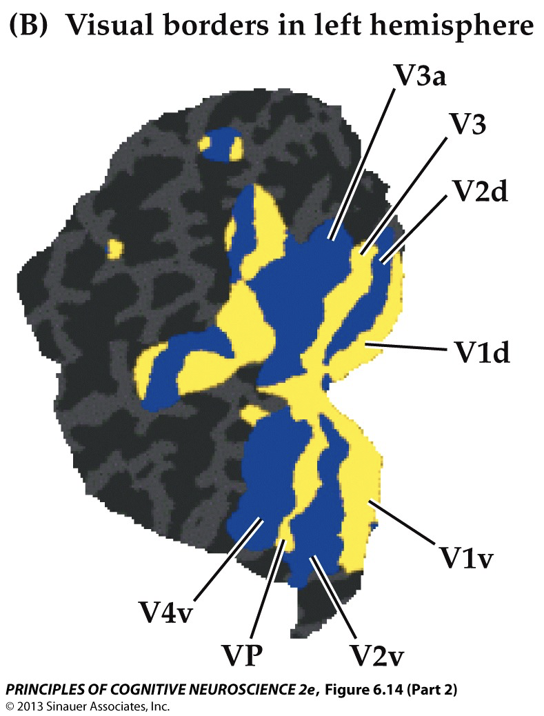
Visual Attention: Modulation of V1
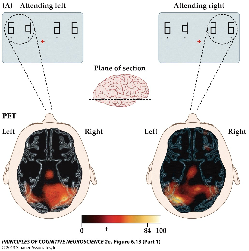
fMRI study shows location of attention effects w/ respect to the retinotopic organization of the low-level visual areas in occipital cortex… Effects of attention follow retinotopic organization! Due to limited temporal resolution, we cannot infer early or late selection
i.e. attending to the R side of bilateral stimuli presented in the Upper visual field produces attention-related enhancements in the low-level visual cortical region in L ventral occipital cortex
Reentrant process
Attention-related activity returns to the same low-level sensory areas that were initially activated in the ascending pass through the system, presumably reflecting enhanced late processing of stimulus information in those areas
LGN & V1
attention can even influence LGN activity, though not as much as V1 (relay station in thalamus- receives input from retina & sends it to V1)→ attention operates extremely early in the visual processing stream — even before the cortex gets involved. This means the brain isn't passively waiting to receive input — it’s proactively shaping perception from the very first stages
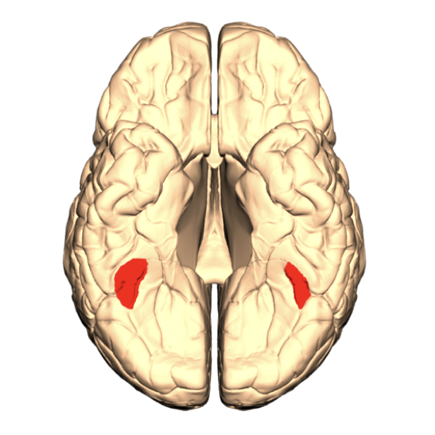
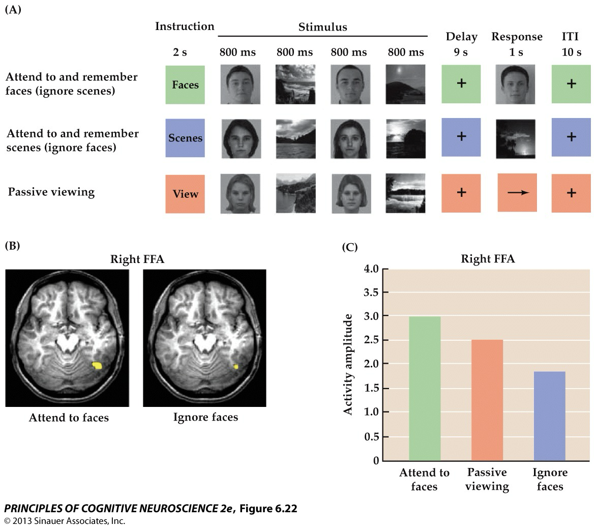
Fusiform Face Area (FFA)
part of the inferior Temporal Lobe that is needed to perceive unchanging aspects of human faces; Right FFA typically more active; part of the Ventral Stream
FFA- related finding
Activity in the face-sensitive FFA in ventral occipital cortex was higher when attending to faces relative to just passively viewing stimuli + lower than passive viewing when ignoring faces
Visual attention modulates neural processing in early visual areas including:
LGN, V1 (follows retinotopic organization)… concerned with where we direct our visual attention and when attention influences brain activity
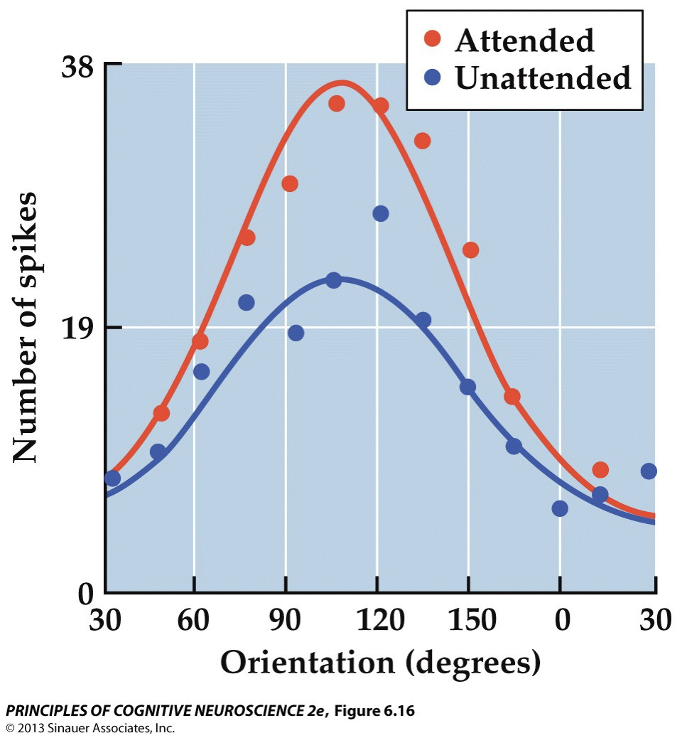
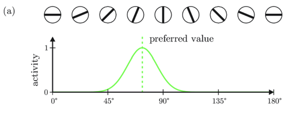
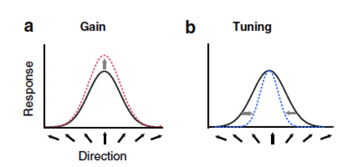
Tuning curves
demonstrate selectivity of response to a certain stimulus parameter; tells us the preferred stimulus for a neuron and how sensitive or selective that neuron is to variations in that stimulus
Gain
Differences maximal at peak response. Imagine attention as a multiplier (response of 1*1.1 = 1.1 vs response of 2*1.1 = 2.2)

Tuning
Differences maximal at weaker responses. Attention enhances selectivity.
V1 & V4
Primary visual cortex (V1) & Ventral stream (V4):
Increased response when attention engaged
shows increased activity in visual areas;
V4 had larger increase
critical for object recognition
Tuning curve did not change
multiplicative scaling
it caused gain, but not tuning!
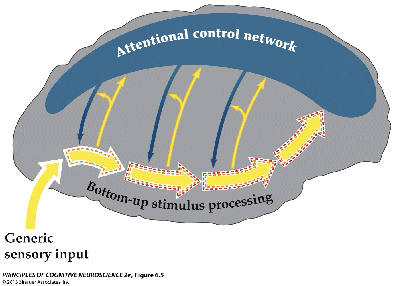
Findings imply that the dorsal frontoparietal network exerts modulatory control over stimulus processing in the sensory cortices, & thus over concomitant behavioral performance
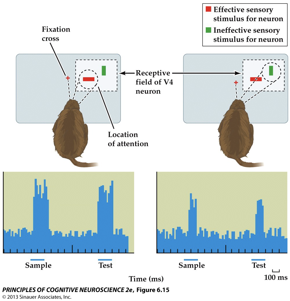
When both an effective stimulus & an ineffective stimulus were presented within the receptive field & the money attended to the effective stimulus, responses were robust; however, when the money attended to the ineffective stimulus, the responses were much reduced, despite presence of ES in receptive field
i.e. When attention was directed to a location outside the receptive field of the cell, the response to the effective stimulus within the receptive field was not modulated
Later stages of visual processing in ventral pathway, different results
More nuanced view:
Attention biases processing in favor of some stimuli; not a strict filter
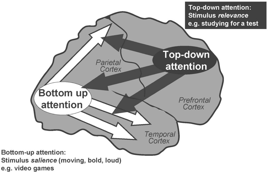
Drivers of attention:
Salience (bottom-up— in visual attention this means starts with lower-level processing of specific sensory characteristics→ higher order processing, exogenous)
Cognitive control (top-down— in visual attention means that higher order processing ➔ specific sensory characteristics, endogenous)
Novelty & surprise (can be top-down & bottom-up→ context; cool breeze, seeking new experiences
Schematic representation— yellow & blue vertical arrows indicate interactions between sensory processing & control networks at various levels
Endogenous control
Dorsal fronto-parietal; IPS/ SPL→ Intra-parietal sulcus/ Superior parietal lobule
FEF (frontal eye field)→ a region in the frontal cortex of the brain that controls eye movements but is also involved in attention, cognition, working memory…
Involves dorsal aspects of the frontal & parietal cortices
Involved in top-down control of visual processing
IPS & FEF also modulated by stimulus-driven control

Exogenous control
Fronto-parietal & temporal exogenous; involves ventral aspects of the frontal & parietal cortices (plus superior temporal gyrus, which is ventral relative to the dorsal network)
VFC = Ventral Frontal Cortex→ ventromedial prefrontal cortex (vmPFC) comprises a set of interconnected regions that integrate information from affective sensory and social cues, long-term memory, & representations of the 'self’
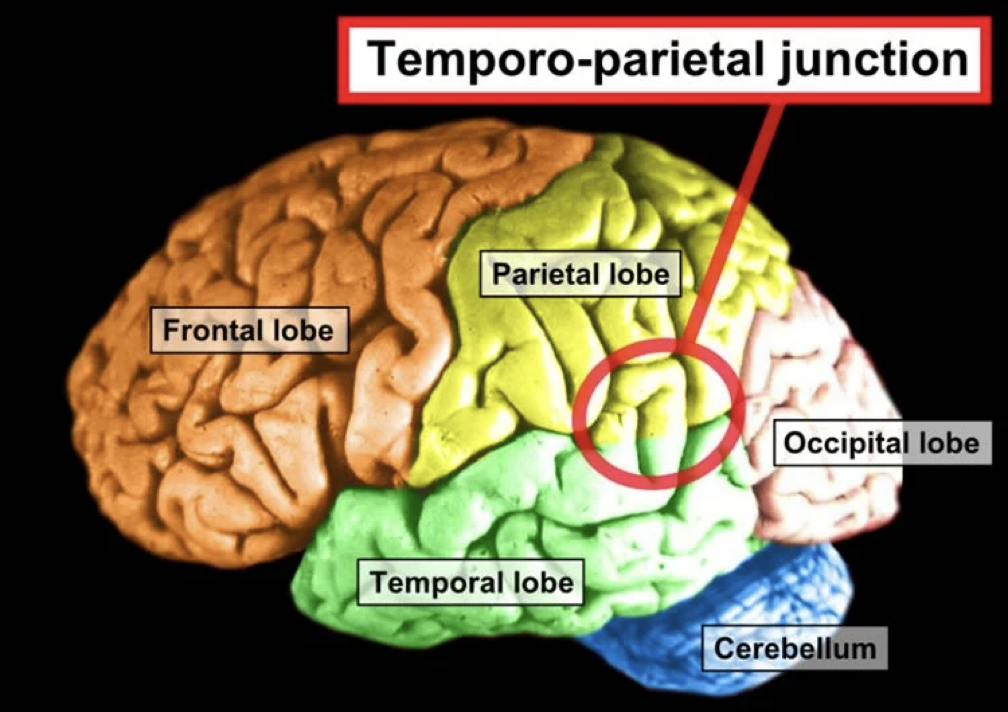
TPJ→ Tempro-parietal Junction
Involved in stimulus-driven control
Exogenous system more apparent on the right hemisphere; stimuli in both fields activates R TPJ
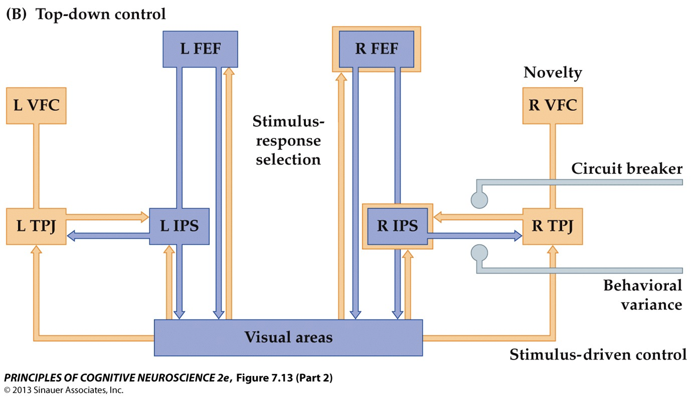
Circuit breaker/ behavioral variance
connections between the IPS & TPJ can interrupt ongoing top-down control when unattended salient stimuli are detected
Brain perturbation studies support importance of a frontoparietal network: Lesion in the PFC
reduced ability to use attention for sensory modulation
Brain perturbation studies support importance of a frontoparietal network: TMS to enhance frontal cortex on one side
increased ipsilateral visual cortex & enhanced ability to pay attention in corresponding visual field
Brain perturbation studies support importance of a frontoparietal network: TMS to disrupt right parietal lobe
neglect-like symptoms
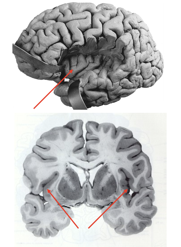
Hemispatial Neglect
Most commonly a result of damage to the right parietal cortex; left visual field is affected
Issue of attention, not vision
It is essentially a failure to pay sufficient attention to sensory input
Matches ventral better than dorsal (e.g., right sided ventral system)
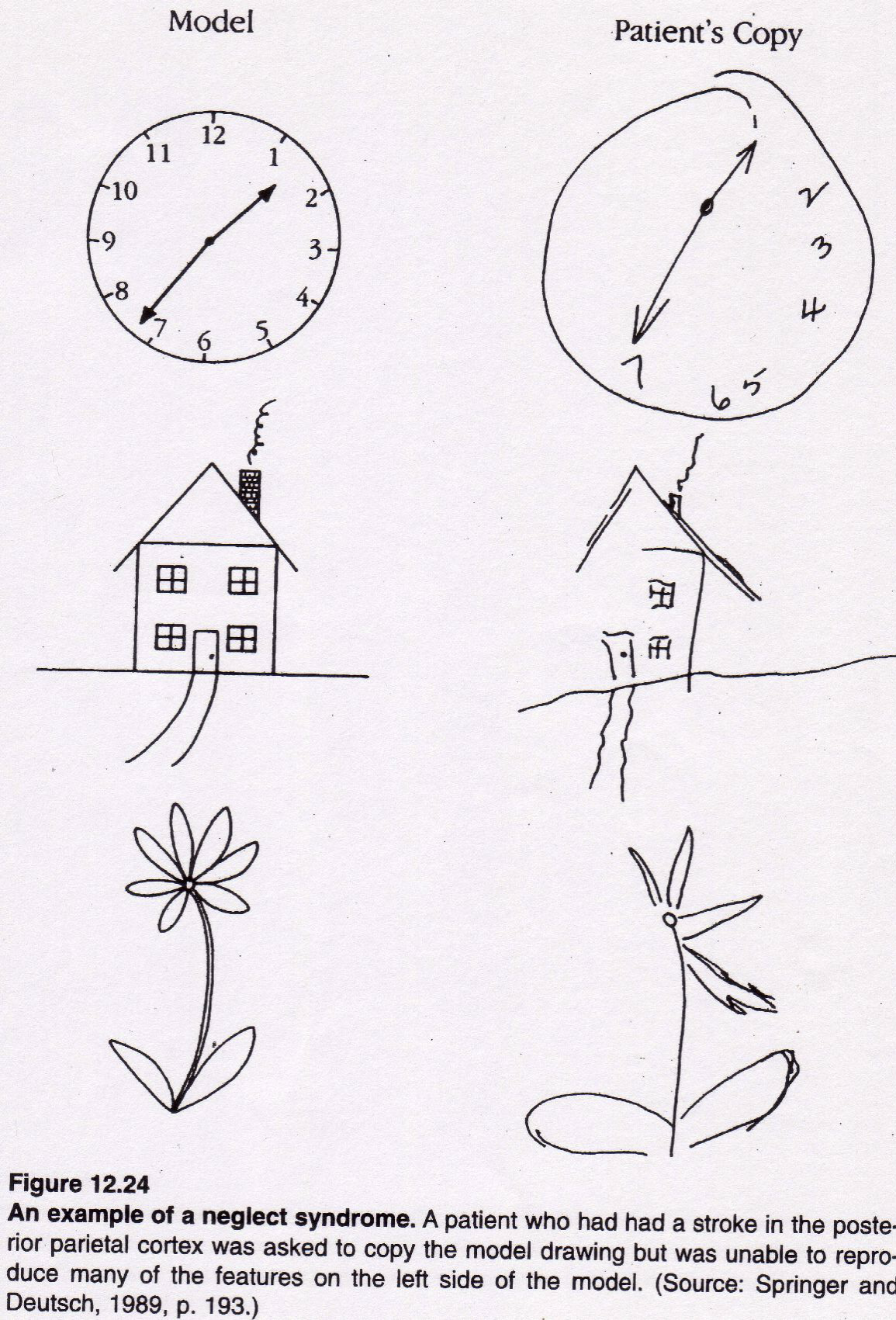
Attention Deficits: Hemispatial neglect
Patients fail to respond or orient to contralesional stimuli & events
Sometimes even their own body/body parts (paralyzed or not)
Most commonly due to right hemisphere parietal injury
Symptoms may occur in multiple sensory modalities yet are…
Independent of the existence of primary sensory or motor loss
What about ADHD?
Prefrontal Cortex – important for top-down attention regulation
Reduced size & functional activity of the Prefrontal Cortex
Disorganized white matter tracts emanating from PFC & weaker PFC connectivity
Default Mode Network – more active when attention is idling (DMN activity is anticorrelated with frontoparietal attentional control networks)→ overactive & overconnected DMN
more DMN activity in ADHD (DMN includes PCC, ventral ACC, & mPFC)
Therapeutic doses of stimulants improve PFC functions & enhance the efficiency of PFC activity in normal, young adult subjects. A similar, but much more pronounced profile is observed in subjects with ADHD. Thus, stimulant actions in ADHD are not paradoxical, but simply more apparent.
ADHD associated w/ creativity/ divergent thinking & ‘hyperfocus’
Bottom-up attention: stimulus salience (moving, bold loud, i.e. video games)
Top-down attention: stimulus relevance (i.e. studying for a test)
Memory
Series of processes which allows NS to:
Encode information acquired from experience
Store this information over time,
Retrieve it to guide behavior & plan actions
Fundamental to every aspect of human cognition→ emotion, action, language, decision-making, attention, sensation
Amnesia
Memory loss:
Retrograde— Loss of memories from before the injury or brain damage; You forget things that already happened.
Anterograde— Inability to form new memories after the injury or brain damage; You can’t create new memories moving forward
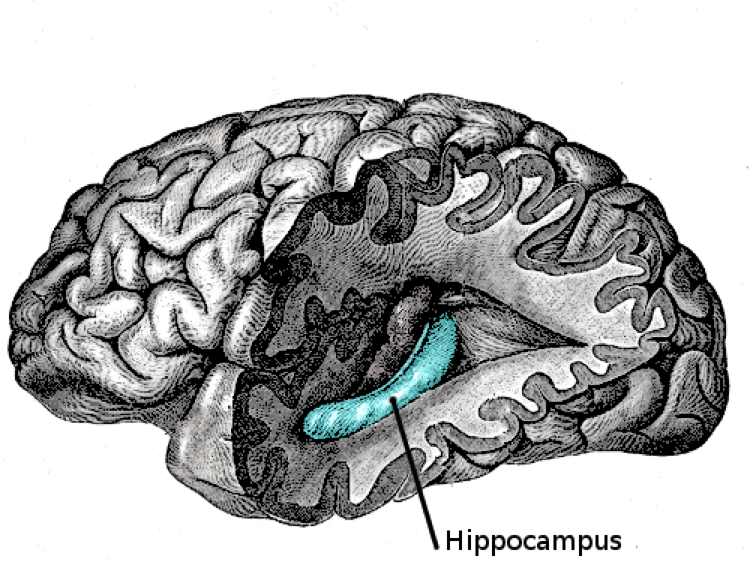
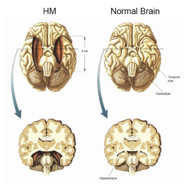
Hippocampus
Found deep within the medial temporal lobe— there are 2 hippocampi, one in each cerebral hemisphere. Its name comes from the Greek for "seahorse" because when it is removed from the brain, it resembles the equine fish.
Case Study: H.M.
Bilateral Medial Temporal Lobe (MTL) resection in treatment of intractable epilepsy at 27 years old (in 1953)
Spared working memory
Could maintain a 3-digit # for over 15 minutes if no distractions (if given a distraction, # was lost)
Could maintain a digit span of up to 6 digits at a time
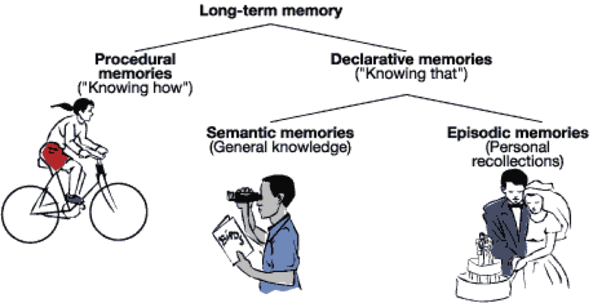
Spared nondeclarative memory
Skill learning→ could acquire new skills (i.e. mirror-drawing task)
Priming→ showed a normal priming effect
Conditioning→ could be classically conditioned
* Did not remember any of these things happening to him (no explicit memory of them)
Primary deficit: Anterograde amnesia – cannot make/ form new memories for events & facts after surgery
Some retrograde amnesia— loss of memory around 3 years prior
HM – AA caused by loss of tissue in the medial temporal lobe, including the hippocampi (surgery removed amygdala, most of H, & surrounding cortex from both temporal lobes)
Surgical patients with similar surgery but an intact hippocampus are able to form new memories but sometimes have different types of memory issues…
Temporo-Parietal Cortex
Case Study: K.C.
Lost hippocampi, motorcycle accident, could recall facts but no personal life events
Kent’s hippocampal damage accounts for his AA but not for the loss of his autobiographical memory
Brian-imaging studies show that semantic & episodic memories are processed & stored in different locations; postmortem examinations of KC’s brain also revealed the complex nature of his brain damage
Could not retrieve personal memory
(2 distinct subtypes of declarative memory)
Retrograde amnesia for episodic memory
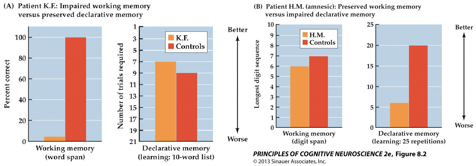
Working vs. L-T Memory: comparing Case Studies
Double-dissociation:
KF (Left temporoparietal damage):
Impaired working memory but normal long-term memory
HM (Bilateral hippocampal damage):
Normal working memory but impaired long-term memory
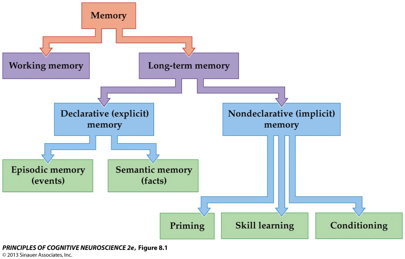
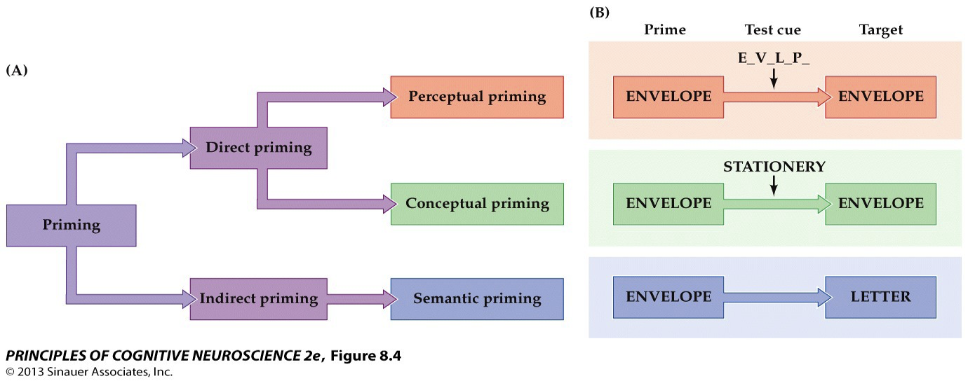
Priming
Change in the processing of a stimulus due to a previous encounter w/ the same (or closely related) stimulus
Can happen in just 1 exposure
Can be direct or indirect
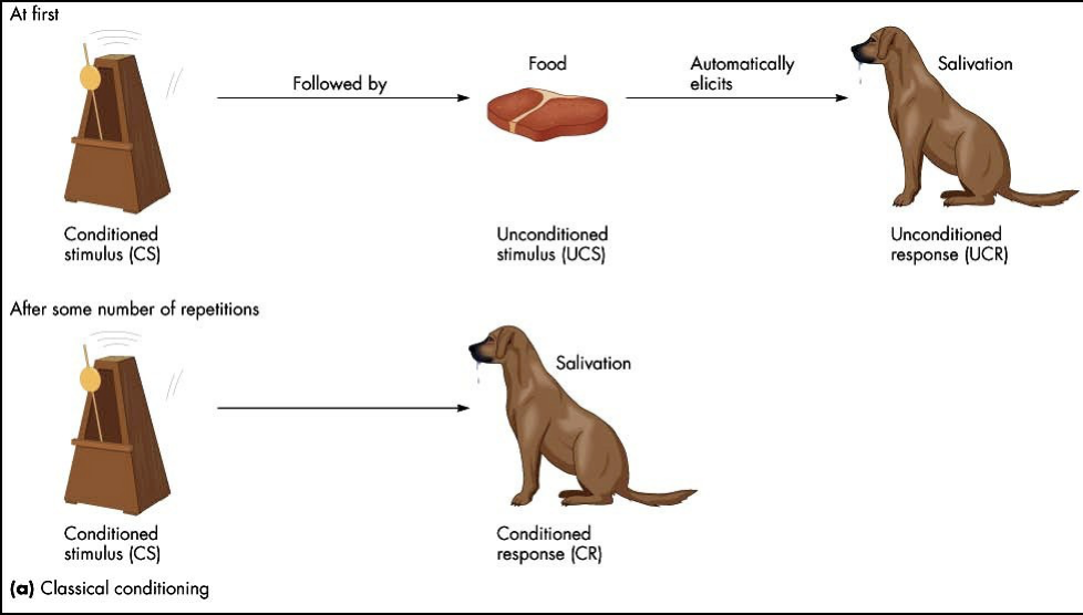
Classical Conditioning
Change in reflexive behavior due to reliable association w/ a novel environmental event (Pavlov); usually requires multiple exposures
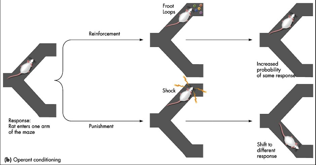
Operant Conditioning
Probability of a behavioral response altered by association w/ reward (or punishment)
Skill Learning
Gradual improvement in performance due to repeated practice
Fits what we intuitively think of when we think of “learning”; takes hours to years
Motor Sequence Learning
tasks focus on incremental acquisition of movements into a well-executed behavior
Serial Reaction Time (SRT) Task
Preserved in MTL damaged patients
Impaired in Basal Ganglia damaged patients
Results→ even when participants are unaware that there is a sequence, the sequence repetition leads to faster reaction times as the task progresses (implicit learning!)
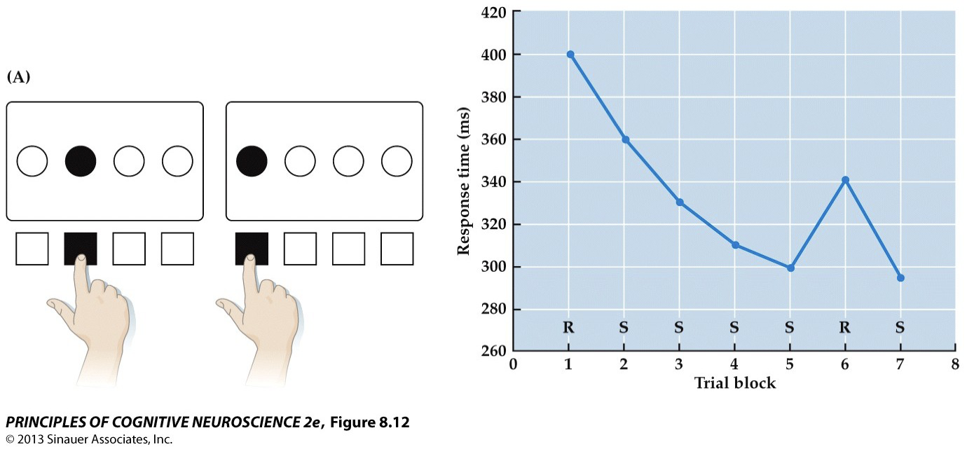
Cellular Mechanisms of Learning & Memory: Habituation
Decrease in response due to repeated presentation; decreased neurotransmitter release
Neural circuit in the abdominal ganglion of the sea slug→ touching the animal’s siphon elicits withdrawal of the gill; siphon repeatedly touched, response progressively decreases (H); conversely, a shock to the tail enhances the habituated gill withdrawal response (S)
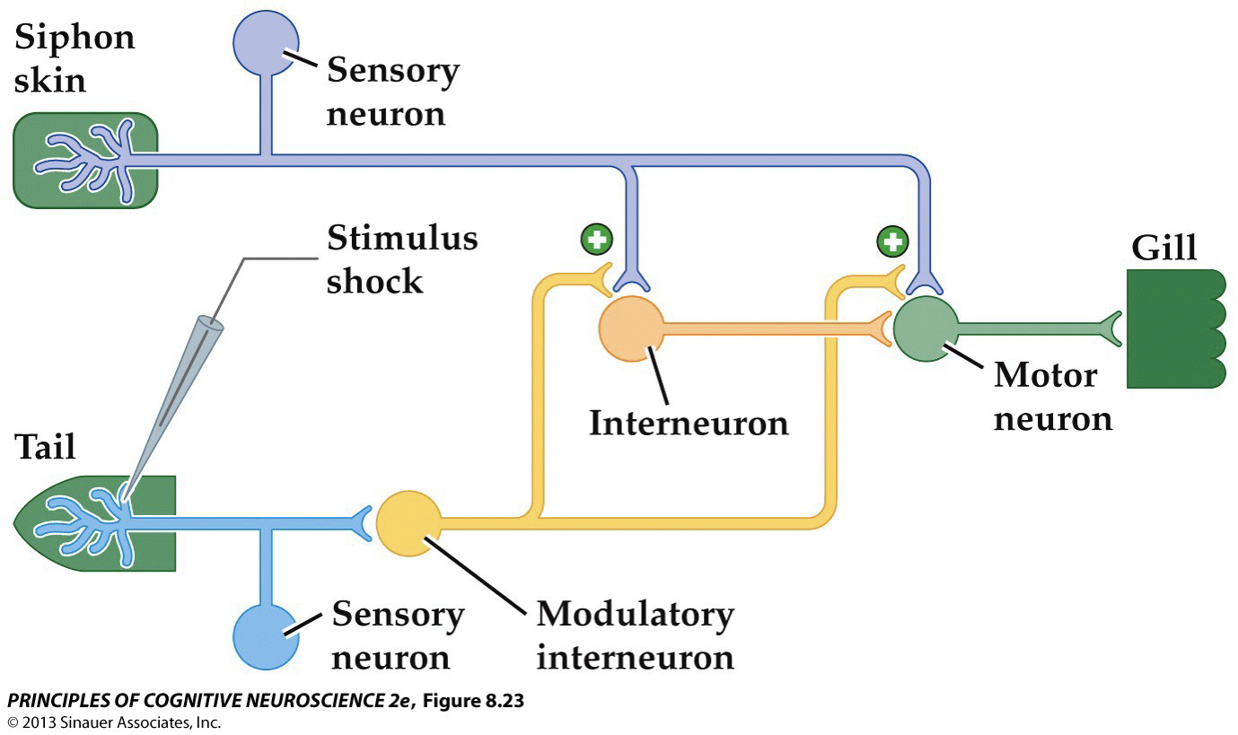
Cellular Mechanisms of Learning & Memory: Sensitization
Increased response to habituated stimulus when paired with aversive stimulus; increased neurotransmitter release

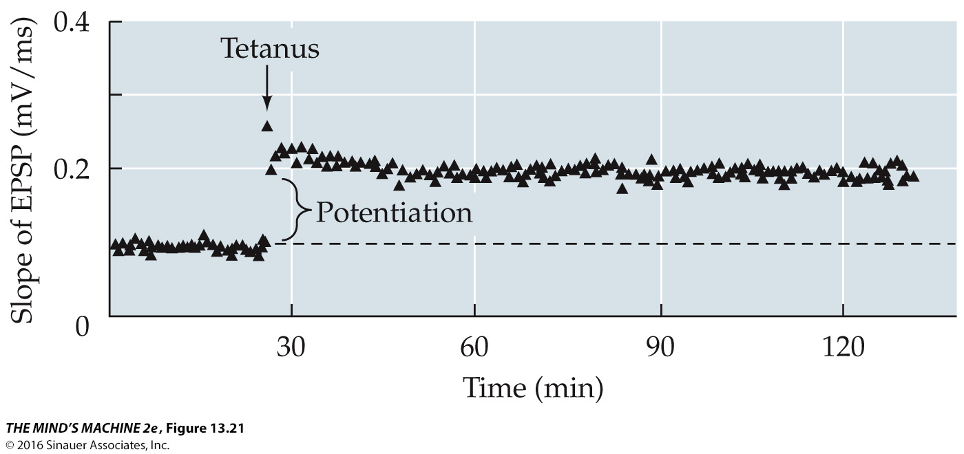
Long-Term Potentiation
“When an axon of cell A is near enough to excite a cell B & repeatedly or persistently takes part in firing it, some growth process or metabolic change takes place in one or both cells such that A’s efficiency, as one of the cells firing B, is increased” (Hebb, 1949)
Neurons that fire together, wire together
Prolonged increase in synapse responsiveness
Stable & enduring increase in the effectiveness of synapses
Some mechanisms of increasing or strengthening connections in neuroplasticity:
Increase in receptor response
More NT released
Increasing # of synaptic connections
Commonly used neural pathways take over neural territory from less used pathways
In experiments, can be electrically induced
Tetanus
An experimentally induced repeated electronic stimulation
Many action potentials in short amount of time
Causes a form of LTP
Afferent neurons
Carry information from sensory receptors of the skin & other organs to the CNS (brain & spinal cord)
Efferent neurons
Carry motor information away from the CNS to the muscles & glands of the body
Hebbian Learning - LTP/LTD
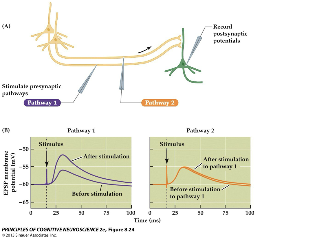
Researchers stimulated different afferent pathways & recorded responses in a postsynaptic N (PPs)
Because this focal enhancement lasted a long time, they called the phenomenon long-term potentiation (LTP
High-frequency stimulation of one pathway (Pathway 1) caused a long-lasting strengthening of that specific synaptic connection (LTP→ didn’t affect other, unstimulated pathways); can be triggered by just one burst of high-frequency stimulation (explaining how some memories form instantly; only active synapses are strengthened, other nearby synapses are not, which mirrors the specific nature of memories; if a weak & a strong pathway impinging on a target neuron were activated at the same time, then both pathways would tend to become associated via the output of the target cell→ consistent with Hebbian learning (“neurons that fire together, wire together”)
To prevent saturation (where all synapses just keep getting stronger), the brain also uses long-term depression (LTD)- founded late 1970s
LTD weakens synaptic connections through low-frequency stimulation over a longer period
Like LTP, LTD is specific to active synapses + can last for hours; serves to balance the effects of LTP
LTD may also help fine-tune learning by blocking the formation of associations that don’t follow Hebb’s rule
Declarative (explicit) memory
What we intuitively think of when we think of “memory”
Ability to consciously (explicitly) remember events & facts over time
Consists of episodic memory (events) & semantic memory (facts)
Episodic Memory
Conscious memory of events that an individual has experienced personally
Recollection: Remembering contextual details
Familiarity: Having vague feelings of knowing
Semantic Memory
Conscious memory for knowledge about the world (ex: language, facts learned in college etc.)
Prefrontal regions mediate…
both semantic/linguistic processes & executive functions that directly contribute to encoding & retrieval
During encoding, these processes are thought to help link incoming stimuli w/ preexisting knowledge & organize them into meaningful units that can be more easily remembered
During retrieval, the prefrontal regions help guide the memory search, evaluate the information recovered, & underlie decisions about its validity
Posterior parietal regions support attentional control processes, which are critical for selecting information during encoding & for guiding the memory search during retrieval. Prefrontal & posterior parietal regions are less critical during storage, but they may play a role during consolidation.
Neural Model of Declarative Memory: Encoding
Different aspects of events (“memory traces”) stored throughout the cortex in the regions involved in perceiving them
Memory traces stored through synaptic changes
Hippocampus in MTL stores summary representation via indices pointing to cortical locations
Phase: Sensory information from an event is converted into neural representations— involves active processing in various perceptual regions (visual, auditory, motor, etc.), where synaptic changes are initiated.
Is not immediate; requires a process of synaptic consolidation (involves changes at the level of individual synapses—the junctions where neurons communicate. When you learn something new, synaptic connections strengthen through processes like long-term potentiation (LTP)). Shortly after learning (minutes to hours), molecular events such as protein synthesis occur at the synapse. These changes help "lock in" the memory by making the synaptic connections more stable. This process ensures that the memory trace is stable enough for short-term retention & sets the stage for further consolidation; like "cementing" the memory at the cellular level immediately after learning. System Consolidation is like "organizing a library" over time, moving information from a temporary holding area (the hippocampus) to a more permanent archive (the neocortex). Understanding both processes helps explain why some memories remain vivid & retrievable for years, while others fade if they don’t undergo these stabilization and reorganization steps. This dual-stage view of memory consolidation is a foundational concept in human neuroscience, highlighting how both immediate cellular changes & long-term brain network reorganization work together to support our memory functions— involving gene expression, protein synthesis, & synaptic plasticity
In the encoding of declarative memory, 2 types of representations are created: distributed cortical traces and hippocampal indices

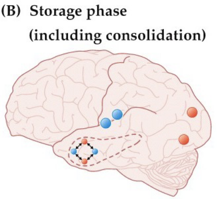
Neural Model of Declarative Memory: Storage
Some memories strengthened & become long-lasting while others presumably lost
Memories strengthened through process of consolidation
The strengthening process is known as consolidation. The assumption that some memories are lost during storage is difficult or impossible to prove in complex organisms because the fact that a particular memory cannot be retrieved does not imply that the memory does not exist; the memory may be available but not accessible. In animal models in which learning can be linked to synaptic changes, alterations in synaptic form & function reverse over time in parallel w/ behavioral measures of forgetting, suggesting that some memories do become lost.
Depends on system consolidation, which involves formation of direct connections among cortical traces; crucial for making memories more permanent & accessible over the long term, allowing them to be integrated with our broader knowledge & experiences; a slower process that reorganizes where & how a memory is stored within the brain's networks. At first, new memories depend heavily on the hippocampus, a structure within the medial temporal lobe, to bind different aspects of an experience together. Over days, months, or even years, these memory representations are gradually transferred to the neocortex. This means that older memories become less dependent on the hippocampus & more integrated with other cortical areas.
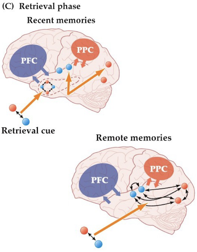
Neural Model of Declarative Memory: Retrieval
Triggered by retrieval cue (a piece of information associated w/ a particular aspect of the original event— may be external or internal) – information associated w/ aspect of original event
Recent memories: Activation of hippocampal index representations leads to reactivation of cortical traces
Remote memories: After enough consolidation, hippocampus no longer needed
The retrieval process may be involuntary or voluntary. For example, seeing a person at work (external cue) who was at a recent party may spontaneously remind you of the party (involuntary retrieval). Alternatively, you can intentionally try to remember the party (voluntary retrieval) by generating an image of the house where the party was held or of the host (internal cue). In episodic memory tests in the lab, the cues are typically external (e.g., the word house provided by the experimenter), & retrieval is usually voluntary (e.g., trying to recall the word that was paired with house).
In the case of recent memories, access to cortical traces is assumed to be mediated by the hippocampus: the retrieval cue accesses the hippocampal index, which leads to access of the distributed cortical memory traces, & in turn to the experience of remembering the original event. In the case of remote memories, however, because direct associations have been created among distributed cortical traces, these traces can be accessed directly by retrieval cues, bypassing the hippocampus.
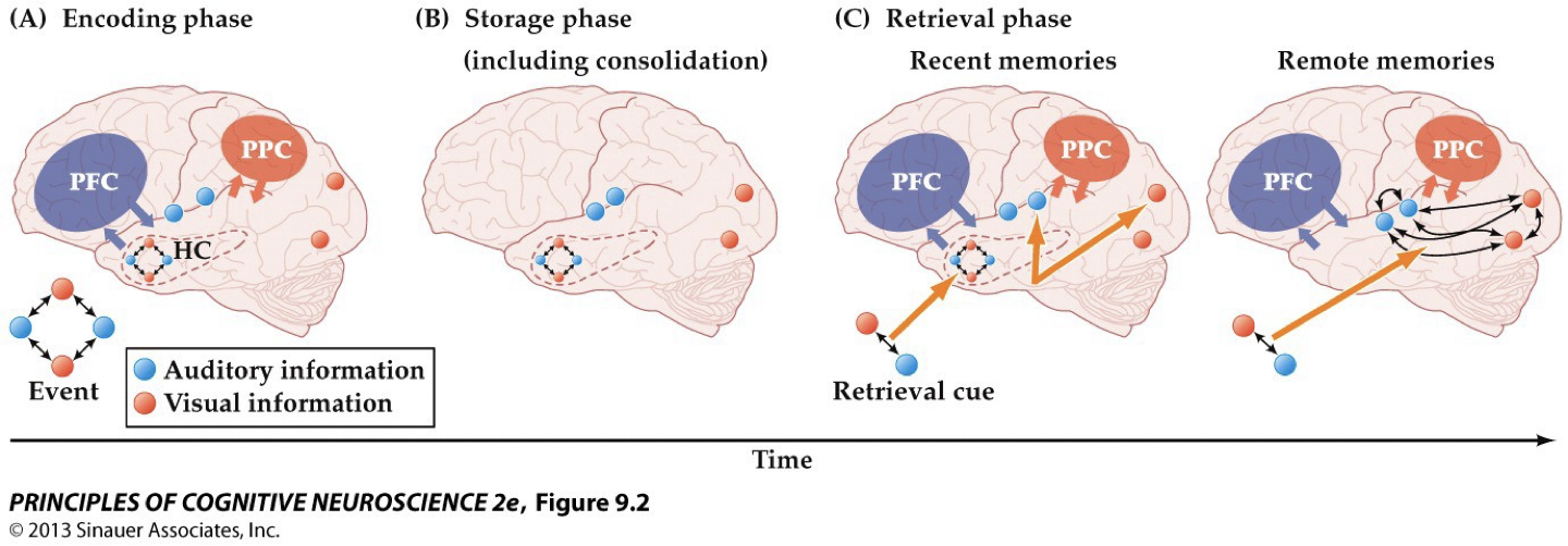
Simple model of episodic memory encoding, storage, & retrieval
A) Encoding— each aspect of an event is stored in the cortical area involved in processing (perceiving) this aspect of the event (visual information is stored in visual cortex, auditory info in AC, so on…) The hippocampus is assumed to store a summary representation, or index, pointing to the locations of memory distributed over the cortex
B) Storage— traces for some memories are stabilized- a process known as consolidation
C) Retrieval— a retrieval cue may access the hippocampal index, which leads to simultaneous access of the various cortical traces & the experience of remembering the OG event
Medial Temporal Lobe
Primary subregions:
Para-hippocampal cortex
Perirhinal cortex
Hippocampus
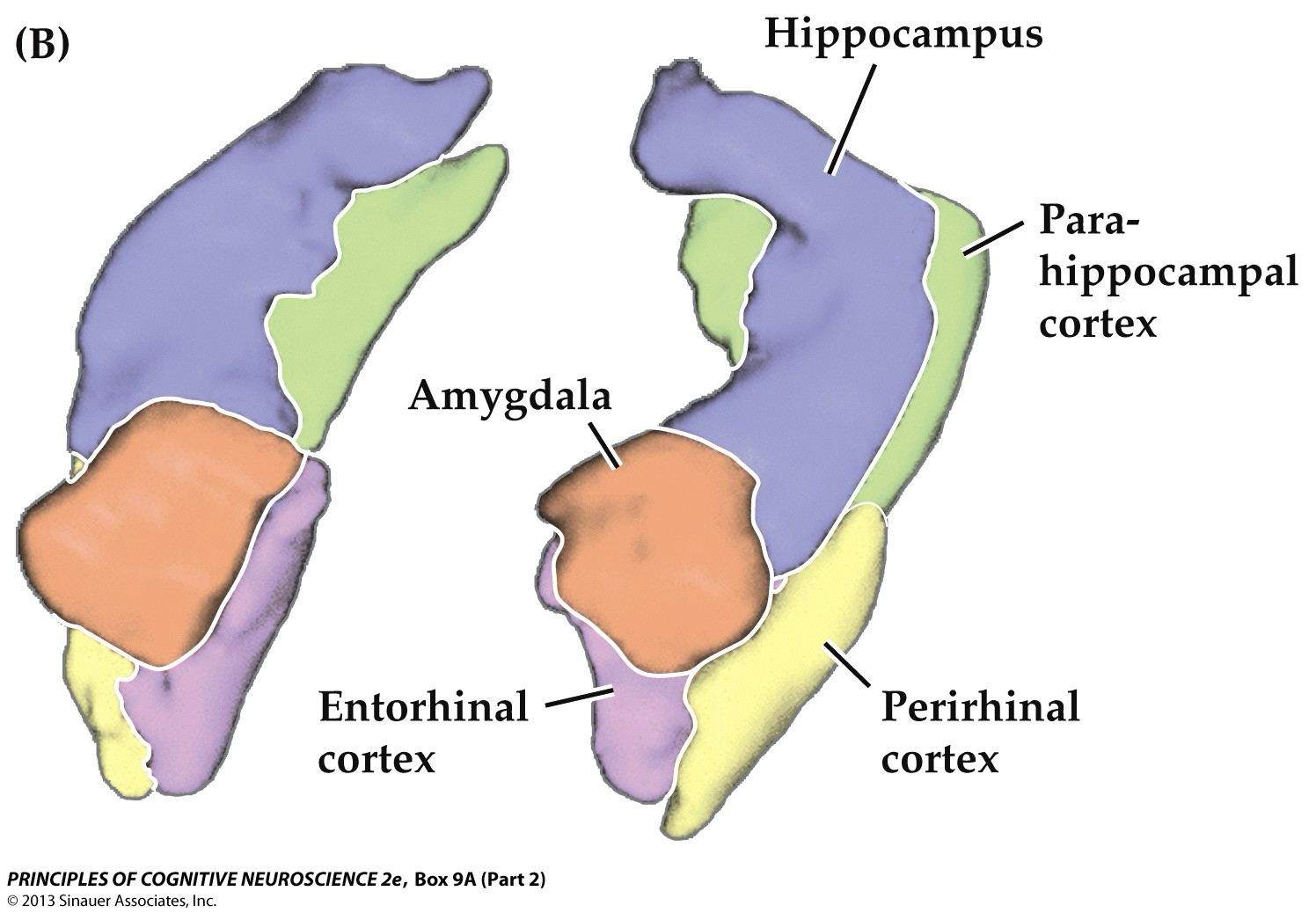
Para-hippocampal cortex
Memory for spatial layouts; receives most visual input from dorsal stream
Perirhinal cortex
Memory for objects; receives most visual input from the ventral stream
Hippocampus
Integrates both types of information (memory for objects & spatial layouts)
Domain-general relational memory: refers to the ability to form & retrieve memories that involve relationships between pieces of information, regardless of the specific type of information or domain
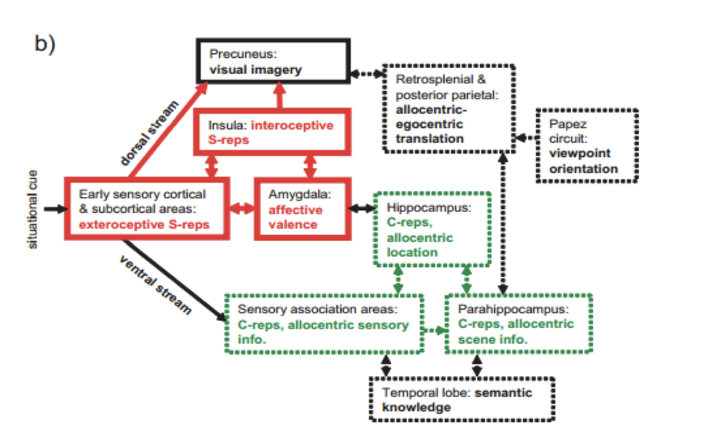
A model of healthy memory formation
Insual subjective emotional experience; James-Lange
The 1st functional area is the V1→ contains a low-level description of the local orientation, spatial-frequency & color properties within small receptive fields. PV1 projects to the occipital areas of the ventral stream (visual area V2 & visual area V4), & the occipital areas of the dorsal stream—visual area V3, visual area MT (V5), & the dorsomedial area (DM).
The Ventral stream is known for the processing the "what" in vision, while the dorsal stream handles the "where/how." VS provides important information for the identification of stimuli that are stored in memory. With this information in memory, the dorsal stream is able to focus on motor actions in response to the outside stimuli.
To allow imagery, the allocentric BVC representation (specifying layout in terms of North, South, etc.) must be translated into egocentric coordinates (i.e., left, right relative to the head). This translation is proposed to occur in posterior parietal & retrosplenial cortices, where neurons encode locations in combined egocentric & allocentric reference frames. The process requires the current (or imagined) viewing direction, which is specified by the head-direction cells found along Papez's circuit. The final egocentric image is supported by medial parietal areas
The place cells then reactivate the complete representations of boundaries & visual textures via reciprocal connections back to parahippocampal & perirhinal cortices.
The Perirhinal cortex = a cortical region in the MTL that is made up of Brodmann areas 35 & 36. It receives highly processed sensory information from all sensory regions, & is generally accepted to be an important region for memory.
A schematic model of memory & imagery, showing the approximate regions and pathways involved in, & supporting, abstracted contextual representations (C-reps, in green) & sensory bound representations (S-reps, in red). Scenes with strong affective content create enduring S-reps supported by amygdale, sensory areas and insula. S-reps are normally reactivated voluntary via a corresponding C-rep, but strong S-reps with weak association to C-reps can cause involuntary intrusive imagery.
The imposition of a viewpoint location (via place cells) & viewing direction (via head-direction cells) on the products of retrieval from long-term (allocentric) memory stores allows the creation of spatially coherent (egocentric) scenes in medial parietal areas
The prevailing view is that the perirhinal cortex is particularly specialized for representing objects, the parahippocampal cortex is specialized for representing spatial “contexts” or places, and the hippocampus binds together outputs from both these areas to represent episodes as a whole (the objects in the correct spatial contexts).
One specific proposal of this type, that looks beyond purely mnemonic processing, is that the hippocampus is specialized for the formation of associations between disparate elements of an episode, occurring across space and time and in the flexible manipulation of learned relationships among items (the relational memory theory; Cohen and Eichenbaum, 1993; see also Olsen et al., 2012 and Zeithamova et al., 2012). A wealth of studies have demonstrated that the hippocampus is necessary for spatial memory in humans
It should be noted that damage to the hippocampal system alone would not result in intrusive imagery. For this to occur, an impoverished C-rep must be accompanied by the creation of an enduring S-rep through an extreme event such as a trauma cortices
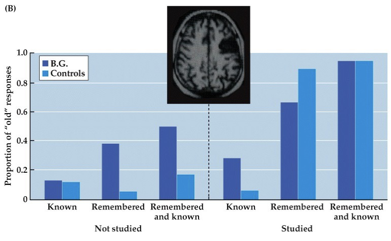
Prefrontal Cortex
Cortical regions mediate control processes during encoding & retrieval
Frontal lobe damage leads to source errors (errors in monitoring) in recalling previously known information
Patient BG: Right dorsolateral prefrontal lesion produced high proportion of incorrect “old” responses
"Old" = Item seen before in the lab
"New" = Item never seen before
"Known" = Feels familiar (vague)
"Remembered" = Specific memory of learning it
Source errors happen when we misattribute where a memory came from (thinking it came from a friend but it came from a book)
Caused by failures in source monitoring, which tracks when, where, & how info was learned
During Encoding:
Help focus attention, organize info, and connect it to what we already know.
Example: grouping or relating new info to existing knowledge
During Retrieval:
Help search for the right memory, suppress distractions, & use cues effectively
Brain Areas:
Involve the prefrontal cortex and related cortical regions.
These areas manage memory like a “control center,” directing attention, filtering info, and guiding recall
Korsakoff’s syndrome
dementia arising from severe alcoholism; because of source monitoring deficits, individuals w/ Korsakoff's syndrome may generate spontaneous confabulations
Spontaneous confabulations
High confidence reports of personal events that never happened; false or distorted memories that the person believes to be true, not created intentionally to deceive, but rather as a consequence of the brain “filling in” missing information.
When a memory is weak or incomplete, the brain may inadvertently draw on unrelated information or even create new details, leading to a confabulated account.
The prefrontal cortex is critical for executive functions, including evaluating and verifying the source of memories. Damage to this area undermines the ability to monitor the accuracy of memory details, leading to source errors. This impaired source monitoring can contribute to the spontaneous confabulations observed in Korsakoff's patients, as the brain struggles to correctly attribute the origins of its memories.
Memory Consolidation: Encoding
is not immediate; requires a process of synaptic consolidation, involving gene expression, protein synthesis, & synaptic plasticity
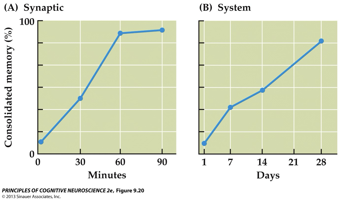
Memory Consolidation: Storage
depends on system consolidation, which involves formation of direct connections among cortical traces

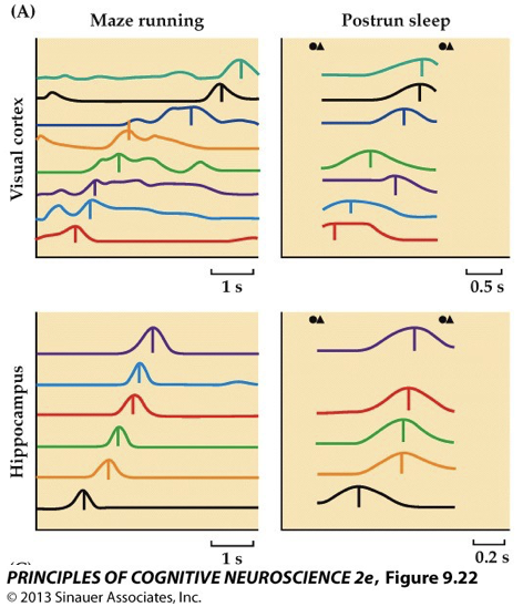
Memory Consolidation: Reactivation & Sleep
Consolidation promoted by memory reactivation during retrieval
memories reactivated/ replayed during sleep
Strategies to Aid Memory
To aid consolidation
Practice retrieval
Sleep well
To encode more deeply
Relate material to what you already know
Topics from class that you already understand & rememnber
Outside interests
Anything else
Add emotion
Organize information
Meaningful & manageable units
Relate those units of information to each other
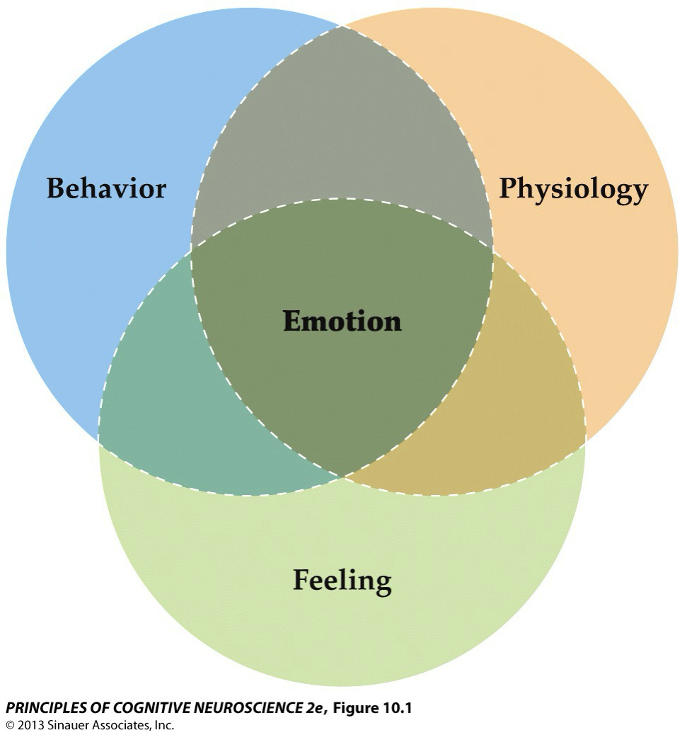
Emotions
Feelings accompanied by:
Expressive behavior (facial response)
Physiological responses (heart rate, pupils)
Behavior (fight or flight)
Brain activity
Guide our actions…
Evaluate situation, based on sensory data about what is going on in the world & in your body
What is happening?
Is it good or bad for me?
What should I do?
Generate appropriate behavior
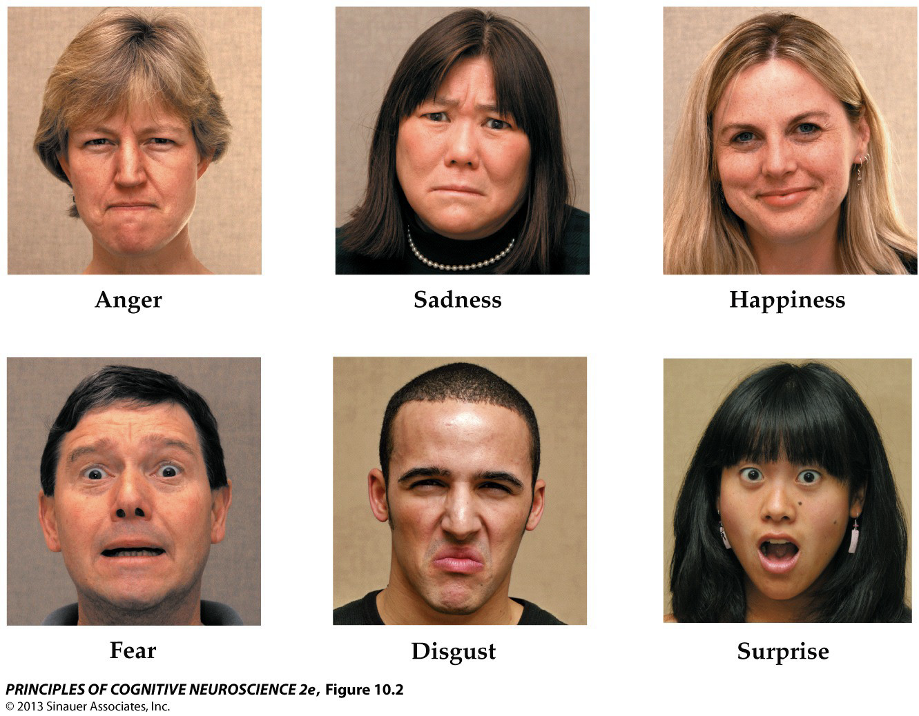
Classifying Emotions Categorically: Basic emotions
Innate, universal
Classifying Emotions Categorically: Complex emotions
Species-specific, culture-specific, learned
e.g., moral disgust, schadenfreude
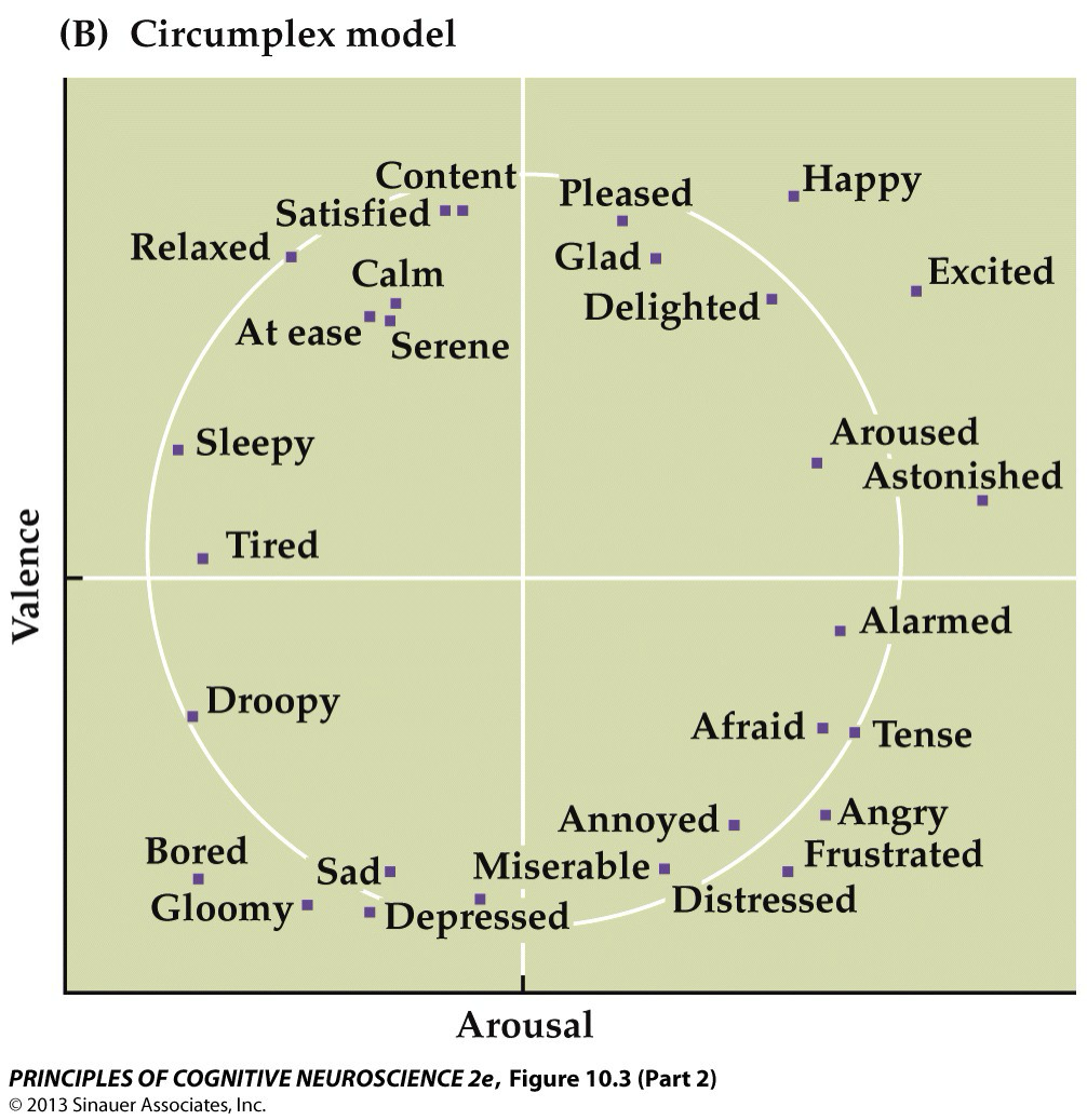
Classifying Emotions Dimensionally
Emotions vary continuously
2 key dimensions:
Valence→ pleasantness of emotion (+/-) x-axis
Arousal→ intensity of emotional feeling or response, y-axis
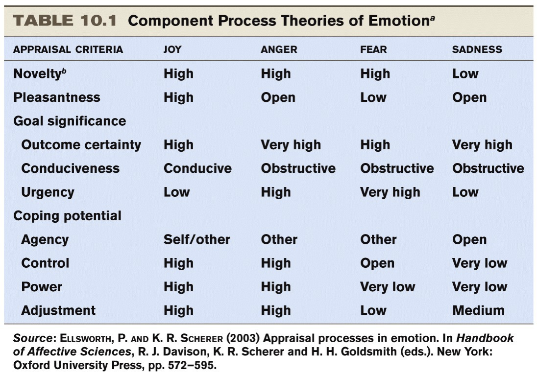
Classifying Emotions: Component Process Theories
Emotions vary continuously along multiple dimensions; focus on emotion appraisal
Rather than viewing emotions as fixed states, component process theories attempt to capture the fluid nature of emotions, which require flexible interactions of multiple component processes. These theories emphasize processes that are involved in cognitively appraising the emotional meaning of events, as well as the link between the appraisal outcome and a behavioral and physiological response. In this view, emotions are related according to the similarity of the appraisal processes engaged.
Some investigators argue that important appraisals include the sense of urgency for action in response to the elicitor, how well a person can cope with the presence of the elicitor, and whether the elicitor facilitates or impedes progress toward a goal (Table 10.1). In this view, emotions are organized in the brain according to their overlap in recruiting specific appraisal mechanisms, which may vary across individuals, depending on social context and cultural influences. Because of their complexity, component process models have not been extensively tested with neuroscientific methods.
Theory of Constructed Emotion
Posits that emotions are not universal, but vary from culture to culture
Posits that emotions “are not triggered; you create them. They emerge as a combination of the physical properties of your body, a flexible brain that wires itself to whatever environment it develops in, & your culture and upbringing, which provide that environment”
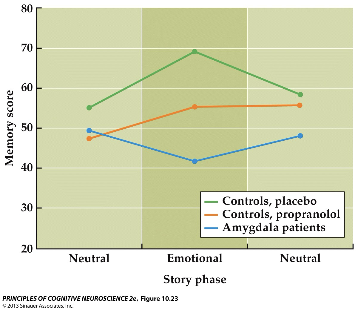
Memory for Emotional Content
Emotions influence memory
Compared to non-emotional content, emotional content is:
remembered better for controls
remembered equally well after propranolol (“beta blocker”)
remembered worse for amygdala patients
Audiovisual narrative containing 3 parts— 1st & last emotionally neutral, middle— arousing (car accident); healthy adults (green) show L-T memory enhancement for the emotionally arousing portion of the study— advantage lost when propanol, beta blocker, administered to HAs before story is heard, as well as patients w/ bilateral amygdala damage without drug administration
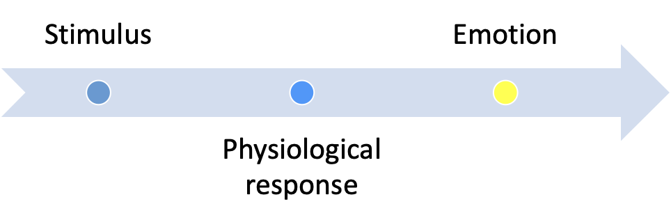
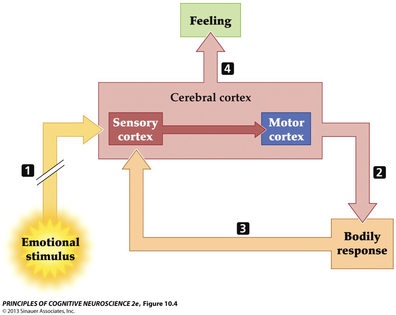
Theories of Emotion: James-Lange Theory
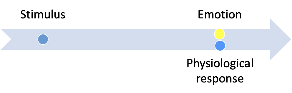
Theories of Emotion: Cannon-Bard Theory
Neural Basis of Emotional Behavior: Classical theories→ James-Lange Feedback Theory
Body ➔ Emotion
An eliciting stimulus (1) directly causes a bodily reaction (2), which feeds back into the brain (3) to generate an emotional feeling (4). According to this theory, no specialized brain region for processing emotion exists, and bodily reactions and feelings have a one-to-one correspondence
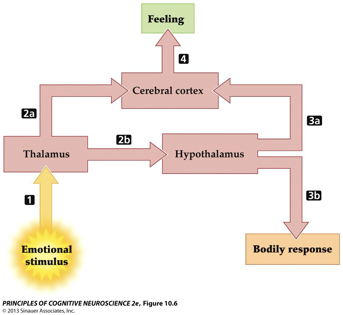
Neural Basis of Emotional Behavior: Classical theories→ Cannon-Baird Diencephalic Theory
Input from emotion-provoking stimuli (1) is directed in parallel from the thalamus to the cortex directly (2a) or indirectly (2b, 3a) for the generation of feelings (4), and from the thalamus to the hypothalamus (2b) for bodily expression (3b).
ANS responses not differentiated enough to explain wide array of emotions
also, neuroendocrine responses slower than emotion onset
also, hormonal changes alone do not cause a specific emotion
decorticate animals could exhibit integrated emotional reactions, such as defensive behavior, only if the transection occurred above the level of the diencephalon (hypothalamus and thalamus). These findings were bolstered by the studies of Walter Hess, a contemporary of Cannon and Bard who showed further that electrical stimula-tion of the hypothalamus elicits emotional reactions in the cat, including sham rage, in which hissing, growling, and attack behaviors are directed randomly toward innocuous targets.
Parallel processing theory (one of the first examples)
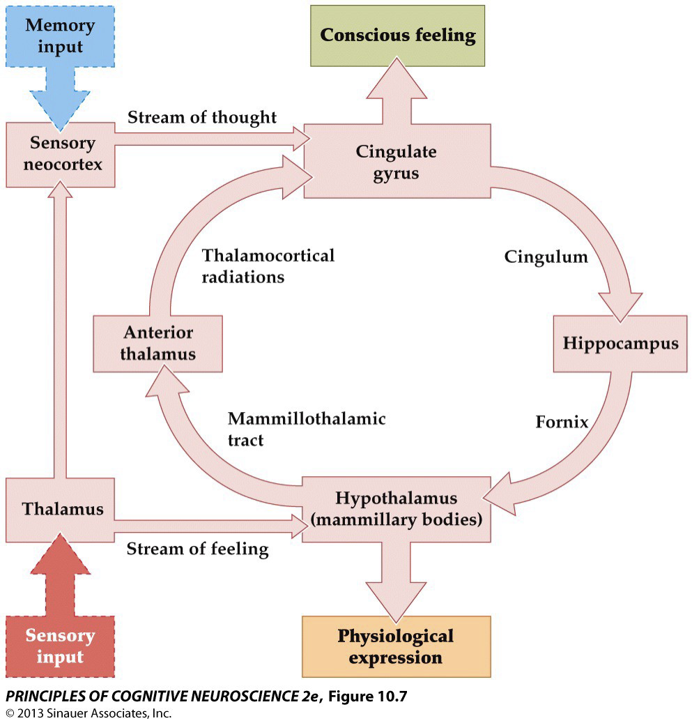
Neural Basis of Emotional Behavior: Classical theories→ Papez circuit
Papez’s rationale in identifying this circuit was based on brain structures directly connected with the hypothalamus, the locus most central to the Cannon-Bard theory
Basing his work on known anatomical projections (white matter tracts) to the hypothalamus and interconnected structures, Papez described a fundamental circuit for generating emotions involving portions of the limbic forebrain and diencephalon. The cingulate gyrus was assumed to signal conscious emotional feelings. Papez believed that it was crucial for integration.
Kluver-Bucy Syndrome— inability to evaluate the emotional & motivational significance of objects in the environment, particularly by sight
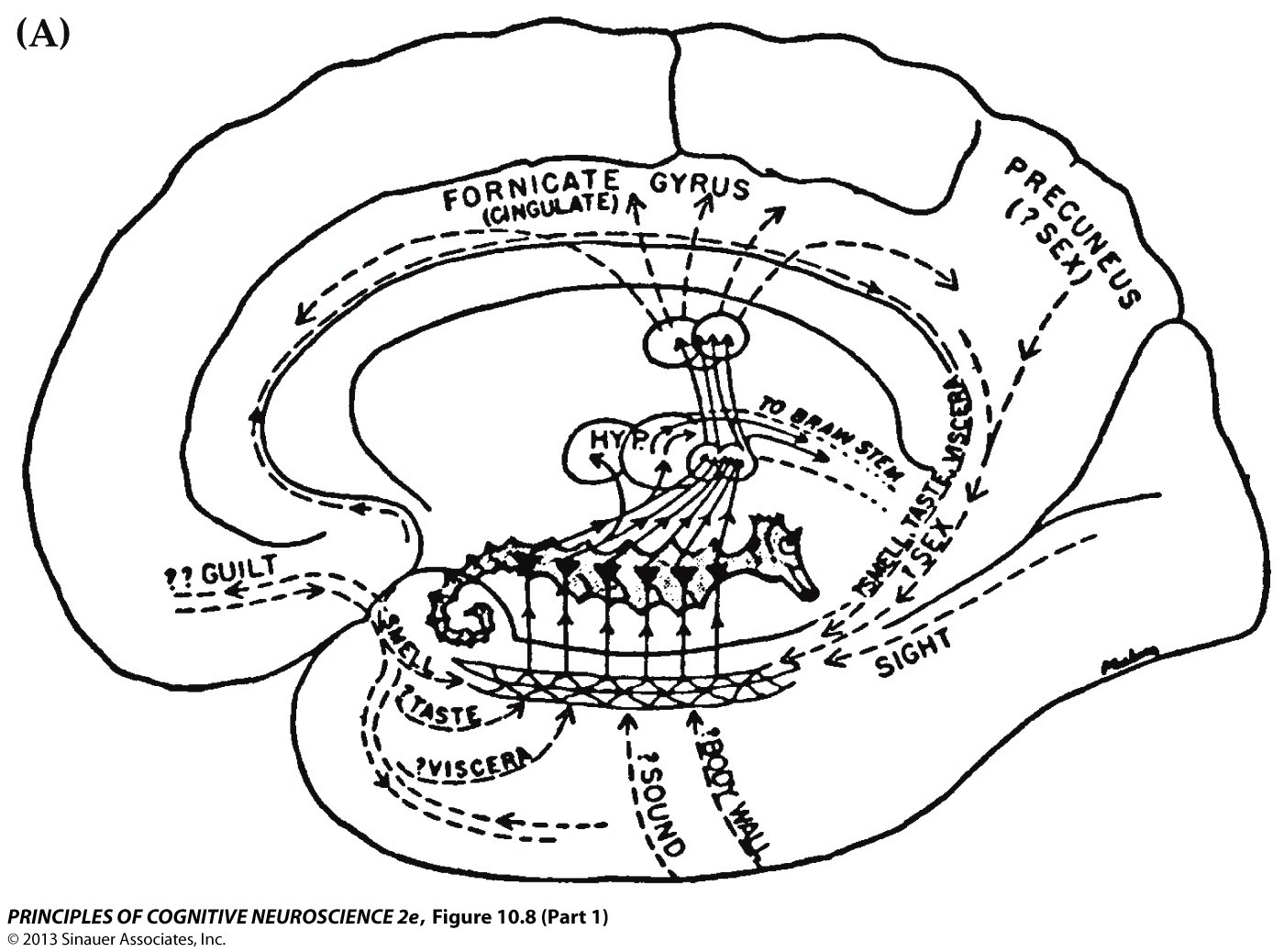
Neural Basis of Emotional Behavior: Classical theories→ Limbic System Theory
MacLean’s original 1949 drawing of the visceral brain is reproduced here. In MacLean’s interpretation, the cornerstone of this system was the hippocampus (“seahorse”) and its connections to the hypothalamus. (B) More contemporary views of emotion emphasize the role of anterior sectors of this circuit (shaded in green), including the amygdala, orbitofrontal cortex, ventromedial and orbital prefrontal cortex, and hypothalamus. These components were added later by MacLean, and their inclusion may help explain the longevity of the limbic system concept. By contrast, posterior components (shaded in blue)—including the hippocampus, posterior cingulate cortex, and their diencephalic connections—are more critical for other cognitive functions.
Issues – lacking functional or structural coherence; centrality of hippocampus not supported
Key structures involved in emotional processing
Amygdala
vmPRC
Insula
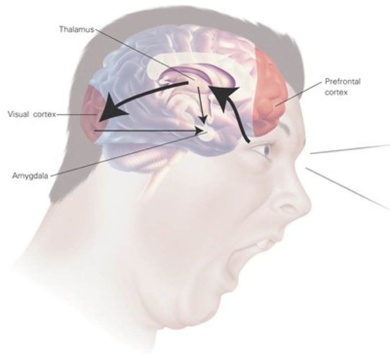
Amygdala
Central role in evaluating emotional significance of stimuli
“Low road” (fast) & “high road” (slower) input routes to amygdala
Critical for acquisition & expression of fear (essential for fear conditioning)
& the unconscious perception of fear (eye whites)
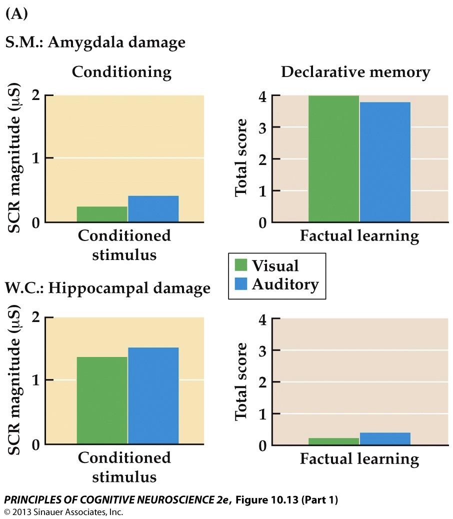
Compare: S.M. Amygdala damage vs. W.C. Hippocampal damage
Double dissociation between conditioned fear learning & declarative knowledge in patients w/ selective damage to the amygdala & hippocampus; patient S.M. (bilateral AD) shows intact factual knowledge of the predictive relationship between the CS & US, but fails to exhibit conditioned skin conductance responses (SCRs); in contrast, patient W.C. (bilateral HD) show the opposite pattern— that is, intact SCRs to conditioned fear cues but no factual knowledge about the predictive relationship between the CS & the US
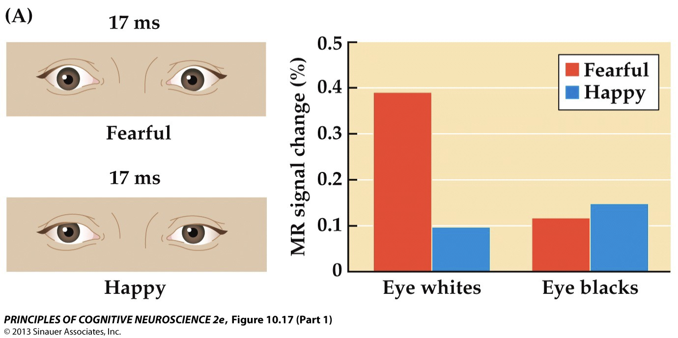
Amygdala & the unconscious perception of fear
The amygdala signals visually fearful facial expressions, even when stimuli are presented briefly/ masked to limit conscious awareness; fear conveyed solely by the whites of the eyes (increases during fear as a result of sympathetically mediated contraction of the subcutaneous muscles in the skin surrounding the eye); amygdala— greater activity for fearful > neutral facial expressions, even when participants reported seeing only the house (when fearful face was presumably processed subcortically as a result of rivalry
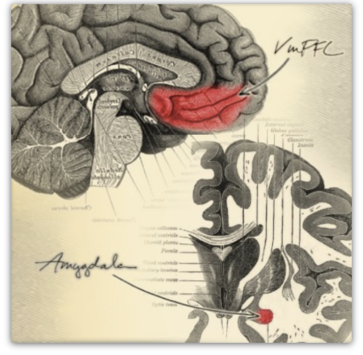
vmPFC
Inhibition of amygdala, fear extinction
Emotion regulation (helps regulate and modulate emotional responses/ involved in dampening excessive emotional reactions & ensuring that our decisions are informed by both rational thought & emotional context)
Social & moral reasoning (contributes to social cognition by evaluating the emotional aspects of social interactions- role in moral reasoning & empathy means that it influences how we perceive & react to the emotional states of others)
Integration of emotion & decision-making (key in linking emotional experiences with decision-making processes/ integrates affective information (such as personal values, emotional memories, and social cues) to guide behavior and judgments)
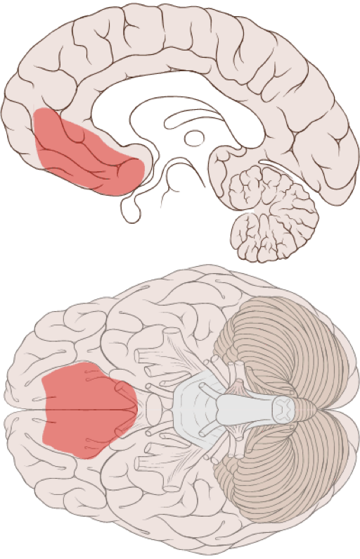
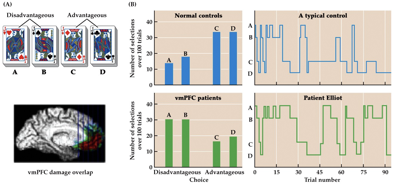
Iowa Gambling Task &
vmPFC lesions
Emotions — via bodily signals (somatic markers) — guide decision-making by marking certain options as “good” or “bad” based on past outcomes.
Participants choose cards from 4 decks (A, B, C, D) over 100 trials.
Decks A & B = high short-term reward but big losses → net loss
Decks C & D = smaller wins but less risk → net gain
Healthy Participants:
Start off exploring.
Over time, they learn to avoid risky decks (A & B).
Show anticipatory skin conductance responses (SCRs) before risky choices — a bodily signal saying, “bad idea.”
mPFC-Damaged Patients:
Keep choosing risky decks.
Show no anticipatory SCRs before bad choices.
Don’t learn from negative outcomes.
Lack the emotional "gut feeling" to guide behavior.
Without somatic markers, decision-making becomes cold, logical, and often ineffective (e.g., H.M. or patient Elliot).
Damasio’s theory supports the idea that emotion enhances reasoning, rather than disrupting it.
The vmPFC helps us make good decisions by attaching emotional “gut feelings” (somatic markers) to past experiences. Without it, as seen in vmPFC patients, we don’t feel risk and struggle to learn from bad outcomes, even when we know the facts.
Insula
Crucial for interoception: sensing the state of your body; activation in response to disgust expressions; stimulation leads to induction of disgust; damage leads to difficulty recognizing facial/ vocal expressions of disgust