Diagnostic Imaging 2- Exam 2- Extremity Trauma #1 start to elbow notes
 Call Kai
 Learn
Learn Practice Test
 Spaced Repetition
 Match
Match Flashcards
 Knowt Play
1/143

There's no tags or description
Looks like no tags are added yet.
Name | Mastery | Learn | Test | Matching | Spaced | Call with Kai | Chat |
|---|
No analytics yet
Send a link to your students to track their progress
144 Terms
When looking at a trauma there is a minimum of how many views that need to be taken
2 at right angles
Most common location for a fracture that you can't see on X ray at first
Scaphoid
Tension results in what type of fracture
Transverse fracture
Compression results in what type of fracture
Oblique
Rotation results in what type of fracture
Spiral
Angulation results in what type of fracture
Transverse
Angulation and compression result in what type of fracture
Transverse and oblique fracture
What type of fractures run at right angles to the long axis and result from direct blows or tensile forces?
Transverse fractures

What type of fractures are common in pathological bone conditions
Transverse fractures
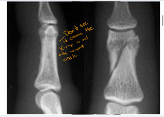
Example of a transverse fracture

What type of rapture runs about 45 degrees to the long axis of the bone and is from Angulation or angulation and compression type of injury
Oblique fractures

Oblique fractures sometimes have a small _________ component
Transverse
Example of a transverse fracture
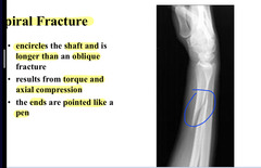
Fracture that encircles the shape and is longer than an oblique fracture that results form torque and axial compression? The ends are pointed like a pen
Spiral fracture
What describes the closeness of the bony contact at the fracture site
Apposition
When takin about apposition of a fracture, how do you describe it?
Describe the offset of the distal in relation to the proximal
What are some ways that you can describe apposition
Overriding, distraction, no apposition
Example of displaced vs non-displaced fractures
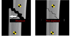
Examples of fractures loosing apposition

What describes the position of the distal fragment in relation to the proximal in the longitudinal axis
Alignment
Alignment deals primarily with
The direction and degree of Angulation
Alignment describes the Angulation of the
Distal fragment
Example of a loss of alignment posteriorly

What is described by being produced by a twisting force along the longitudinal axis
Rotation
Rotation is more easily determined one
Clinical evaluation
When assessing rotation it ideally should include
Joints above and below the fracture
In males the highest rate of fractures is when? When will it decrease and then increase again?
2nd and 3rd decades
Decreases 45 yrs and then raises after 65
In females the highest rate of fractures is when? When will it decrease and then increase again
Up to 20 years
Decreases at 45 then steadily increases after that
Between 10-16 yers of age___________ are the most common as this is the weakest area of teh bone
Epiphsyeal
Common sites of fractures in the elderly
Thoracolumbar pain
Proximal femur
Distal radius
Proximal humerus
Indirect signs as a diagnostic clue of a fracture
Joint effusions
Double cortical line
Cortex buckling
Displacement or obliteration of fat stripe
Rates of fracture repairs in infants
4-6 weeks
Rates of fracture repair in adolescents
6-8 weeks
Rates of fracture repair in adults
10-12 weeks
Sometimes 16-20 weeks
What is it when the callus is sufficiently developed to allow weight bearing or similar stress
Clinical union
Factors influencing the rate of repair
Decree of trauma
Age
Vascularity of fragments
Separation
Inadequate immobilization
Presence of pathologies
Complications of fracture
Non-union
Delayed union
Arrest/stimulation of growth
Malunion or angular deformities
Avascular necrosis
DJD
fractures that are healed but with malunion
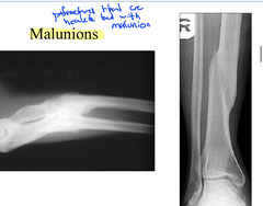
Fractures that are healed with non-union

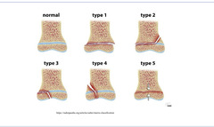
What are injures involving the active physics
Salter Harris fractures
Salter Harris fracture that is a complete shear injury of the physis
Salter Harris type I
Salter Harris fracture that is through the physis and metaphysis
Salter Harris type II
Salter Harris fracture that is through the physis and epiphysis
Salter Harris type III
Salter Harris fracture that is through the physis, metaphysis and epiphysis
Salter Harris Type IV
Salter Harris fracture that is compression fracture of the physis
Salter Harris type V
What is the most common type of salter Harris fracture
Type II
What salter Harris fracture are intra articulator
Type III and Type IV
Salter Harris fracture types diagram
Salter type I fracture example
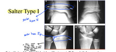
What does a salter Harris type I fracture look like
Wide growth plate and soft tissue swelling
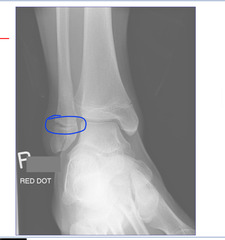
Another salter type I fracture example
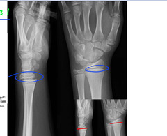
Salter type I fracture in the shoulder example

Salter type I fracture in the hip example
This example also shows a SCFE- slipped capitus femoral epiphysis (from resistive soft tissue trauma)

What line can be used in the pelvis to find a SCFE in the hip
Klein`s line
What sign can be seen with a Salter type II fracture
Thurston-Holland sign
What is the thurston-holland sign
Metaphyseal fragment

Salter type II in the thumb

Another example of a salter Harris type II fracture
Don't forget the thurstand holland sign

Osteopetrosis and salter type II in the hip

Another example of salter Harris type II fracture in the lower leg
Don't forget about the thurstand Holland sign
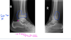
Salter Type III is what kind of fracture
Intra-articular fracture
Salter type III in the ankle

Another example of salter type III fracture

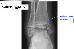
Salter Harris fracture that includes the eipiphysis, physis and metaphysis
Salter type IV
Example of salter Type IV fracture

Another example of salter type IV fracture
What is the least common type of Salter Harris fracture as well as the hardest to see
Salter type V
Example of Salter type V

Routine vies of the shoulder
AP internal
AP external
Y view
How do you get acromio-clavicular joint injuries
Fall on shoulder or outstretched hand (FOOSH)
Mild sprain of the AC joint is what type
Type I
Moderate sprain of the AC joint is what type
Type II
Severe sprain of the AC joint is what type
Type III
When evaluating AC sprains, what do you look at
Look at bottom of clavicle to see if its elevated as a well as the distance between coracoid and clavicle
Also look at both AC joints

When you have elevation of the clavicle it is at least what grade with an AC separation
At least grade 2
Normal AC width is
Less than 5mm
You're allowed how much bilateral difference in the AC width
Less then 2-3 mm
Normal distance between the coracoid and clavicle is
11-13mm
You're allowed how much bilateral difference between the distances of the clavicle and coracoid
Less than 5mm
What grade AC separation is typically radiographically normal
Grade I
With a grade 2 AC separation the _______ ligament is torn white the __________________ ligaments remain intact
Acromioclavicular ligament torn
Coracoclavicular ligament remain intact
What ligaments are torn in a grade 3 AC seperation
Acromioclavicular and coracoclavicular
Grade 1 AC separation

Grade 2 AC separation

Grade 3 AC separation

If there is elevation of the clavicle between 11-13 mm on an AC separation than it is what grade? What if it is greater than 13?
Between 11-13 than Grade 2
Greater than 13 than Grade 3
AC joint separation leading to a distance greater than 5 mm without elevation of the clavicle is what grade
Grade 1
What is the most common fracture to occur during birth
Clavicle fracture
Most common location of a clavicle fracture
Middle third
Complications of a clavicle fracture
Malunion
Non-union
Neuromuscular injury
DJD
Post traumatic osteolysis
What is post traumatic osteolysis that can occur with a clavicle fracture
Reabsorption of distal end after many micro traumas
Transverse midclavicular fracture example

In a clavicle fracture the proximal fragment is elevated by the _____________ and the distal fragment is depressed by______
Elevated= SCM
Depressed= by weight of the shoulder
Another example of a transverse mid clavicle fracture

Undisplaced midhsaft clavicular fracture example

Routine radiographs of the clavicle
Frontal and AP axial
Oblique mid clavicle fracture example

Old transverse mid clavicle fracture with non union example

Old transverse mid clavicle fracture with malunion example

Complications of clavicle fractures
Extra callus may cause neurovascular compromise
Laceration of subclavian artery or brachial plexus