Muscle plasticity
1/19
There's no tags or description
Looks like no tags are added yet.
Name | Mastery | Learn | Test | Matching | Spaced | Call with Kai |
|---|
No analytics yet
Send a link to your students to track their progress
20 Terms
Review of skeletal muscle structure
Heirarchy:
Belly → Fascicle → Fibres (myocyte) → Myofibrils (series of sarcomeres)
epimysium - wraps around mutiple fasciles to form the belly
endomysium → wraps around multiple fibres to form a fascicle
Myocytes = Fibres
peripheral nuclei and striations
Components of myofibril: interact to allow contraction
actin
myosin
proteins that drive muscle contraction
Mechanical coupling
protein-protein interactions
depolarisation of sarcoplasmic reticulum
release of Ca2+ stores
voltage sensor → Dyhydropyridine receptor (DHPR)
conformational change of Ca2+ channel → Ryanodine (RyR)
AP travels down T-tubule
triggers Ca2+ channels
Ca2+ channel opens btw myoplasm and sarcoplasmic reticulum
voltage sensor DHPR senses this calcium
RyR changes shape in response
Calcium transient - Ca2+ released from sarcoplasmic reticulum
Ca2+ release initiates cross-cycling process
Cross-bride cycle for muscle contraction
involves excitation-contraction coupling
Resting state
myosin binding site on actin blocked by tropomyosin
Excitation-contraction coupling
Ca2+ binds to troponin
tropomyosin moves, revealing the myosin binding site
Binding
myosin head binds to actin → cross bridge
Powerstroke
myosin heads flex (- sarcomere shortens)
causes detachment of ADP+Pi
Detachment
ATP binds to myosin head, causing detachment
if Ca2+ still on troposin → powerstroke
no Ca2+ → resting state
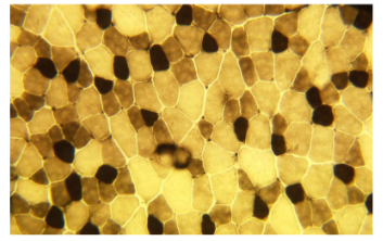
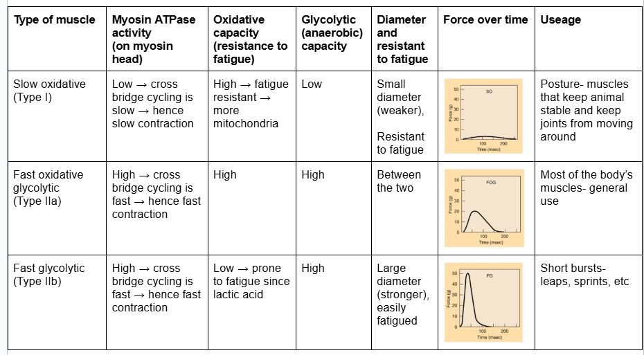
Muscle fibre classification
every muscle has a mixture of fibre types
(patchwork arrangement)
reverse stained images for myosin-ATPase
Fast fibres - big and pale (high myosin-ATPase)
white meat (chicken) - pectoral = fast glycolytic muscle
Slow fibres - small and dark
leg - dark meat = slow oxidative
different muscle role - change in proportion of fibre types
Alternative muscle dye
SDH = succ-inate dehydrogenase
stains mitochondria
more blue if more mitochondria
slow oxidative - blue
more mitochondria to drive citric acid cycle
fast glycolitic is light
Muscle fibre type distribution
semitendinosus
faster fibres peripherally
central fibres = slow
breed - genetic variation
same distribution
sections different sizes
greyhounds have greater number of type II muscles peripherally → adapted for fast burst of activity
individual variation → strong heritability and lifestyle
species variation
ambush predators → more fast fibres
pursuit predators → travelling long distance → more slow fibres
Muscle adaptation
Area change
hypertrophy (NOT hyperplasia) → building muscle bulk due to fibres enlarging
Length change
hyperplasia (more muscle fibres
NOT hypertrophy
Satellite cells
along surface of muscle fibres
peripheral myocyte nuclei and satellite cells → similar appearance
stem cell like properties → differentiate into other cell types
important in hypertrophy
response to injury and repair
muscle hypertrophy + other adaptation processes
stimulated by IGF (insulin-like growth factor) and other growth factors
migrate → form myotubes → fuse to existing myofibres (myocytes)
Triggers for muscle adaptation [5]
muscle is plastic and adapts in response to triggers
Normal development (Growth)
Exercise
Detraining
Aging
Injury/Surgery
Normal developmental changes
heavier → increases load → hypertrophy → increased muscle force
taller → chronic stretch → hyperplasia (sarcomeres added to ends of muscle fibres)
Lifestyle changes
need to locomote
runner etc → linked to exercise
Impact of Exercise on muscle summary
increased loading/contraction → selective hypertrophy
repeated exercise (chronic, long duration 8-24hrs), low frequency stimulation of fast muscle
fibre plasticity: fast → slow for endurance exercise
(eletrical stimulation in lab)
chronic stretch (4wks) → hyperplasia
Detraining/Immobilisation
Fibres return to typeII
Fibres convert to type IIa (intermediate)
occurs x2 quickly as training
anti-gravity (weight-bearing) muscles more at risk
post immobilisation rehab - restore fibre type (then strength training)
Muscle memory
first time training → first time fusion of satellite cells
hypertrophy occurs myotube formation
detraining occurs → decreases fibre diameter (more type IIa and I than IIb)
nuclei still present so re-training is faster
= muscle memory
Nutrition
muscle will not adapt if no nutrients
Protein
skeletal muscle mass maintained if no excessive breakdown
need plenty of protein in diet
Glycogen
exercise capacity linked to glycogen store
sugars drive the cross-bridge cycling process
glycolysis
Lipid (triglyceride)
more in oxidative (I and IIa)
Aging
loss of muscle function
Many reasons
Decreased satellite cells
reduces hypertrophic ability
decreased muscle size and performance
Decreased growth hormone
Denervation → muscle atrophy
Decreased blood supply → less nutrients, build up of waste products → function drops
Increased fibrous connective tissue
prevents efficient contraction
less muscle
influences passive and decreased contractile properties
Injury [7]
healing is fast when inflammation is not as high
return to pre-injury status within 10-20 dyas
No inflammation - no TGF-beta1→ myoblast → myocyte formation
Inflammatory response to injury
Satellite cells activated by inflammation
Satellite cells differentiate into myoblasts
Myoblasts differentiate into myocytes
Myocytes fuse to form a myofibre
Persistent exposure to inflammation triggers myocytes to differentiate into myofibroblasts = fibrotic tissue
regulate inflammation to prevent fibrotic tissue
Myostatin and TGF (transforming growth factor)-beta1
prevents → myoblast → myocyte formation
promote myofibroblast formation
myostatin inhibits macrophages
Selective hypertrophy
increased loadinf/contraction
Fast fibres respond twice as fast
Fibre type plasticity
Default → become slower with increased enndurace capacity
unless strength training is used → leads to selective hypertrophy (lifting weights)
training - amount of exercise (endurance) and type (strength) → weights vs supporting own body weight
Muscle length changes
Chronic stretch (tension load) (4wks) increase sarcomere length by 20%
Loads of muscles
weight → hypertrophy
tension → hyperplasia
time → endurance → muscle plasticity
Muscle fibre distribution summary [3]
varies within and btw muscles
varies btw individuals (activity + genetics + nutrition + age), breeds, species
modulated by exercise and training