Lab 1 — Histology & Introduction to Microscopy
 Call Kai
 Learn
Learn Practice Test
 Spaced Repetition
 Match
Match Flashcards
 Knowt Play
1/27

There's no tags or description
Looks like no tags are added yet.
Name | Mastery | Learn | Test | Matching | Spaced | Call with Kai | Chat |
|---|
No analytics yet
Send a link to your students to track their progress
28 Terms
What are the two major functional halves of a compound microscope?
Image magnification: oculars + objectives
Illumination: condenser + light source
The stage separates these two halves

What does the condenser do?
It is part of the illumination system below the stage and is adjusted to properly illuminate the specimen
2× → condenser LEFT
10×–40× → condenser RIGHT

What does the field iris diaphragm control?
→ Controls the size/diameter of the illuminated field

What does the condenser height adjustment knob do?
→ Raises or lowers the condenser
Key adjustment for Köhler illumination

What does the condenser centering knobs control?
→ Control the horizontal position of the condenser/light beam so the illumination is centered in your field of view
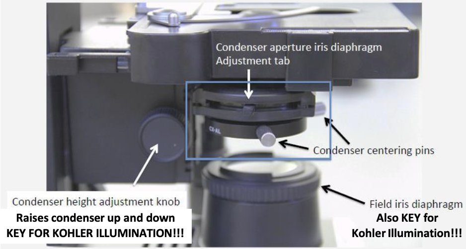
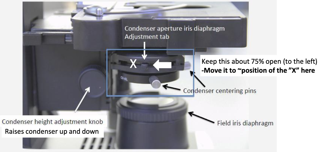
What does the condenser iris diaphragm control?
→ Controls contrast/light cone
Key adjustment for Köhler illumination
It is part of the illumination system and should initially be approximately 75% open when setting up the microscope
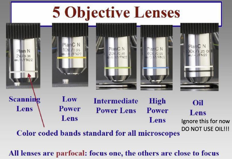
What does parfocal mean?
Once the specimen is focused with one objective, the other objectives will be close to focus when switched into position.

Why is being parfocal useful?
You do not have to completely refocus the specimen every time you change objective lenses
What are the basic steps for initially setting up the microscope?
Turn microscope on; light intensity ~3
Place slide on stage coverslip UP
Completely open light-source/field diaphragm
Set condenser iris diaphragm ~75% open
Slide condenser RIGHT
Put 10× objective into place
Focus specimen
Proceed to Köhler illumination
What is Köhler illumination?
A method for aligning and adjusting the microscope's light source to produce an optimally illuminated image.
Why is Köhler illumination important?
It is critical for aligning and adjusting the light source to provide optimal contrast
What microscope structures are especially important for Köhler illumination?
Field iris diaphragm
Controls size/area of illuminated field
Condenser height adjustment knob
UP/DOWN → Focuses the illumination
Condenser centering knobs
SIDE-TO-SIDE → Centers the illumination
Condenser aperture/iris diaphragm
Controls the light cone/contrast

What is the step-by-step sequence for Köhler illumination?
Focus the specimen first using the 10× objective.
Close the field iris diaphragm to see the smaller illuminated opening.
Focus the condenser by moving it up/down until the edge of that diaphragm looks sharp.
Center the light using the condenser centering knobs.
Open the field iris diaphragm until its edge just disappears outside the field of view.
Adjust the condenser iris diaphragm for the best balance of contrast and resolution
Why can the same tissue structure look different in different histologic sections?
Histology shows a 2-D slice of a 3-D structure
The appearance depends on the plane/orientation at which the structure was cut
How can different planes of section change the appearance of a straight tube?
Depending on the cut, a tube may appear:
Circular → perpendicular/transverse cut
Oval → oblique cut
Long/rectangular → longitudinal cut

Why might one structure appear to be several separate structures on a histology slide?
Bent, branching, or complex 3-D structure can intersect the plane of section multiple times, producing several apparently separate profiles

How does an oblique section affect the appearance of simple columnar epithelium?
The cells/nuclei can appear arranged differently than in a true perpendicular section, potentially making interpretation more difficult

How can Depth Of Sectioning change the appearance of tissue?
Different depths through the same 3-D tissue can reveal different portions of the structure

Why may multiple sections at different depths be necessary when evaluating a biopsy?
Because one section may not show the entire lesion or its relationship to the tissue margin

What does a Tumor Margin represent?
It evaluates the amount of tissue surrounding the tumor that is free of tumor and helps determine whether the tumor was completely removed
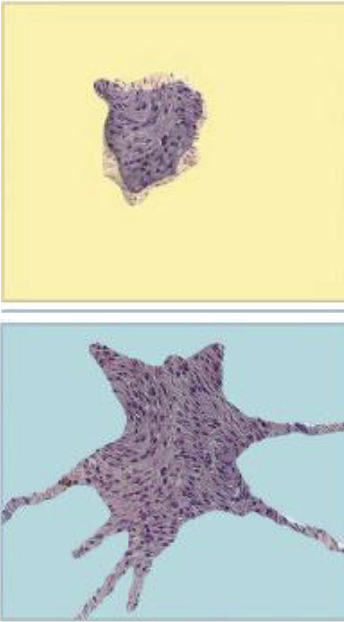
What is a NEGATIVE/CLEAN Tumor Margin?
Tumor does NOT extend to the examined margin → consistent with complete excision in the examined section

What is a POSITIVE/DIRTY tumor margin?
Tumor extends to the examined margin → consistent with incomplete excision in the examined section

Why are Tumor Margins an estimate rather than an absolute assessment of the entire tumor?
Only selected 2-D sections of the 3-D tissue are examined
Different planes and depths can reveal different relationships between tumor and surrounding tissue
If the microscope image is dark, which side of the microscope should you troubleshoot?
The illumination side BELOW the stage
Condenser slider in wrong position
Light output too low
Field diaphragm closed
Condenser iris diaphragm closed
Light beam out of adjustment
Köhler illumination needs adjustment

If the microscope image is fuzzy, which side should you troubleshoot?
The image-formation side
Dirty slide
Dirty lens
Ocular out of adjustment
Slide upside down
Objective not fully clicked into position
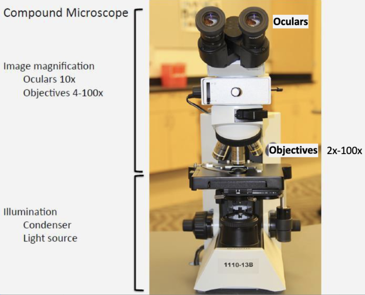
Dark image vs. fuzzy image — what is the key distinction?
Dark = illumination problem → BELOW stage
Fuzzy = image-formation problem → ABOVE/at specimen & optics
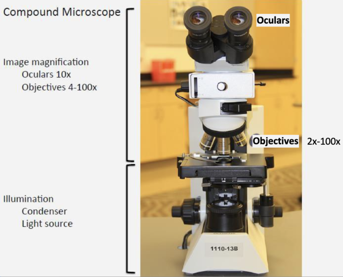
2× vs. 10–40×: where should the condenser be positioned?
2× = LEFT for LOW
10×–40× = RIGHT for HIGH

What are the three major ideas to remember when interpreting a histologic section?
Plane + depth + 3-D structure