anatomy exam 2 part 1
 Call Kai
 Learn
Learn Practice Test
 Spaced Repetition
 Match
Match Flashcards
 Knowt Play
1/86

There's no tags or description
Looks like no tags are added yet.
Name | Mastery | Learn | Test | Matching | Spaced | Call with Kai | Chat |
|---|
No analytics yet
Send a link to your students to track their progress
87 Terms
Main function of endocrine system
Regulate body processes using hormones as chemical messengers.
Role of hormones
chemical messenger released by a cells in one part of the body that affect cells in other parts of the body (travels through the blood)
Endocrine–nervous interaction
Nervous system detects changes; endocrine system releases hormones to adjust physiology.
What types of structures detect changes in our internal and external environment? Do those structures belong to the endocrine system or nervous system?
receptors that belong to the nervous system
Endocrine vs nervous communication
Endocrine: hormones, slow, long-lasting, widespread. Nervous: electrical signals, fast, short-lived, targeted.
Endocrine vs exocrine glands
Endocrine secrete hormones into blood; exocrine secrete substances through ducts.
Do all cells respond to hormones
No; only cells with specific receptors respond
Water-soluble vs lipid-soluble hormones
Water-soluble bind membrane receptors (hydrophilic)
act quickly, short lived, ex) insulin, glucagon
Lipid-soluble bind intracellular receptors (hydrophobic)
act slowly, long lasting, ex) cortisol, estrogen, T3, T4
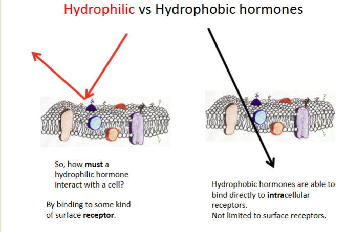
Receptors for hydrophobic hormones
Inside the cell because lipid-soluble hormones cross membranes.
action of hydrophobic hormones
Hydrophobic hormones bind to and activate receptors within cells.
The activated receptors alter gene expression, resulting in the formation of new proteins.
The new proteins alter the cell’s activity, causing the physiological response of the hormone
Receptors for hydrophilic hormones
On the cell membrane because water-soluble hormones cannot cross membranes.
action of hydrophobic hormones
The binding of a hydrophilic hormone to its membrane receptor sets off a cascade of events within the cell
hormonal interactions
The responsiveness of a target cell to a hormone may depend on the influences exerted by other hormones
can have permissive effects, synergistic, or antagonist
Permissive effect
One hormone is necessary for another to produce an effect
• Enables another hormone to fully function
ex) thyroid hormone enables epinephrine
Synergistic effect
Two hormones amplify a response (estrogen + progesterone).
Antagonistic effect
Hormones oppose each other (insulin vs glucagon).
Three ways hormone secretion is stimulated
Neural: any input that activates neurons, triggering electrical or chemical signaling within the nervous system
hormonal: release of a hormone triggered by another hormone
humoral stimulation: regulation of hormone release in response to changes in the composition of bodily fluids, such as blood, including ion or nutrient concentrations.
Location of hypothalamus
In diencephalon; regulator of the endocrine system, controlling hormone production and maintaining homeostasis through its interaction with the pituitary gland.

Location of pituitary gland
Below hypothalamus; anterior pituitary secretes hormones, posterior stores/releases hypothalamic hormones.
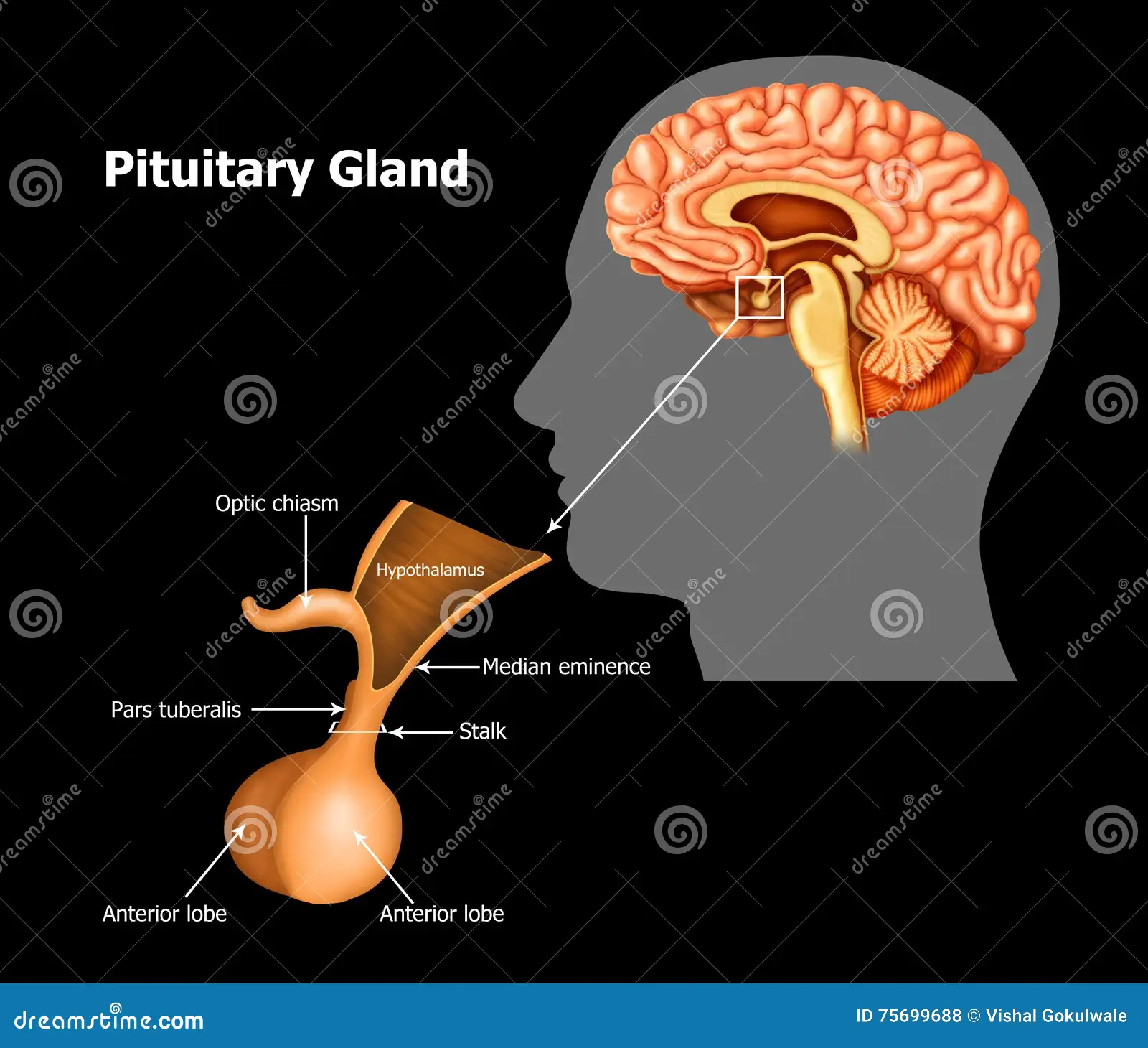
Why posterior pituitary is neurohypophysis
hormones are produced in hypothalamus and travel down axons to posterior pituitary gland. action potentials from hypothalamus trigger release
ADH
Oxytocin
Why anterior pituitary is adenohypophysis
True glandular tissue that produces and secretes hormones.
Portal system
two capillary beds connected by veins; allows targeted hormone transport.
Hypophyseal portal system
Carries hypothalamic hormones to anterior pituitary to be released
Posterior pituitary hormones
ADH: target kidneys, increases BP
oxytocin: target uterus, increases contractions
both made in hypothalamus
Negative vs positive feedback
Negative reverses change; positive amplifies change.
seven anterior pituitary hormones
TSH, ACTH, GH, PRL, FSH, LH, MSH; target thyroid, adrenal cortex, liver/bone, mammary glands, gonads.
melanocyte stimulating hormone (MSH)
stimulated by: UV exposure and leptin
function: stimulates melanocytes to produce melanin, regulate energy balance
target organs: melanocytes
follicle stimulating hormone (FSH)
stimulated by: gonadotropin-releasing hormone (GnRH) from hypothalamus
function: stimulated follicle/ovum development and estrogen production, sperm development
target organs: ovaries (female), testes (male)
luteinizing hormone (LH)
stimulated by: gonadotropin-releasing hormone (GnRH) from hypothalamus
function: stimulated ovulation, androgen production
target organs: ovaries (female), testes (males)
prolactin
stimulated by: suckling of nipples
function: stimulates mammary gland formation in puberty, stimulated production of milk
target organs: mammary glands
growth hormone (GH)
stimulated by: growth hormone releasing hormone (GHRH)
function: stimulates release of growth factors from liver, breakdown of triglycerides in adipose tissues, enhances cellular proliferation
target organs: liver, bones, adipose tissue, nervous system cells, immune system cells
thyroid stimulating hormone (TSH)
stimulated by: TRH
function: stimulates release of thyroid hormones T3 and T4
target organs: thyroid gland
adrenocorticotropic hormone (ACTH)
stimulated by: corticotropin releasing hormone (CRH) from hypothalamus
function: stimulates release of cortisol and androgens from adrenal cortex
target organs: zona fasciculata of adrenal cortex
Releasing hormones function
Stimulate anterior pituitary secretion.
Inhibiting hormones function
Reduce anterior pituitary secretion.
Tropic hormones
Hormones that stimulate other endocrine glands.
Pineal gland
Located in epithalamus; secretes melatonin.

Function of melatonin
Regulates sleep–wake cycles.
Thyroid histology
Follicles filled with colloid; follicular cells surround colloid; parafollicular cells between follicles.
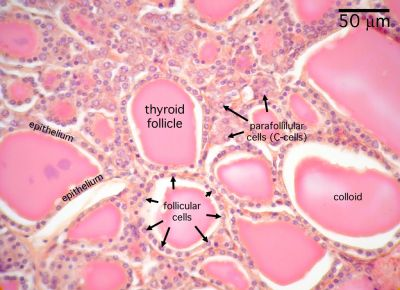
Follicular cells secrete
T3 and T4.
Parafollicular cells secrete
Calcitonin.
Functions of T3/T4
Increase metabolic rate, heat production, heart rate, oxygen consumption.
Most abundant thyroid hormone
T4 (90%); active form is T3 (10%).
Thyroid hormone solubility
Hydrophobic; transported bound to proteins.
how are thyroid hormones transported in the blood
Most thyroid hormones are attached to transport proteins
• Only free T3 can be utilized by the cells
Transport of iodide
Active transport into follicular cells.
Hypothyroidism
Low thyroid hormone; causes fatigue, weight gain, cold intolerance.
Hyperthyroidism
Excess thyroid hormone; causes weight loss, heat intolerance, tachycardia.
Primary vs secondary hypothyroidism
Primary: thyroid problem; secondary: pituitary problem.
Primary vs secondary hyperthyroidism
Primary: thyroid overactivity; secondary: excess TSH.
Goiter
Enlarged thyroid; often due to iodine deficiency or excess TSH.
Calcitonin
Lowers blood calcium; secreted by thyroid.
How calcitonin lowers Ca2+
Increases bone deposition; decreases osteoclast activity.
Parathyroid glands location
Posterior thyroid; secrete PTH when blood calcium is low.
Function of PTH
responds to low blood calcium, Raises blood calcium via bone resorption, kidney reabsorption, vitamin D activation.
Calcium homeostasis
Calcitonin lowers Ca2+; PTH raises Ca2+.
Why pancreas is endocrine & exocrine
Exocrine pancreas – (Acinar cells) release enzymes related to digestion
Endocrine pancreas – (Pancreatic Islets) responsible for controlling blood sugar
Pancreatic islets
Clusters of endocrine cells; alpha cells produce glucagon, beta cells produce insulin.
Hyperglycemia
High blood glucose.
Hypoglycemia
Low blood glucose.
How insulin lowers glucose
Increases uptake into cells; stimulates glycogen formation.
How glucagon raises glucose
Stimulates glycogen breakdown and gluconeogenesis.
Insulin + glucagon homeostasis
Opposing hormones maintain stable blood glucose.
Diabetes mellitus
Chronic hyperglycemia due to insulin issues.
Type I vs Type II diabetes
Type I: no insulin production; Type II: insulin resistance.
Normal fasting blood glucose
About 70–99 mg/dL.
Gestational diabetes cause
Pregnancy hormones from placenta cause insulin resistance; usually resolves after birth.
(hypothalamic-pituitary adrenal) HPA axis stimulus
Stress (physical, emotional, metabolic) triggers hypothalamus → CRH → pituitary → ACTH → adrenal cortex → cortisol
Effect of cortisol
Increases glucose, suppresses immune system, helps stress response.
Adrenal cortex hormones
Mineralocorticoids, glucocorticoids, androgens.
Adrenal medulla hormones
Epinephrine and norepinephrine.
Zona glomerulosa
Outer layer; secretes aldosterone (mineralocorticoids)
Zona fasciculata
Middle layer; secretes cortisol (glucocorticoids)
Zona reticularis
Inner layer; secretes androgens
Mineralocorticoids
steroid hormones that regulate electrolytes; main one is aldosterone. increase reabsorption of Na in kidneys
Glucocorticoids
steroid hormones that regulate metabolism and stress; main one is cortisol. increases rate of protein catabolism. cushings syndrome/addison’s disease
Main androgen
testosterone
Aldosterone
Increases sodium retention and potassium excretion.
Aldosterone & blood pressure
Raises blood volume via kidney sodium retention.
renin-angiotensin-aldosterone system (RAAS) pathway
renin → angiotensinogen → angiotensin I → ACE converts it into → angiotensin II.

what is RAAS stimulated by
low blood volume or low Na+ concentration stimulate renin
ACE
Converts angiotensin I to II.
ACE inhibitor
Blocks angiotensin II formation; lowers blood pressure.
ADH
organ secreting: produced by hypothalamus but stored and released from the posterior pituitary gland
stimulus: high blood osmolarity (too much solute), dehydration, low BP, low blood volume, angiotensin II
target tissue: kidneys
function: absorption of water increases blood volume and BP
part of HPA axis
Atrial natriuretic peptide
organ secreting: atria (top chambers of heart)
stimulus: increased stretch on atrial wall due to increased blood volume or pressure
target tissue: kidneys
function: decrease sodium and water reabsorption, reduce blood pressure and volume
Renin
organ secreting: kidneys
stimulus: low BP
function: initiates RAAS
Erythropoietin
organ secreting: kidneys
stimulus: increased hypoxia in cells
target tissue: bone marrow
function: increase red blood cell production