L1: The Lungs
 Call Kai
 Learn
Learn Practice Test
 Spaced Repetition
 Match
Match Flashcards
 Knowt Play
1/77

There's no tags or description
Looks like no tags are added yet.
Name | Mastery | Learn | Test | Matching | Spaced | Call with Kai | Chat |
|---|
No analytics yet
Send a link to your students to track their progress
78 Terms
what are the margins of the thorax
the superior to inferior thoracic aperture.
what does the thorax contain
the heart
trachea
oesophagus
lungs
thoracic duct
great vessels
what protects the thorax
the thoracic wall
what is the mediastinum
the area between the R and L lungs
what are the superior and inferior thoracic apertures
the two openings at the bottom and top of the thorax.
they allow passage for structures such as: trachea, oesophagus, blood vessels and nerves.
what is a thoracotomy
surgical procedure where a surgeon makes an incision between the ribs to access the organs in the chest such as:
lungs, heart or oesophagus for treatment or diagnosis.
what are the 4 divisions of the mediastinum?
superior mediastinum
anterior mediastinum
middle mediastinum
posterior mediastinum

where is the superior mediastinum?
its above the heart from the thoracic aperture/inlet until T4-T5
what are the contents of the superior mediastinum?
thymus
aortic arch
brachiocephalic veins
trachea
esophagus
vagus and phrenic nerves
where is the anterior mediastinum
in front of the pericardium
what are the contents of the anterior mediastinum
fat
lymph nodes
remnants of the thymus
internal thoracic vessels
where is the middle mediastinum?
contains the heart and pericardium
what are the contents of the middle mediastinum
heart
ascending aorta
pulmonary trunk
superior vena cava
phrenic nerves
where is the posterior mediastinum
behind the pericardium in front of the spine
what are the contents of the posterior mediastinum?
esophagus
descending aorta
thoracic duct
azygos system
vagus nerves
where does the trachea run to and from?
from C6 to T4 and then it divides into the main R and L bronchi.
what is the trachea covered in?
15-20 U-shaped hyaline cartilages
what is the function of the oesophagus
fibro-muscular tube connecting the pharynx to the stomach
where does it exit the thorax?
at T10 and pierces the muscular right crus of the diaphragm through the oesophageal hiatus
what are the 3 narrowings of the oesophagus
cricopharyngeus
left bronchus
gastro-oesophageal junction
describe lobes and fissures of the R lung
3 lobes
separated by horizontal and oblique fissures
describe the lobes and fissures of the L lung
2 lobes separated by an oblique fissure
what are the 3 surfaces of the lungs
costal - facing the ribs
mediastinal - faces the heart and mediastinum
diaphragmatic - rests on the diaphragm
what are boarders of the lungs
anterior, posterior and inferior
these are important for auscultation and imaging interpretation
what is auscultation
listening to sounds produced in the body
what are the 2 blood supply for the lungs
pulmonary circulation: for gas exchange
bronchial circulation: supplies lung tissue
what is the parasympathetic innervation to the lungs
vagus nerve - bronchoconstriction and increased secretion
what is the sympathetic innervation to the lungs?
thoracic sympathetic branch - bronchodilation

label the sternum

what are the true ribs and their features
ribs 1-7
they have costal cartilages which attach to the sternum
what are the false ribs and their features
ribs 8-10
have costal cartilages which attach to the CCs above
what are the floating ribs and their features
ribs 11 and 12
have no costal cartilages
what are the atypical ribs
1
2
11
12
why is rib 1 atypical
only has one articulation with the vertebral body
short, broad and most curved
why is rib 2 atypical
less curved that 1
has a rough tuberosity for serratus anterior
why are 11 and 12 atypical
1 articulation w the vertebral body
no anterior articulation
no tubercle for articulation with the transverse process
what are the typical features of a rib (3-10)
head 2 articulations
neck
tubercle
curve
costal groove
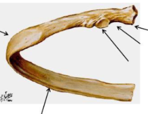
what are the 3 sternal joints
sternoclavicular
manubriosternal joint
xiphisternal joint
describe the sternocostal joints
between the sternum and costal cartilages
1st costal joint - primary cartilaginous
2-7 are synovial
describe the costochondral joints
between the rib and costal cartilage
primary cartilaginous
no ligaments
describe intercondral joints
between costal cartilages
synovial
no ligaments
describe costovertebral joints
synovial joints between ribs and thoracic vertebrae
head articulates with the bodies of the vertebrae
ribs 2-10 have 2 facets where as 1,11 and 12 only have one facet
the tubercles articulate with the transverse process of the same vertebra (costotranverse joint)
what are the muscles of respiration
diaphragm
intercostal muscles
accessory muscles
what is the structure and function of the diaphragm
dome shaped musculotendinous structure
separates the thoracic cavity from the abdominal cavity
responsible for expiration
what is the nerve supply to the diaphragm
phrenic nerve
C3,4,5 keeps the diaphragm alive
what are the diaphragmatic aperatures?
T8 - IVC hiatus and (R phrenic nerve)
T10 - oesophageal hiatus
T12 - the aortic aperature: thoracic duct, azygos vein , hemiazyos vein
what is a diaphragmatic hernia?
condition where abdominal organs move into the chest cavity through a defect in the diaphragm - often requires surgery
what are the attachments of the diaphragm
landmarks of the inferior thoracic aperture
left and right crus
what are the 3 types of intercostal muscles
external
internal
innermost
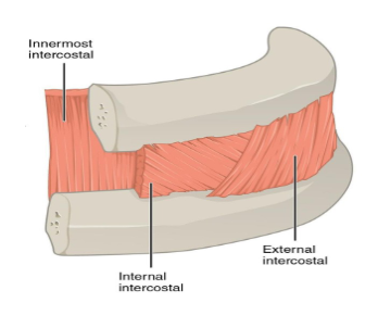
what are is the origin and insertion of the external intercostal muscles?
from the tubercles of ribs 1-11
to the cartilages of ribs 2-12
how do the external intercostals terminate?
as the external intercostal membranes
what is the orientation of the external intercostal muscles
infero-anteriorly
what is the action of the external intercostals?
they elevate the ribs for inhalation
what is the origin and insertion of the internal intercostal muscles
from the sternum between the costal cartilages of the true ribs
to the outer costal cartilages of the false ribs
what is the orientation of the internal intercostal muscles
fthey face infero-posteriorly
what do the internal intercostal muscles terminate as
the posterior intercostal membranes
what is the function of the internal intercostal muscles terminate as
they assist forced expiration
what is the function of the innermost intercostal muscles
they assist expiration by narrowing the intercostal spaces
where are the innermost intercostal muscles located
between the ribs
when do the accessory muscles of respiration act
exercise
respiratory distress
what are the 6 accessory muscles
scalene muscles
SCM
trapezius
pec major
pec minor
serratus anterior
what is the costal groove described as?
a neurovascular bundle
what is the groove van?
V- vein
A-artery
N-nerve
they lie between the innermost layer of the intercostal muscles in the groove
what is the blood supply and drainage of the thoracic wall
posteriorly: descending aorta
anteriorly: subclavian artery
what are the movements of the thorax
lateral expansion
anterior expansion
descent of the diaphragm
which muscles partake in quiet, deep and very deep inspiration
quiet - diaphragm
deep - diaphragm and external intercostals
very deep - accessory muscles
what muscles partake in quiet and forced expiration
quiet - passive as the diaphragm releases
forced - internal and innermost intercostal and abdominal muscles
what is the lingula
a unique, tongue-like projection of tissue found exclusively in the left lung,
representing a specific segment of the upper lobe

what is the function of the lingula
to facilitate gas exchange
what are the the types of lung pleura
visceral
parietal
between the two lies the pleural cavity

why would a chest drain be inserted
to remove unwanted substances from the pleural space such as air, blood or pus
where are they inserted and why
ypically inserted at the 4th or 5th intercostal space, just anterior to the
mid-axillary line, and always above the rib to avoid damaging the
neurovascular bundle that runs along the inferior border of each rib
what is the hilum of the lung
A triangular depression on the inner (mediastinal) surface of each lung.
Acts as the entry and exit point for structures connecting the lungs to the body (the "root of the lung").
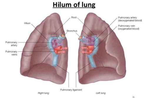
what is the bronchial tree?
trachea - bronchi - lobar bronchi - segmental bronchi - bronchiole - alveolar duct - alveolus
the last 3 have no cartilage

what is the blood supply to the bronchi?
bronchial arteries: arise from the descending thoracic aorta
bronchial veins: usually two on each side, drain into the azygos system of the veins
what is the innervation of the bronchi
pulmonary plexus
describe the innervation of the visceral pleura
Sensitivity: Not sensitive to pain, temperature, or touch.
Innervation: Autonomic only (i.e., no sensory); receives sympathetic and parasympathetic fibers from the pulmonary plexus (Vagus n.).
describe the innervation of the parietal pleura
Sensitivity: Sensitive to pressure, pain, and temperature.
Innervation: Sensory innervation from intercostal nerves and phrenic nerves.