637 Ch22 Pediatric CVP Implications
 Call Kai
 Learn
Learn Practice Test
 Spaced Repetition
 Match
Match Flashcards
 Knowt Play
1/52

There's no tags or description
Looks like no tags are added yet.
Name | Mastery | Learn | Test | Matching | Spaced | Call with Kai | Chat |
|---|
No analytics yet
Send a link to your students to track their progress
53 Terms
When does the respiratory system begin developing?
4th week of gestation
What are the four stages of maturation of the lungs and their key features? When do most of the alveoli develop?
Pseudoglandular period (6-16)
All in place except those involved w/ gas exchange
Canalicular period (16-26)
Lung tissue becomes vascularized
Terminal saccular period (26-birth)
Surfactant starts being produced at 20 and reaches maturation at 34
Alveolar period (32wk-8years)
After birth, surface area & lung tissue grow rapidly, 95% of alveoli develop AFTER birth
When does the heart beat start? When does circulation extend around the fetus’ body? When does the atria divide into R and L? When do all 4 form?
Day 22-23, circulation extends around fetus’ body by day 27
Middle of 4th gestational wk - atria divide
All 4 by 7th gestational wk
What is the process of fetal circulation and the structures involved with it? How is CO2 removed?
Heart pushes most blood past the lungs to the LV w/ foramen ovale
O2 obtained and CO2 is removed through the placenta
Foramen ovale (shunt btwn LA&RA) fuses after 6mo, closes hours after birth
Ductus arteriosus (PA to aorta) fuses after weeks, closes 12-24hr after birth
Pulmonary blood vessel vasodilate after birth to allow enough blood for gas exchange
What does the foramen ovale and ductus arteriosis connect? What kind of blood do they deliver?
Foramen ovale connects R atrium to L atrium
Oxygenated blood bypasses fetal lungs to L atrium then L ventricle and ascending aorta
Ductus arteriosus connects pulmonary artery to aorta
Right ventricle deoxygenated blood bypasses fetal lungs to descending aorta, organs, and back to placenta
What is the etiology of congenital heart disease (CHD)?
Genetic link
Environmental factors - asbestos, lead exposure
Maternal factors - diabetes, smoking, alcohol, folic acid (B9), obesity, diet, depression
What are acyanotic lesions? Sxs? Findings? Examples?
O2SAT MAINTAINED; increase pulmonary blood flow (increased risk of pHTN) and fully oxygenated blood is brought back to lungs and body (L to R shunting)
Sxs: sweating, increased RR, HF, skin well perfused
Findings: reduced PaO2 to periphery, reduced systemic SV, increased CO
EX: ventricular septal defect, atrial septal defect, patent ductus arteriosus, coarctation of the aorta, pulmonary stenosis, aortic stenosis
How is O2sat affected in acyanotic and cyanotic lesions?
Acyanotic - O2sat maintained
Cyanotic - O2sat NOT maintained, reduced
True or false: cyanotic and acyanotic lesions have signs of cyanosis
False, only cyanotic lesions
What are cyanotic lesions? Sxs? Findings? Examples?
O2SAT REDUCED; most of the blood bypasses the lungs, deoxygenated blood flows back to body (R to L shunting). More RBCs produced → polycythemia → increased risk for CVA; more concerning, will need surgery immediately!
Sxs: fatigue, fainting, SOB, chest pain, HF
Findings: increased RBC count, cyanosis, SpO2 70-85%, capillary refill is delayed
EX: hypoplastic left heart syndrome, tetralogy of fallot, transposition of the great arteries, total anomalous pulmonary venous return, tricuspid atresia, pulmonary atresia, truncus arteriosus, pediatric heart failure
What are typical sxs of acyanotic lesions?
Body working harder i.e. difficulty w/ feeding, fussing, crying, sweating
What is an atrial septal defect? Findings? Tx?
ACYANOTIC LESION
Hole btwn R & L atriums that doesn’t close after birth (such as foramen ovale)
Findings: heart murmur & enlarged pulmonary A
Closed w/ surgery if it doesn’t close on it’s own by 2-3y/o
*What is patent ductus arteriosus? What is normally supposed to happen? Findings? Tx?
ACYANOTIC LESION
Pathway of normal fetal circulation btwn descending aorta & pulmonary A doesn’t close after birth
Too much blood enters the lungs (risk of pHTN, destruction of pulmonary tissue, causes scarring → fibrosis)
High incidence in premies
Normal: O2 level increases → reduces prostaglandin E1 → closes PDA (within 5-14 days)
*Findings: tachycardia, respiratory distress, limited weight gain
*Tx: surgery or Indomethacin to reduce prostaglandin prod.
What is ventricular septal defect? ? Findings? Tx?
ACYANOTIC LESION
Hole in septum separating ventricles: small deficits acyanotic, large deficits complications → bacterial endocarditis (fever), pulmonary vascular obstructive disease, aortic regurgitation, lower RTIs (risk of infection is HIGH), CHF (ventricular arrhythmias)
Findings: feeding difficulties (hard to eat and breathe at the same time), rapid breathing, poor weight gain, irritability, restlessness, dehydration
May close 5mo-6y/o w/o intervention; surgery for more complicating defects
*What is atrioventricular septal defect (AVSD)? Findings? Tx?
ACYANOTIC LESION
Failure of formation of embryonic atria/ventricles (one big chamber, no septum development)
Down syndrome
Impacts endocardial cushion development
Findings: pHTN, lung congestion, HF
*Tx: surgery REQUIRED within the first few months (BiPAP/CPAP to prolong if too young)
What is coarctation of aorta? Findings? Tx?
Narrowing of aorta which obstructs LV outflow; >50% must be narrowed, diagnosed w/ echocardiogram NOT ultrasound
Pressure proximal to constriction of aorta is increased = HTN in upper body above level of coarctation, normal-low BP in LE
Many go undiagnosed until later, 50% rate of mortality under 30y/o if not tx
Findings: secondary HTN, systemic congestion (increased HR), pulmonary edema (weight gain fluctuations)
Tx: surgery to remove constriction of aorta, LIKELY TO REOCCUR even if tx
Differentiate the key findings between each of the acyanotic lesions.
Atrial septal defect
Heart murmur
Enlarged pulmonary A.
Can close on it’s own or surgery
Patent ductus arteriosus
Too much blood in lungs
Premies
Too much prostaglandin E1, doesn’t close PDA
Tachycardia, respiratory distress, limited weight gain
Indomethacin or surgery
Ventricular septal defect
Large defect comp.
Fever, high risk of infections, feeding difficulties, tachypnea, poor weight gain, irritability, restlessness
Can close on it’s own or surgery
AV septal defect
1 chamber
Down syndrome
pHTN, lung congestion, HF
surgery REQUIRED
Coarctation of aorta
HTN UE, normal/low LE, systemic congestion, pulm edema
High rate of mortality, surgery tx, may reoccur
What is hypoplastic left heart syndrome (HLHS)? Cause? Fx? Sxs? Tx?
CYANOTIC LESION
Idiopathic cause, underdeveloped LV, aortic & mitral valve stenosis (or atresia), and coarctation of aorta
Diagnosed during pregnancy or shortly after birth
Fx: L side of heart doesn’t form correctly
Systemic blood flow is dependent upon PDA, once closed → severe CHF
Sxs: tachycardia, tachypnea, poor feeding, cyanosis, weakness and cold extremities
Tx: mech vent, surgery soon after birth → not permanent fix, 3 stage palliative surgery or heart transplant
What is tetralogy of fallot? Cause? Fx? Sxs? Tx?
CYANOTIC LESION
Tetrad of defects: pulmonary stenosis, overriding aorta, ventricular septal defect, RV hypertrophy
Degree of cyanosis dependent on severity of stenosis
Idiopathic cause, RF: family hx, virus during pregnancy, alcohol, smoking, or eating poorly during pregnancy, mom >35y/o, down syndrome
Fx
Pulmonary stenosis - obstruction to RV outflow → hypertrophy
VSD: oxy&deoxy blood mixing
Aorta above VSD on RA instead of LV → RV hypertrophy
Sxs: “TET’ spells of cyanosis w eating or crying, SOB, fainting, difficulty w/ weight gain, tachypnea
Tx: surgery 3-6mo
What is transposition of great arteries? Sxs? Tx?
CYANOTIC LESION
Positions of aorta & pulmonary A. are reversed, does not support life, often diagnosed in utero or in first few hours of life
Severity is determined by the amount of blood that is mixing on both sides
Sxs: severe cyanosis if PDA, VSD, or ASD is not available
Tx: surgery, prostaglandin E1 to maintain PDA; may develop coronary stenosis, arrhythmias, or ventricular dysfunction later in life
What is atresia? Describe atresia of the tricuspid valve, pulmonary valve, and truncus arteriosus.
CYANOTIC LESION
Poor/missing development
Tricuspid valve: obstruction of blood flow to RV which is typically underdeveloped w/ some shunting of blood → lungs (ASD and VSD may be present)
Sx: anoxia, R sided HF, dyspnea, severe cyanosis
Tx: surgery
Pulmonary valve: fails to develop leading to an obstruction from R side of heart → lungs; blood flow to lungs is dependent on PDA (ASD and VSD would allow shunting to the body)
Truncus arteriosus: aorta and pulmonary A. do not separate in development and both empty pulmonary and systemic blood flow into one large vessel, single ventricle (VSD is always present)
What is total anomalous pulmonary venous return? Sxs? Tx?
CYANOTIC LESION
Pulmonary veins attach to the R atrium or other veins that attach to the R atrium (ASD might be present which could aide in atrial decompression)
Sxs: cyanosis, HF, pulmonary congestion
Tx: surgery ASAP to join pulmonary veins to LA
What is pediatric heart failure? What are the 3 types? Sxs classification?
Circulation doesn’t meet metabolic needs of body b/c reduced CO or reduced ventricular relaxation (diastolic HF)
Cardiomyopathy: categorized as dilated, hypertrophic, or restrictive conditions
Dilated: most common cause of heart transplant in children >1y/o, enlarged ventricles w/ reduced systolic ejection and CO
More common in males and in infants w/ muscular dystrophy
Hypertrophic: reduced ventricle filling & reduced CO; most common cause of SCD in children
Restrictive: poor ventricular diastolic relaxation but normal ventricular size
Failed palliative of CHDs
Sxs: Ross Classification for Infants & Children
Diaphoresis, tachypnea, dyspnea, tachycardia, hepatomegaly
0-2 (no HF), 3-6 (mild HF), 7-9 (moderate HF), 10-12 (severe HF)
What are the sxs classified in the Ross Classification for Infants & Children? Scoring?
Diaphoresis, tachypnea, dyspnea, tachycardia, hepatomegaly
0-2 (no HF), 3-6 (mild HF), 7-9 (moderate HF), 10-12 (severe HF)
What is a ventricular assist device (VAD)? Goals? Conditions that may need a VAD?
Mechanical device to help blood flow around the body
Goals: increase candidacy for transplant, recovery from illness or bridge for designation tx
50% reduction in transplant waitlist mortality
Used in correlation w/ PT: improvement in fx, exercise capacity, accelerated transplant recovery time
Common conditions: myocarditis, CHD, transplant rejection, cardiomyopathy, heart dysfunction after chemo
What are 8 pediatric respiratory conditions?
Prematurity
Persistent pulmonary HTN
Respiratory distress syndrome
Sudden infant death syndrome
Meconium aspiration syndrome
Bronchopulmonary dysplasia
Cystic fibrosis
Primary ciliary dyskinesia
What is extracorporeal membranous oxygenation (ECMO)? Two types? Fx? Can you mobilize pts on ECMO?
External heart and lung bypass machine
V-V (venovenous) lungs
V-A (venoatrial) lungs and heart
Fx: removes blood, adds O2, removes CO2
Yes, mobilizing pts on ECMO decreases ICU morbidity
What are 6 pediatric conditions with secondary CVP concerns?
Down syndrome - chromosomal disorder, endocardial cushion (AV) defect, VSD, ASD, TOF
Cardiac defects & sleep apnea common
Lower PA, reduced muscle strength, endurance, poor eating habits
VATER association - vertebral defects, imperforate anus, tracheoesophageal fistula, and radial and renal dysplasia
DiGeorge syndrome - immunodeficiency disease w/ significant cardiac changes, TOF, coarctation of aorta, ASD, pulmonary stenosis, hypoplasia left heart, PDA, VSD, truncus arteriosus
Marfan syndrome - connective tissue disease, high prevalence of aortic aneurysm & aortic/mitral insufficiency, increased height and decrease weight for age, scoliosis , excessively long extremities, stooped shoulders, depressed sternum, fragility of blood vessels
Williams syndrome - chromosomal condition associated w/ supravalvular aortic and pulmonary stenosis
Fetal alcohol syndrome - chronic alcohol exposure in utero, facial dysmorphia, poor coordination, hyperactivity, learning disabilities, delays in development
VSD, TOF, pulmonary valve stenosis, PDA commonly seen in this population
What are some activity and breathing impairments associated with pediatric conditions with reduced acitvity and posture?
Muscle weakness, abnormal muscle tone (hypertonicity & hypotonicity), motor learning deficits, medical fragility, motor planning deficits
What are pediatric conditions with pulmonary system impairments?
Cerebral palsy, spina bifida, muscular dystrophy, spinal muscular atrophy, spinal cord injury, severe progressive scoliosis
What are associated impairments with an ineffective cough in pediatric conditions?
Poor saliva control, poor gag reflexes, muscle spasms, altered breathing patterns
CP have increased O2 demands and fatigue faster - take more breaks
What are observations you want to make during a pediatric PT examination?
Environment
Resting position
Vital signs
Pattern of breathing
Shape of rib cage
0-3 months: triangular, ribs horizontal, diaphragm for inspiration
3-6 months: rectangular, ribs horizontal, diaphragm and accessory muscles for inspiration
6-12 months: rectangular, ribs angled downward, diaphragm and intercostals for inspiration
Integumentary condition
Resting posture
What are the typical shapes of rib cage from 0-1y/o?
0-3 months: triangular, ribs horizontal, diaphragm for inspiration
3-6 months: rectangular, ribs horizontal, diaphragm and accessory muscles for inspiration
6-12 months: rectangular, ribs angled downward, diaphragm and intercostals for inspiration
Describe some items on a medical chart review you would like to know for a pediatric patient.
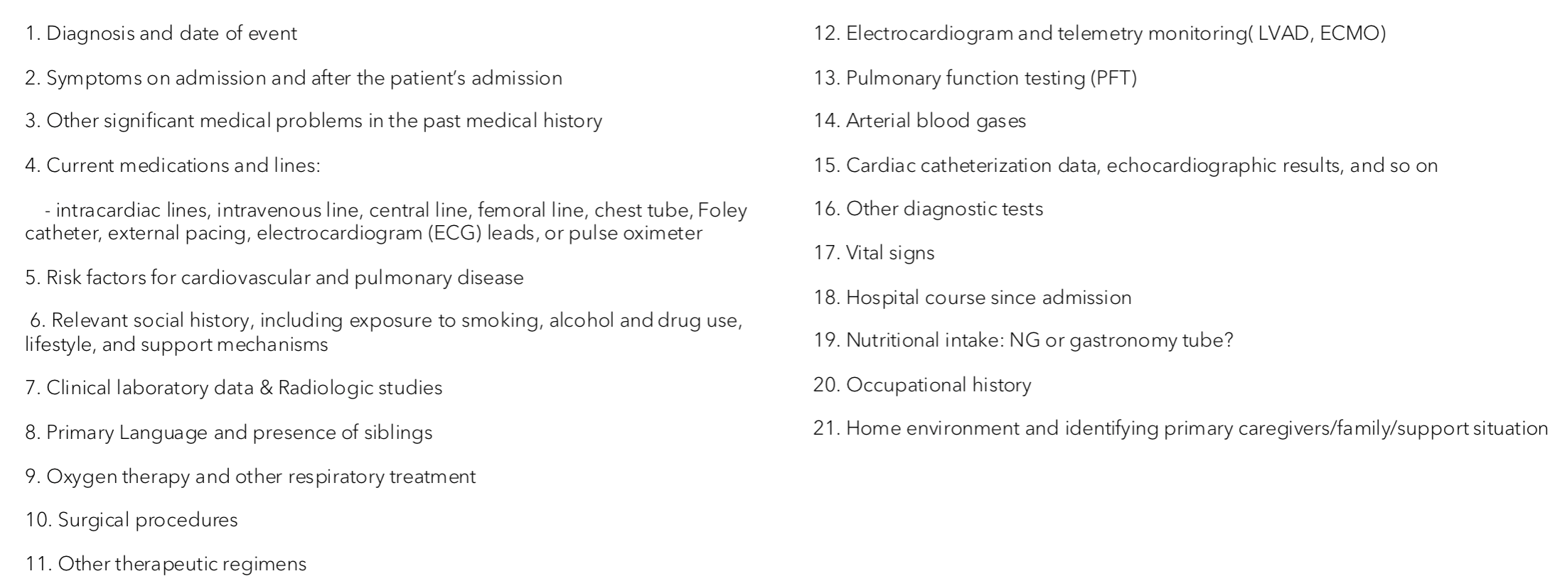
What should you look at during a pediatric PT evaluation?
Rib cage mobility
Flexibility
Dynamic posture
Use of accessory muscles
Breathing pattern: shallow, irregular breathing
Thoracic expansion w/ tape measure (4th IC space 1”)
Monitoring vital signs (norms w/ aging below)
Weight increases
HR decreases
RR decreases
BP increases
Is it ok for a baby to be using accessory muscles during breathing? Head turns?
Accessory muscle use up to 6mo OK (2y/o?)
No head turns w/ breathing
What are two prevention goals for the CVP pediatric population?
Maximize O2 transport and aerobic activity
Reduce impact of lifestyle conditions
Describe 12 PT interventions for the CVP pediatric population.
Rib cage mobility
Flexibility
Strength
Posture
Breathing pattern
Airway clearance techniques
Positive expiratory pressure (PEP) device
Blow toys
Huffing
Assisted cough
Functional mobility
CV/endurance training
Describe rib case mobility as a pediatric intervention.
In supine, prone, S/L (special consideration for osteopenia)
Manual release techniques for Q/L and intercostals, scar tissue massage for thoracic incision
Describe flexibility as a pediatric intervention.
S/L trunk rotation for intercostals; laying supine over a ball, corner pectoralis stretch, pelvic tilt, stretches for Q/L, lats, and abs
Describe strength as a pediatric intervention.
Body weight exercises
Global - games that involve UE and LE body weight and dynamic stabilization (wheelbarrow, hopping and jumping, prone on therapy ball)
Spinal - extensors and retractors (good for hypotonicity
Inspiratory - inspiratory muscle trainer (ex program for CP 30% of their max inspiratory pressure for 10-15 breaths over 15 minutes, 2x/day, 6 wks)
Describe posture as a pediatric intervention.
Mirror training, tapping to facilitate spinal extensors, consider position in stroller, car seat and chair, and amount of time spent in each (want to adjust posture for optimal positioning)
Describe breathing pattern as a pediatric intervention.
After 2 y/o child should not demonstrate use of accessory muscles
If kids fatigue quickly, they may use accessory muscles to breathe shallow due to pain from an incision, train diaphragm and intercostals (can use stuffed animals for diaphragmatic breathing; can use inhibitory techniques as a last resort)
Once efficient breathing pattern is established should progress to harder positions like sitting
Describe airway clearance techniques as a pediatric intervention.
PD, vibration, percussion
Trendelenburg position contraindicated in a child younger than 2 y/o, kids w/ CF b/c reflux → vomiting
Can modify for kids w/ CF → 20deg head down
Infants, young children in PT/caregiver’s arms (keep pt awake during PD)
Describe high-frequency chest wall oscillation as a pediatric intervention.
Contraindications: bronchospasm, chest pain, lung contusion, burns or open wounds, recent pacemaker, skin grafts, recent spinal epidural, etc.
Describe positive expiratory pressure (PEP device) as a pediatric intervention.
Provide resistance to exhalation
Bubble PEP: using suction tubing and a container of water
Describe blow toys as a pediatric intervention.
Kid versions of incentive spirometer
Kazoo, noisemakers, pinwheels, bubbles, cotton ball
Describe huffing as a pediatric intervention.
Start w/ low volume and progress to larger force; less likely to risk dynamic airway collapse than strong coughing
Describe assisted cough as a pediatric intervention.
NM weakness
Educate pt and caregiver on manual assisted cough techniques and positioning
Rhythmic rocking for increased tone, dissociating extremities, reducing external stimulation
Should you be getting a child out of bed after surgery? in the PICU?
Yes, ASAP to reduce CP complications after surgery (DVT, respiratory infections, delirium)
Early mobilization in PICU is safe and recommended
How can you help kids overcome fear? Infants?
Dolls and toys, games that increase mobility i.e. scavenger hunts, assistive devices (be mindful of lifting, pushing, pulling restrictions after surgery)
Infants - bring extremities to midline for calming, swaddling w/ baby’s hands near face, facilitate more milestone directed movement w/ support (can initiate infant in prone on parent’s chest w/ parent gradually reclining)
Describe CV/endurance training as a pediatric intervention.
Monitor HR, BP, SpO2, RPE, dyspnea
Goals can include social interactions: walking further to go on a vacation or do an activity w/ friends
Increasing length of time before a rest, increasing overall time of exercise, increasing intensity, decreasing amount of SpO2 needed, decreasing RPE
If child is interested in/excited by the activity → increase performance; HEP → bike riding, fitness video games, playing tag, jumping rope, obstacle course
Educate pt and caregiver on what exercise should feel like and when to stop
*What are sxs to stop exercise?
If the child has any of these sxs → stop exercising and REST. Attempt a lower intensity or duration during the next exercise session.
Chest pain or discomfort
RPE >17
Dyspnea of 4
Dizziness
Visual changes
Pale or ashen appearance
New-onset joint pain
HR >80% of maxHR
O2sat falls <90% (in some cases MD will say it’s OK)