Histology Chapter 1 Objectives
 Call Kai
 Learn
Learn Practice Test
 Spaced Repetition
 Match
Match Flashcards
 Knowt Play
1/31

There's no tags or description
Looks like no tags are added yet.
Name | Mastery | Learn | Test | Matching | Spaced | Call with Kai | Chat |
|---|
No analytics yet
Send a link to your students to track their progress
32 Terms
Histology
The study of the microscopic structure and function of biological tissues.
Cytology
The study of the structure, function, and chemistry of individual cells.
Biopsy methods: Scalpels
Used to sample directly accessible tissues like skin, mouth, and nose.

Biopsy methods: Needles
Used to sample solid internal organs (e.g., percutaneous kidney biopsy).
Biopsy methods: Endoscopic tubes
Used to sample tissue from the alimentary tract or body cavities.
Biopsy methods: Flexible cannulae
Used to access and sample tissue inside blood vessels.
Cell
Basic functional unit of life derived from a fertilized ovum; adaptable and specialized.
Cell adaptations: Hypertrophy
An increase in individual cell size.
Cell adaptations: Atrophy
A decrease in individual cell size.
Cell adaptations: Hyperplasia
An increase in the number of cells via cell division.
Epithelial tissue
Tissue that covers body surfaces, lines internal cavities, and forms glands.

Muscular tissue
Contractile tissue responsible for body movement and generating mechanical force.

Nervous tissue
Tissue specialized for transmitting electrical signals and processing information.

Connective tissue
Supportive tissue that connects, binds, protects, and cushions organs.

Simple tissue
A tissue composed of cells with the same structure and function (e.g., adipose).
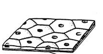
Compound tissue
A tissue containing a mixture of cells with different functions (e.g., nervous tissue).
Organ
An anatomically distinct group of different tissues performing specific functions.
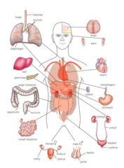
Organ system
A group of organs or widely distributed cells working together for related roles.

Light Microscopy (LM) Tissue Prep Steps
1) Fixation (Formalin)
2) Embedding (Paraffin)
3) Sectioning (Microtome)
4) Mounting & Staining
Paraffin sectioning thickness (LM)
Tissue is cut into sections approximately 5 to 8 µm thick.
Purpose of Fixation in LM
Retards alterations and prevents tissue degradation, commonly using formalin.
Transmission Electron Microscopy (TEM)
Employs a beam of electrons transmitted through a specimen to form an image (requires thin slides)

Scanning Electron Microscopy (SEM)
Scans the surface of solid tissue samples, providing 3D views of cellular structures (does not require thin sections)

Confocal Microscopy
Uses laser light to scan samples and create high-resolution images, often used for thick specimens.
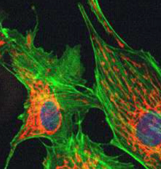
Four major types of histological stains
Empirical, histochemical, enzyme histochemical, and immunohistochemical stains.
Hematoxylin and Eosin (H&E) stain
Most common stain; hematoxylin stains nuclei blue, eosin stains cytoplasm pink.
Periodic acid-Schiff (PAS) reaction
Histochemical stain that detects carbohydrates, glycogen, and mucins (stains magenta).
Giemsa stain
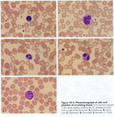
Stain commonly used for blood smears and bone marrow to differentiate blood cells.
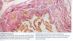
Van Gieson stain
Stain used to differentiate collagen (red) from elastic fibers and muscle (yellow).
Tissue Microarray (TMA)
Robotic technique assembling tiny cores from hundreds of tumor specimens onto one slide.
TEM Tissue Processing Steps
1) Fixation (Glutaraldehyde/OsO₄),
2) Resin Embedding,
3) Ultramicrotomy (30-70 nm),
4) Heavy Metal Staining.
Virtual Microscopy
Digitizing glass slides using high-magnification scanners for computer or smartphone examination.