Cell Neuro Exam 1
 Call Kai
 Learn
Learn Practice Test
 Spaced Repetition
 Match
Match Flashcards
 Knowt Play
1/95

There's no tags or description
Looks like no tags are added yet.
Name | Mastery | Learn | Test | Matching | Spaced | Call with Kai | Chat |
|---|
No analytics yet
Send a link to your students to track their progress
96 Terms
Patellar stretch reflex
Detect the stretch (+ and electrical potential changes)
A sensory neuron, which lives in ganglion (specifically the dorsal root ganglion), detects the stretch
The sensory neuron sends a neurite or process out to the muscle
The muscle stretch activates this neurite and causes it to have an electrical charge (electrical potential across membrane)
When the sensory out in muscle stretched, ion channels open, ions move across membrane (causing depolarization), electrical potential of the sensory neuron changes, and may make an action potential if the depolarization reaches the threshold potential
Action potential is generated in the sensory neuron and travels in a nerve
The action potential reaches the CNS
The signal passes across a synapse because it must be transmitted to another neuron
The sensory neuron is the presynaptic synapse/neuron and the next neuron is the postsynaptic neuron
At a synapse, the presynaptic neuron releases neurotransmitters into the postsynaptic cleft, where it diffuses to the postsynaptic neuron membrane. The neurotransmitters open ion channels to change the electrical potential on the postsynaptic neuron.
Synapse takes place at the receiving end of the next neuron, at its dendrites
The postsynaptic cell (in this case) is called a motor neuron, which synapses with muscles and makes them contract
The motor neuron generates an action potential, which travels along the axon of the motor neuron, which is inside a bundle of axons (nerve).
The motor neuron (presynaptic) makes a synapse on the muscle (postsynaptic)
The muscle opens ion channels and the membrane potential changes. The muscle responds to the change in membrane potential by contracting
Leg swings out
Patellar stretch drawing
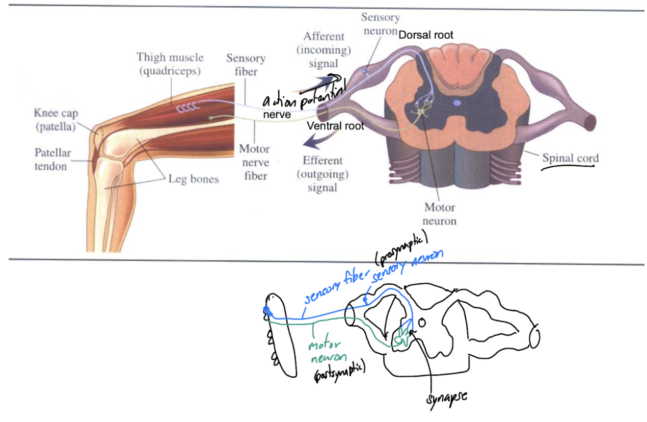
Presynaptic and postsynaptic of patellar stretch
The presynaptic is the sensory neuron and the postsynaptic is the motor neuron
Ganglion
localized, dense cluster of neuronal bodies that acts as a processing or relay center
Nerve
a bundle of neurons
Neurite
any projection or extension from the cell body of a neuron, which can develop into either an axon or a dendrite
Motor neuron
synapse with muscles and make them contract
Afferent
signal or neurite going towards the cell body (to the CNS)
Efferent
signal or neurite going away from the cell body (or CNS)
Neurons
cells. They have all the typical organelles, but their membranes are designed to send and receive electrical signals.
Their shapes are highly variable depending on their purpose
Categories of neurons
sensory neurons, motor neurons, interneurons
Sensory neurons
respond to environmental signals such as light and sound
Motor neurons
send signals to muscles
Interneurons
send signals between sensory, motor, and other interneurons
5 major types of glial cells
astrocytes, radial glial, microglia, oligodendrocytes, Schwann cells
Astrocytes
they are physical support for neurons and regulate K+ concentrations in the extracellular space. They have some nutritive function and remove neurotransmitters from the extracellular space.
What are the most common glial cells int he brain?
astrocytes, which are important fro the blood brain barrier
Radial glial
Important in development. They form a scaffold for neurons to climb during development
What glial cell is most common in neurodegenerative diseases like Parkinsons?
Microglia. It is not entirely clear if these are good or bad
Microglia
cleanup cellular debris if there’s damage and show up when there is an infection or problems as part of immune function. Made in bone marrow. They remove extra synapses during development.
Myelin producing glial cells
Oligodendrocytes and Schwann cells
Oligodendrocytes
Found in the brain and spinal cord (CNS). They wrap around axons of nerve cells with myelin
Inhibit actual nerve regrowth but support myelin repair
Schwann cells
Found in peripheral nerves (not CNS). Whole schwann cells wrap around an axon. If there is nerve damage, they can form a guide for axons to regenerate. Can promote neuronal outgrowth by secreting something
Only glial cell not found in the CNS
Schwann cells
Invertebrate nerve net
a simple, decentralized nervous system made of interconnected neurons spread across the body without a brain or central control center
The simplest neuronal organization (ex: in jellyfish) with no CNS or PNS
Spreads out like a wave
Different from ganglion
Myelin
The cell membrane of glial cells. Glial cells wrap many times around the axon tightly to squeeze out cytoplasm, so myelin is basically many layers of the cell membrane stacked on itself. It is an insulator, speeding up action potentials.
Insulator
allows for faster propagation of action potentials
Nodes of Ranvier
gaps in the myelin that contain action potentials to recharge and boost the action potential as it moves along
Rostral, anterior
towards the nose
caudal, posterior
towards the tails
dorsal
along the back
ventral
towards the belly
lateral
away from the midline
medial
towards the midline
proximal
close to the structure noted
distal
away from the structure noted
distal
away from the structure noted
superior
above (like dorsal in a cat) - brain specific
inferior
below (like ventral in a cat) - brain specific
sagittal section
makes two mirror images
frontal or coronal section
cuts between the front and back
horizontal section
splits into top half and bottom half
How does the nervous system develop?
Nervous system begins as a tube anteriormost and becomes the forebrain. The middle region is the mid brain and the posterior parts of develop the hindbrain.
Image of nervous system development
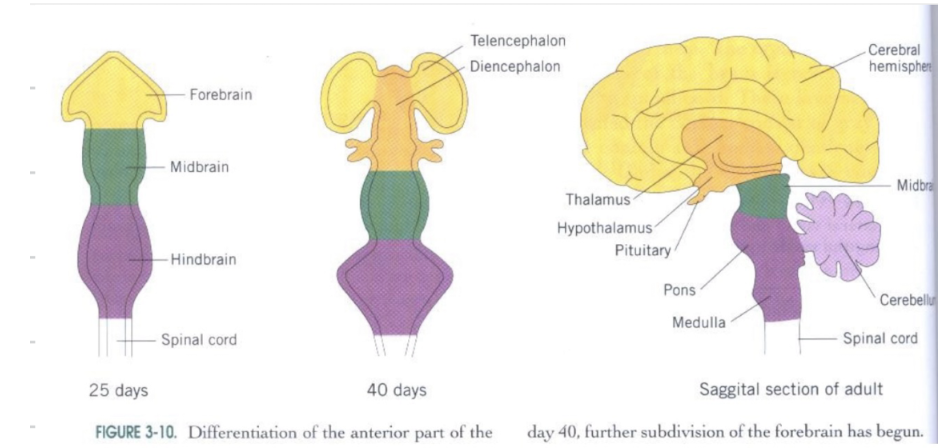
Forebrain components
Telencephalon and Diencephalon
Telencephalon purpose
performs higher mental functions, philosophizing, processing sensory information, and motor control
Telencephalon components
Cerebral cortex (includes 4 lobes), basal ganglia, limbic system - amygdala and hippocampus, nucleus accumbens, olfactory bulb
Brain lobes
Frontal lobe (higher thoughts and self control)
Parietal lobe
Occipital lobe (visual processing)
Temporal lobe (hearing and speech)

basal ganglia
important for planning movement and postural adjustmnets
limbic system
Amygdala: emotional behaviors like rage, fear, repulsion
Hippocampus: memory making and storage
Story of HM, Henry Molaison
HM had seizures so they took out his hippocampus and from then on he could not make memories
Amygdala
Emotional behaviors like rage, fear, repulsion
Hippocampus
Memory making and storage
Nucleus accumbens
reward/pleasure circuit
Olfactory bulb
Processes information about smells
Diencephalon purpose
Major relay, integration, and control center
Diencephalon components
Thalamus - lateral and medial geniculate, and hypothalamus
thalamus
processes and sorts sensory and motor information before sending it to the brain-telencephalon
includes lateral and medial geniculate
lateral geniculate
first step in visual processing from the retina
medial geniculate
Processing from the ears
Hypothalamus
regulates hormone secretion by the pituitary gland
Important for homeostasis - triggers behaviors to keep consistent (temp, bp, hydration, salt)
Midbrain
Mesencephalon
Mesencephalon components
inferior and superior colliculi, and substantia nigra
Inferior colliculus
Auditory information
Superior colliculi
Visual information
Substantia nigra
for posture and motor control (damaged in Parkinsons)
Hindbrain components
Metencephalon and Myelencephalon
Metencephalon components
Pons and cerebellum
Pons
Superhighway of pathways. It communicates with cerebellum
Major tract of axons especially going to and from the cerebellum
Includes collections of ganglia that are important in basic functions like sleep, wakefulness, and respirations
Cerebellum
Coordinating movement
Highly convoluted and important for muscle tone, balance, carrying out movements smoothly, and motor memory
Takes information from the telencephalon (what to do) then it corrects what you did to make motor memory
Myelencephalon components
medulla
Medulla
Continuous with the spinal cord and controls basic functions like bp, coughing, laughing, and heart rate
Cerebellar cells
Purkinje cells, mossy fibers, and climbing fibers
Purkinje cells (input and output?)
Input: receive information on what was the intended movement (mossy fibers) and what was the actual movement (climbing fibers)
Output: corrections if needed of movement to match what was intended - correct motor error
Output is inhibitory of deep cerebellar nuclei and from there ends in the cerebral cortex
Mossy fibers
Axons coming from the pons and bring information on intended movement
Synapse with granule cells and then to parallel fibers to reach purkinje
Climbing fibers
come from medulla and bring information on the actual movement
Purkinje cell drawing

Spinal cord transverse section

Gray matter
cell bodies of neurons, including the dorsal horn and ventral horn
dorsal horn
sensory
ventral horn
motor
white matter
myelin surrounding axons
central canal
Canals/ventricles are filled with cerebrospinal fluid (CSF)
Illustrates the tubular origins of the CNS
In the brain the central canal expands and gets distorted but is there as the brain ventricles
Meninges
supports the CNS, has places to pull out CSF and return fluid to the circulatory system, and contains the choroid plexus
Choroid plexus
full of capillaries that produce the CSF
Endothelial cells of capillaries
In the innermost layer of a blood vessel and joined by tight junctions - BBB
Blood Brain Barrier
tight regulation of what goes form the blood to neural tissue of the brain
What can cross the blood brain barrier?
Lipid soluble items, and items attached to glucose to pass through glucose pumps, and a new method includes attaching to nanobeads
Astrocytes and the meninges
Astrocytes extend “feet” that sit on capillaries. These can secrete molecular signals to capillary cells to tighter or loosen the tight junctions.
Cerebrospinal fluid
Circulates within and around the CNS held there by a set of meninges
Contains ions/salts in proper concentrations for good neural function, also a few proteins
Components of the peripheral nervous system
somatic and autonomic
components of autonomic nervous system
sympathetic, parasympathetic, enteric
sympathetic nervous system
Fight, flight, fright. Short preganglionic fiber terminates in sympathetic ganglion, using acetylcholine (ACh) as a neurotransmitter. It makes a synapse with postganglionic fiber cell, which extends out to the organ or tissue where it makes a synapse using norepinephrine (NE) neurotransmitter
Parasympathetic nervous system
Cell bodies in brain or lower spinal cord
Send a long preganglionic fiver to a ganglion out near the organ innervated synapse with the postganglionic fiber neuron, using ACh as the neurotransmitter. The postganglionic fibers neuron releases ACh at the organ
Enteric nervous system
Network on the gut with many ganglia
Large number of neurotransmitters and many different types
Subject to regulation by sympathetic and parasympathetic nervous systems
Ion channel proteins
Leakage channels: open most of the time
Some open under certain conditions
Ligand gated channels: a molecule binds to them
Voltage gated channels: membrane has a voltage change
Stretch sensitive channels: open when they are physically stretched or moved
Ion channel proteins twist and squirm due to thermal energy and they will randomly open and close at all times
So when we say the channels are open, we mean that the probability that one of these channels is open is very high