physics ch 1 terms
1/18
There's no tags or description
Looks like no tags are added yet.
Name | Mastery | Learn | Test | Matching | Spaced | Call with Kai |
|---|
No analytics yet
Send a link to your students to track their progress
19 Terms
sonography
the use of ultrasound in medical anatomic and flow imaging
doppler ultrasound
the detection, quantization, and evaluation of tissue motion and blood flow by using the doppler effect with ultrasound
ultrasound
sound that is higher in pitch than the range of human hearing
sonographer
an allied health professional who acquires the image
transducer
probe; generates the ultrasound pulses and recieves the returning echoes
intrument
system
sonologist
physician who interprets the images
image
a reproduction, representation or imitation of the physical form of a person or object
gray-scale image
produced by the brightness of each dot corresponds to the echo strength
operating principle 1
describes how ultrasound images are formed line by line sending out a sound pulse and recieving itsechoes one at a time, creating a dot on the screen that develops a scan line
scan line
a series of dots; one line of echo information
linear image
an anatomic image presented in a rectangular display
sector image
an anatomic image presented in a pie-slice format
how to display linear image
cross sectional images are produced with vertical parallel scan lines that are so close together that they cannot be identified individually
linear image
what is this?
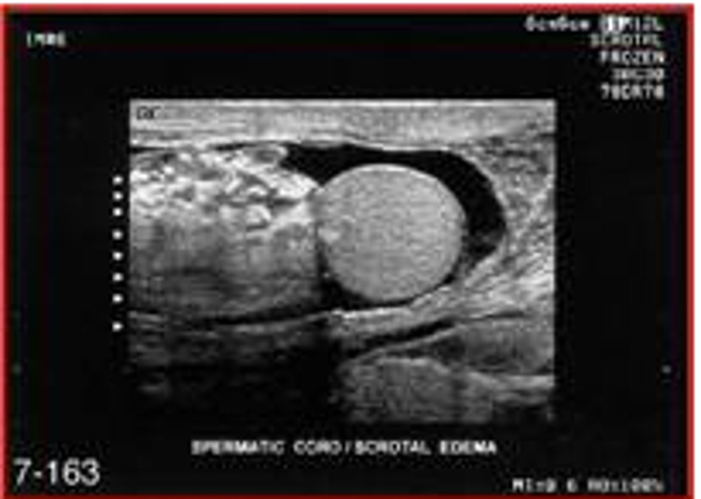
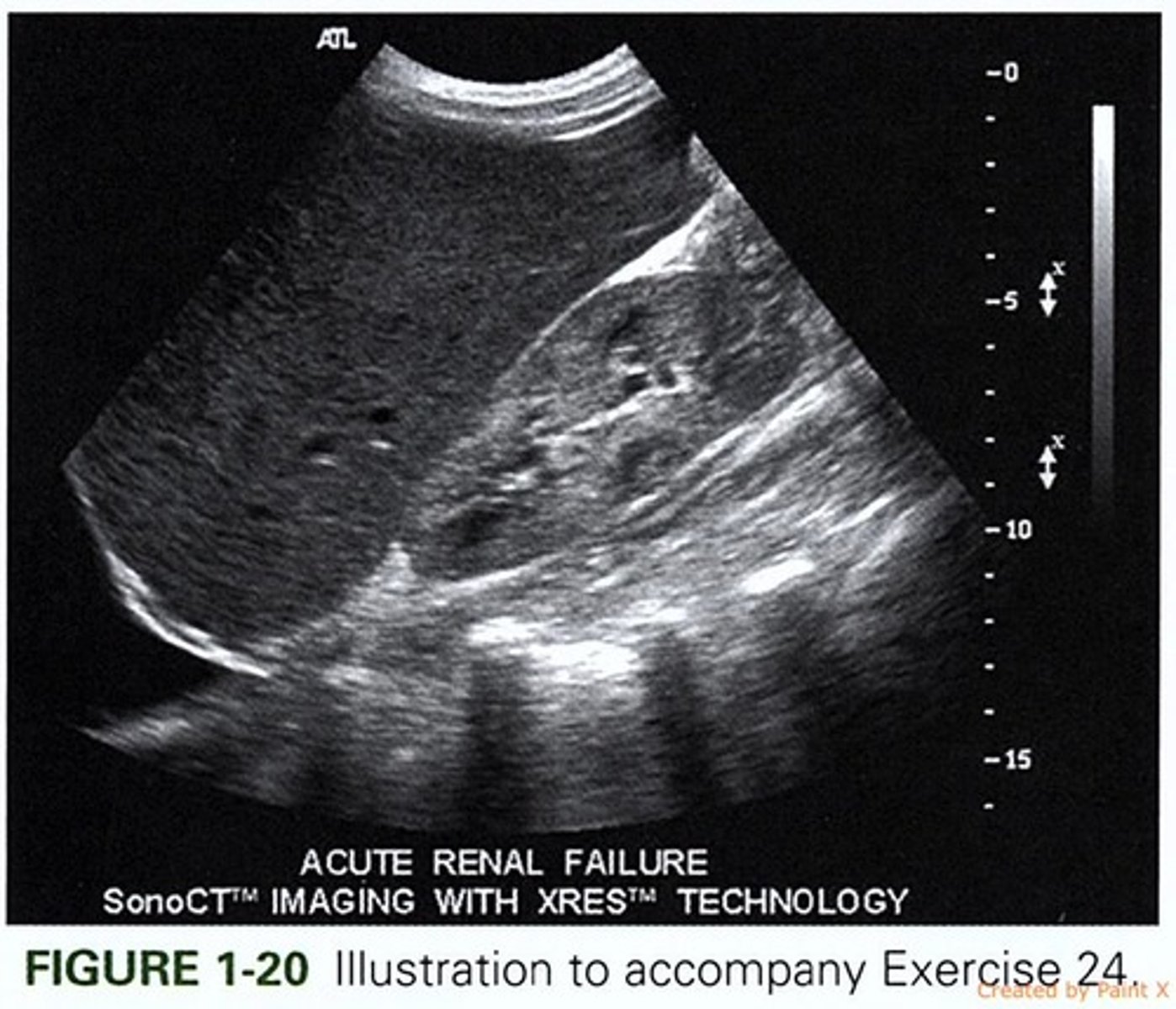
sector image
what is this?
operating principle 2
involves the transducer sending unfocused sound pulses into the body and then receiving the echoes to form an image; more sophisticated
volume imaging
the conversion of 2D imaging into 3D
doppler information is presented in ....
audible, color-doppler, and spectral doppler forms