Lecture 19: Eye Retina Transduction
1/35
There's no tags or description
Looks like no tags are added yet.
Name | Mastery | Learn | Test | Matching | Spaced |
|---|
No study sessions yet.
36 Terms
What is the dioptric system?
All the transparent media b/w outside world + retina.
Form the image.
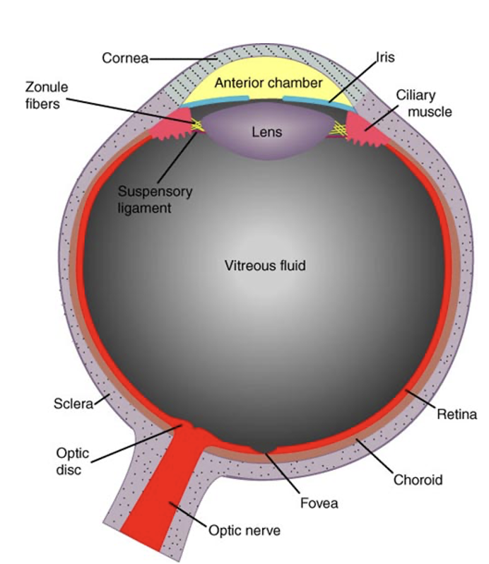
What is the goal of the dioptric system?
To bring parallel light rays to focus on retina.
What 2 important structures does the dioptric system include + function?
Cornea = greatest amount of light refraction occurs at cornea.
Crystalline lens = changes shape (focus power) by contraction/relaxation of ciliary muscle.
What are 2 specializations of the retina?
Fovea = point of highest acuity
Optic disc = blind spot; where axons of the optic nerve (axons of retinal ganglion cells) penetrate.

Draw the cell body layers of the retina.
What does the outer nuclear layer, inner nuclear layer, and ganglion cell layer include?
Outer nuclear layer = cell bodies of rods/cones
Inner nuclear layer = cell bodies of bipolar cells, horizontal cells, amacrine cells
Ganglion cell layer = cell bodies of ganglion cells
What are the synaptic layers of the retina?
Outer plexiform layer = synapses formed by photoreceptor axon terminals, dendrites of bipolar cells, processes of horizontal cells
Inner plexiform layer = synapses b/w bipolar + ganglion cells
What does the fovea contain?
ONLY CONES
All cell bodies of inner plexiform + ganglion cell layer are swept to the side.
Where do foveal cones synapse?
Foveal cones synapse on bipolar cells to the sides of the fovea.
Those bipolar cells synapse on ganglion cells along edges of fovea.
Why are images falling on fovea seen in great detail?
In part bc of high density + small size of cones in fovea.
But also requires the specialized midget cone circuitry.
Outside of fovea, images falling on peripheral retina are terrible acuity bc of low density of cones.
Doesn’t look this way bc we are always moving our eyes + brain creates illusion of continuity of detail
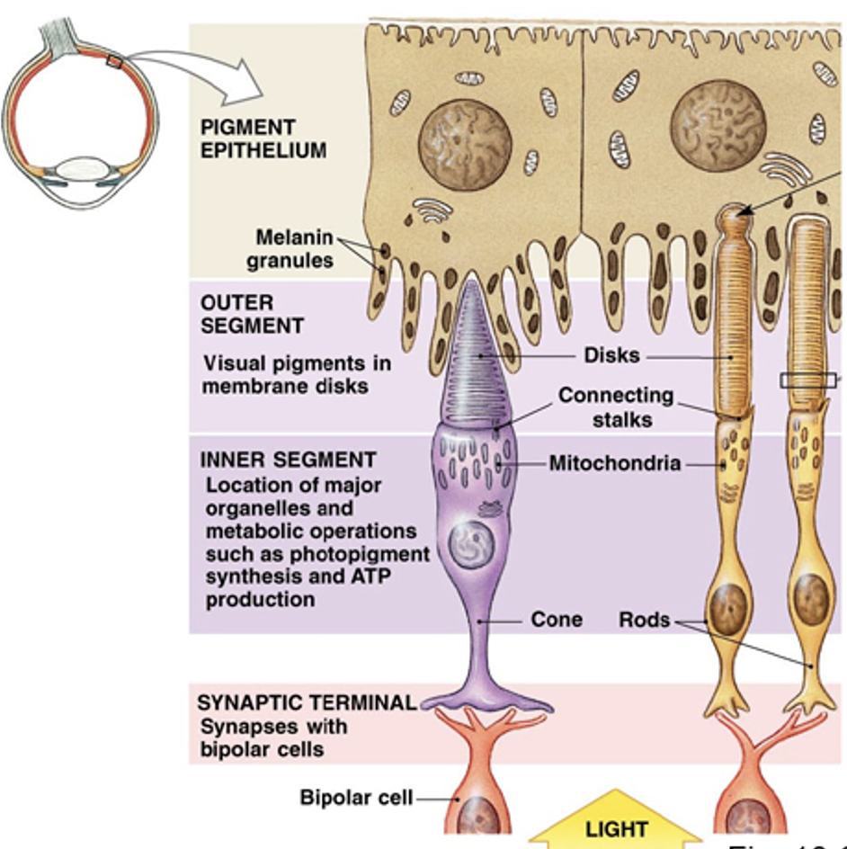
What do outer segments do + contain?
All photon capture occurs in outer segments of rods + cones.
Contain stacks of discs (containing opsins) of photoreceptors.
What are the discs of rods like?
Free-floating, independent structures.
Rod outer segments are longer to increase probability of photon capture.
What are the discs of cones like?
Invaginations of the plasma membrane

Cone density in retina?
Peak at fovea + declines to very low levels for rest of retina.
Rod density in retina?
Non-existent in fovea + peak in a ring 20° away from fovea + decline slowly for the rest of retina.
Rod-rich ring has higher density than fovea, why not better acuity?

What is the duplex retina?
We have 2 retinae for the price of 1:
Fovea (cone-rich)
Rest of retina (rod-rich ring)
What is the retinal pigment epithelium?
Rods + cones are surrounded by protrusions of the retinal pigment epithelium. They have 3 functions:
Absorbs photons that pass through the retina (keeping them from reflecting through the retina).
Contains the enzymes that permit photoreceptors to continue to respond to light.
Consumes shed discs + prevents them from becoming a barrier to oxygen diffusion.
Explain phototransduction
Light makes photoreceptors hyperpolarize. Dark makes them depolarize.
Dark current = caused by influx of Na+ and Ca2+ through cGMP-gated channels; depol. to -30mV
Photon capture —> cascade that closes cation channels —> hyperpol. (more photons captured, more channels closed)
Short distance b/w outer segment + axon terminal —> change in Vm reaches axon terminal w/ little reduction in magnitude.
How do cGMP-gated channels control photoreceptor responses to light?
In the dark, high [cGMP] keeps CNG channels open, allowing Na⁺/Ca²⁺ influx (dark current). Light lowers cGMP via PDE, causing these channels to close. Because the channels respond over a narrow cGMP range (open at ~100 μM, closed at ~10 μM), small decreases in [cGMP] produce large hyperpolarizations, making photoreceptors very sensitive to light.
How do transduction channels differ in photoreceptors vs. hair cells?
Hair cell channels are mechanically-gated by stereocilia movement, opening directly with sound-evoked tension.
Photoreceptor channels are ligand-gated CNG channels, opened by intracellular cGMP.
Light triggers biochemical steps that hydrolyze cGMP, causing these channels to close, which is why all vertebrate photoreceptors hyperpolarize to light.
What makes up a photopigment in rods and cones?
Photopigment = opsin (a GPCR) + chromophore (11-cis retinal) covalently bound together.
Opsin determines wavelength sensitivity, and the chromophore is the light-absorbing molecule.
How does photon absorption activate the phototransduction cascade?
When 11-cis retinal absorbs a photon, it isomerizes + becomes all-trans retinal, changing the shape of the opsin (a GPCR).
Activated opsin then activates transducin (a G-protein), starting the biochemical steps that ultimately close cGMP-gated channels and hyperpolarize the photoreceptor.
How does the 11-cis → all-trans change activate the photopigment?
Opsin (a GPCR) holds 11-cis retinal tightly in its binding pocket. When retinal becomes all-trans, it no longer fits the same way, forcing opsin to shift into its active conformation (R*).
This active opsin then activates transducin (a G-protein) + the chromophore eventually detaches so it can be recycled by the visual cycle.
What type of proteins are opsins, and what is their basic structure?
Opsins are GPCRs (G-protein–coupled receptors) that span the disc membrane 7 times.
Contain a binding pocket formed by their TM loops, where the chromophore 11-cis retinal is held.
Humans have 4 opsins: rhodopsin (rods) and three cone opsins (S, M, L).
Why do different cone opsins absorb different wavelengths of light?
Differences in amino acids within the opsin’s transmembrane regions determine which wavelengths of light are most easily captured by chromophore.
Produces blue (S), green (M), and red (L) cone sensitivities.
What is the sequence of molecular events in the phototransduction cascade?
Light activates opsin (a GPCR, acts as light-activated GEF) → opsin activates transducin (G-protein) → transducin activates PDE (phosphodiesterase, enzyme) → PDE hydrolyzes cGMP → reduced cGMP closes cGMP-gated Na⁺/Ca²⁺ channels → photoreceptor hyperpolarizes.
How does a single photon produce a large electrical response in photoreceptors (amplification)?
One activated opsin (R*) activates hundreds of transducins, and each transducin–GTP activates PDE, which hydrolyzes many cGMP molecules. The drop in cGMP closes numerous cGMP-gated Na⁺/Ca²⁺ channels, producing a big hyperpolarization from just one photon.
What happens when Ca²⁺ drops during phototransduction, and how does it contribute to light adaptation?
Closure of CNG channels in light reduces Ca²⁺ influx → intracellular Ca²⁺ falls.
Low Ca²⁺ activates guanylate cyclase (↑ cGMP production) and relieves inhibition of GRKs, speeding opsin shutoff.
Result: faster recovery + reduced photopigment activity → light adaptation.
How does Ca²⁺ regulate GRK1, and what is the effect on opsin?
High Ca²⁺ (dark): Ca²⁺–recoverin inhibits GRK1 → opsin remains active longer.
Low Ca²⁺ (light): recoverin releases Ca²⁺ → GRK1 becomes active → phosphorylates opsin → arrestin binds → opsin stops activating transducin.
This shortens opsin’s active lifetime.
What differences in GRKs explain why rods show larger hyperpolarization per photon than cones?
Rods have only GRK1, a slow kinase → opsin stays active longer → more transducin and PDE activated → larger hyperpolarization.
Cones have GRK1 + fast GRK7 → opsin is shut off quickly → fewer transducins activated → smaller response per photon.
What Ca²⁺ exchanger do photoreceptors use, and how does it work?
Photoreceptors use an NCKX exchanger (Na⁺–Ca²⁺–K⁺ exchanger).
Moves 1 Ca²⁺ out of the cell in exchange for 4 Na⁺ in and 1 K⁺ out, using the Na⁺ and K⁺ gradients as the driving force.
This exchanger can remove Ca²⁺ even when Ca²⁺ levels are extremely low (≈100 nM in bright light).
Why do photoreceptors need NCKX instead of a normal NCX exchanger?
A normal NCX exchanger reverses at low Ca²⁺, bringing Ca²⁺ into the cell.
But photoreceptors must keep Ca²⁺ falling during light exposure.
NCKX never reverses, always pumping Ca²⁺ out, allowing Ca²⁺ to drop when CNG channels close.
This Ca²⁺ decline drives light adaptation, activating guanylate cyclase and relieving inhibition of GRKs.
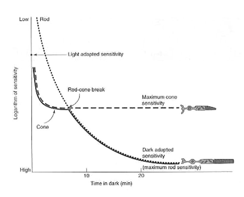
What happens during dark adaptation, and how do rods and cones differ?
Dark adaptation = the increase in visual sensitivity after entering darkness.
Cones adapt quickly but only slightly; rods adapt slowly (≈20 minutes) but achieve much higher sensitivity.
The “rod–cone break” marks the point where rod sensitivity surpasses cone sensitivity.
What biochemical steps are required for photoreceptors to recover sensitivity in the dark?
Both rods and cones must regenerate 11-cis retinal from all-trans retinal after light exposure.
Rods depend on RPE65 in the retinal pigment epithelium to convert all-trans → 11-cis, followed by diffusion back to the outer segment.
This slow chromophore regeneration limits rod recovery time and sets the pace of dark adaptation.
What are Müller cells?
MMost common type of supporting cell in the retina; their cell bodies are in the inner nuclear layer but their processes span the entire retina.
Resemble astrocytic function in brain + SC.
Why do Müller cells speed up cone dark adaptation?
Müller cells supply chromophore.
Take in all-trans retinal, convert it to 11-cis retinol + release that compound.
Only cones have the means to take up 11-cis retinol + have the enzyme to convert it to 11-cis retinal.