Biotech 2 McBride
 Call Kai
 Learn
Learn Practice Test
 Spaced Repetition
 Match
Match Flashcards
 Knowt Play
1/16

There's no tags or description
Looks like no tags are added yet.
Name | Mastery | Learn | Test | Matching | Spaced | Call with Kai | Chat |
|---|
No analytics yet
Send a link to your students to track their progress
17 Terms
ALT & AST tests
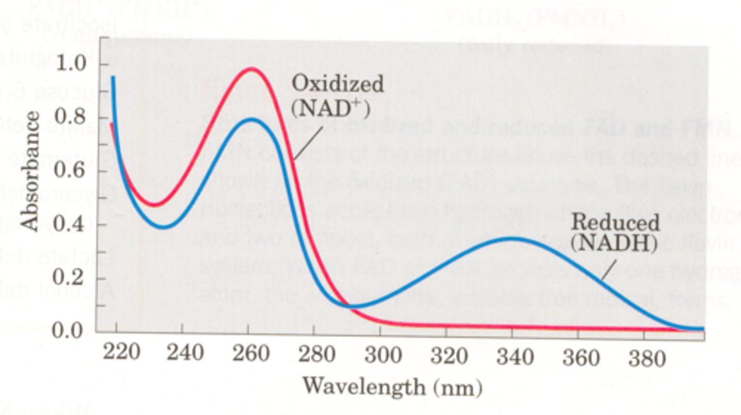
Blood tests. Use NADH -> NAD+ to test how much because it is measurable. NAD has no absorbance, but nadh has absorbance at 340 nm. Aspartate turns into Malate through Malate dehydrogenase and alanine turns into lactate through lactate dehydrogenase
How do centrifuge tests work?
Step 1: disrupt the cell membranes of intact cells to form a homogenate
step 2: centrifuge the homogenate at low speed to yield a pellet consisting of heavy material and lighter supernatant (solution)
NOTE: As you increase the speed/time centrifuging, more and more protiens will become part of pelet on bottom.
Order of pelet creation in centrifuge tests
1) Whole cells, nuclei, cytoskeletons
2) mitochondria, lysosomes, peroxisomes
3) microsomes, small vessicles
4) ribosomes, viruses, large macromolecules
Centrifuge tests (Speed/Velocity)
Start from the top. Different molecules move down solution over different times
Centrifuge tests (Equilibrium)
All mixed in, and steep gradient will separate in terms of density
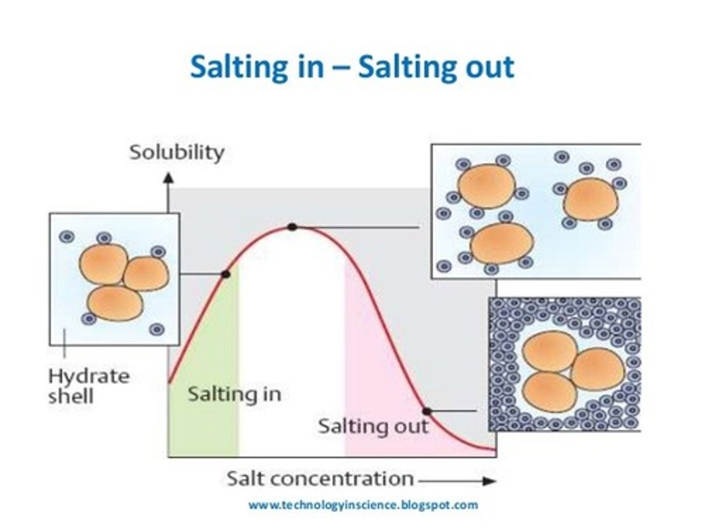
Salting out
Different molecules can be separated at a high enough concentration of salt, because they lose solubility and become insoluble
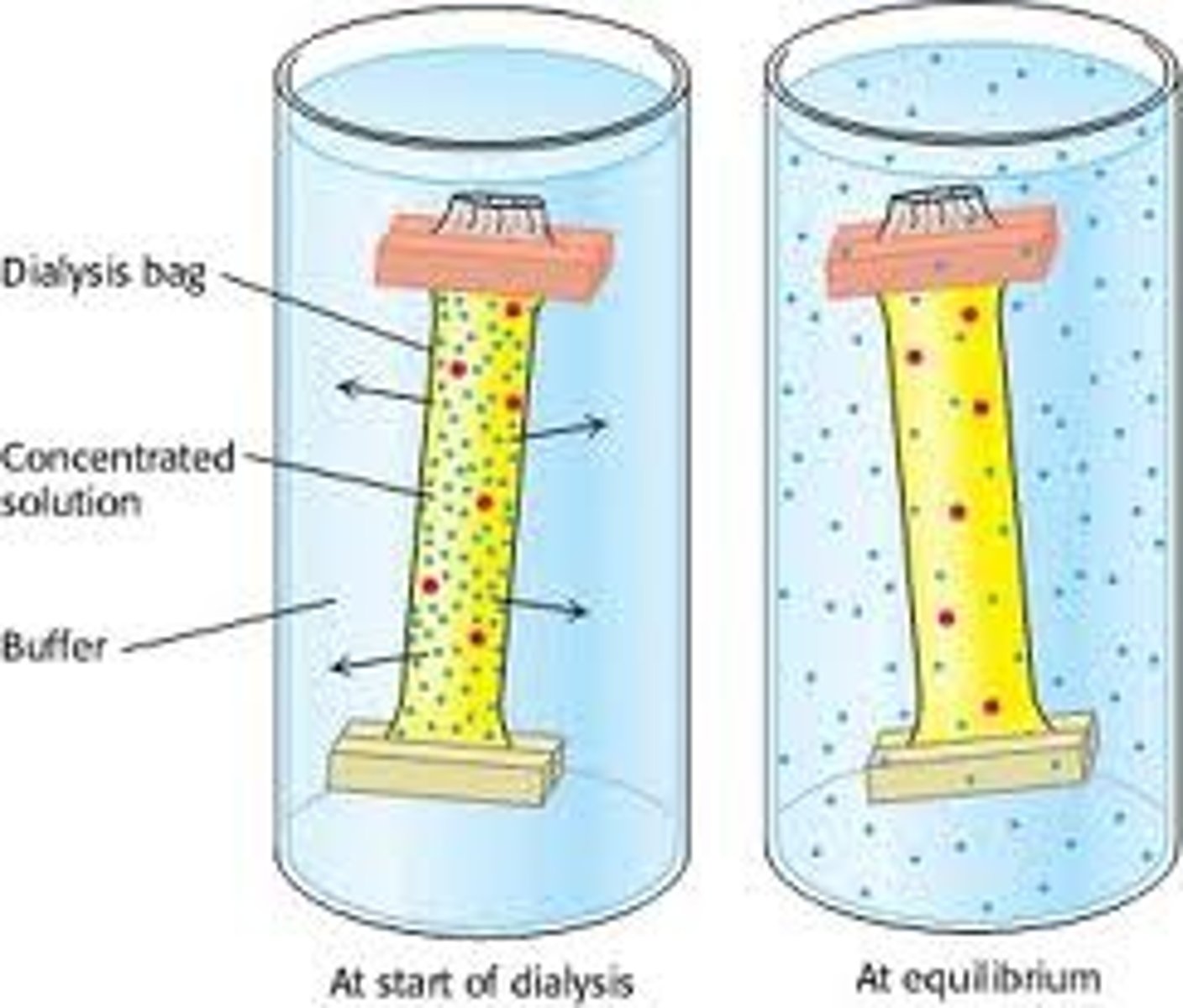
Dialysis
Used to remove a molecule from salt to separate it. Semi-permeable membrane that allows the flow of water and small molecules (like salt) but not large molecules and proteins.
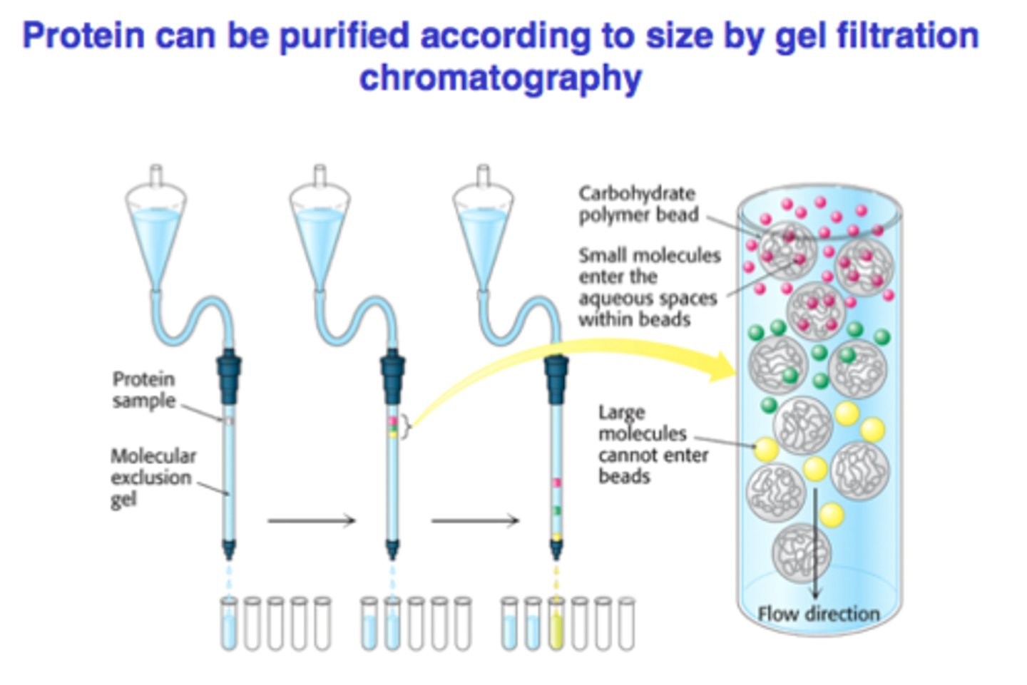
Gel filtration chromatography
Porous beads are put into a column. Small molecules get stuck inside beads and take longer to elute, while larger proteins pass by
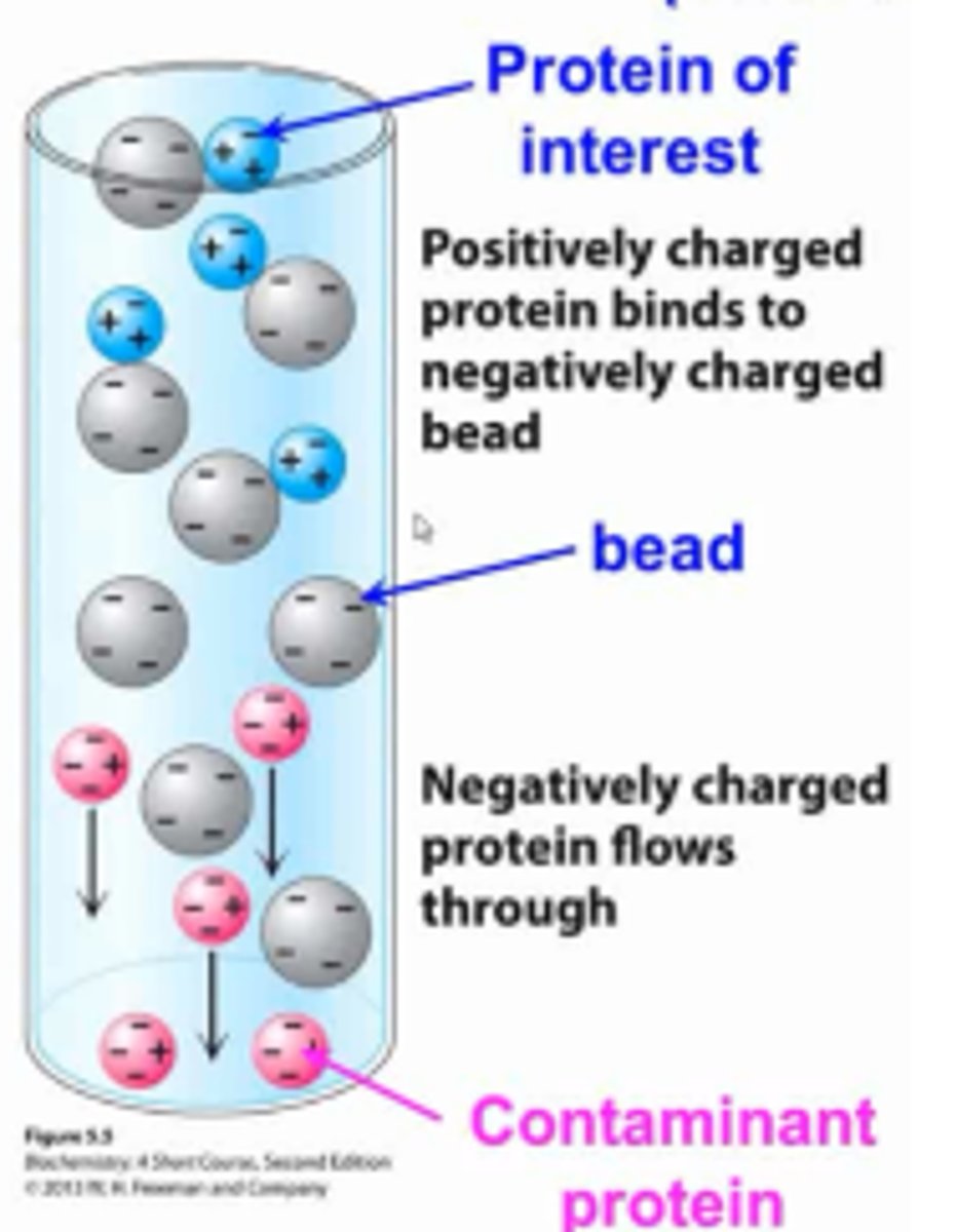
Ion exchange chromatography
Molecules pass through beads with a + or - charge and bind to them. Then, salt out at the end to remove them from the beads
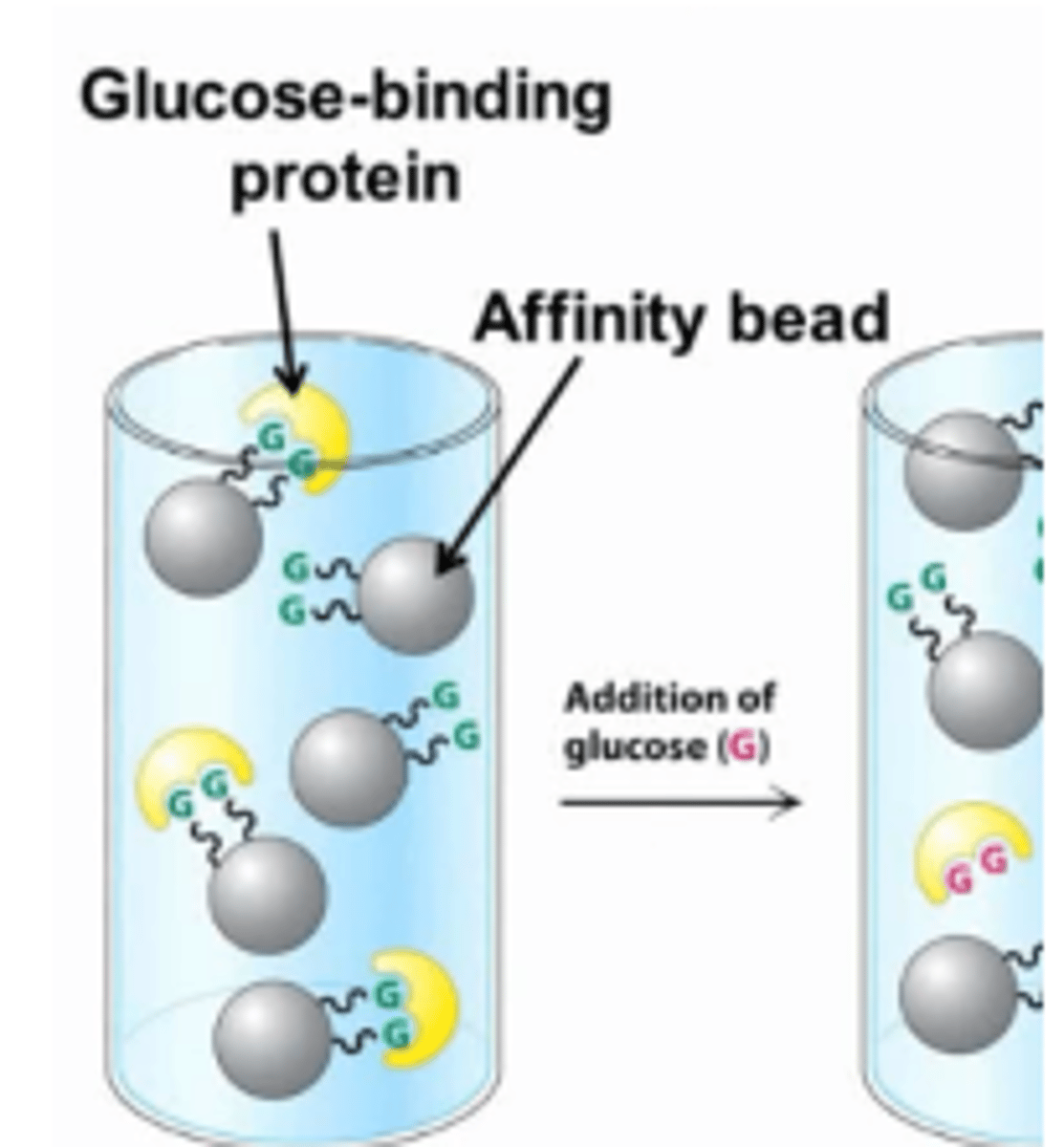
Affinity chromatography
Beads have something that a specific protein would bind to. Afterwards, flush with a ligand to take off the proteins
HPLC
Can decrease time and increase accuracy of all the chromatographies
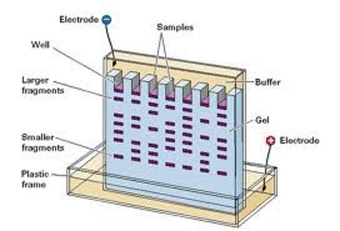
Gel-electrophoresis (Electrical)
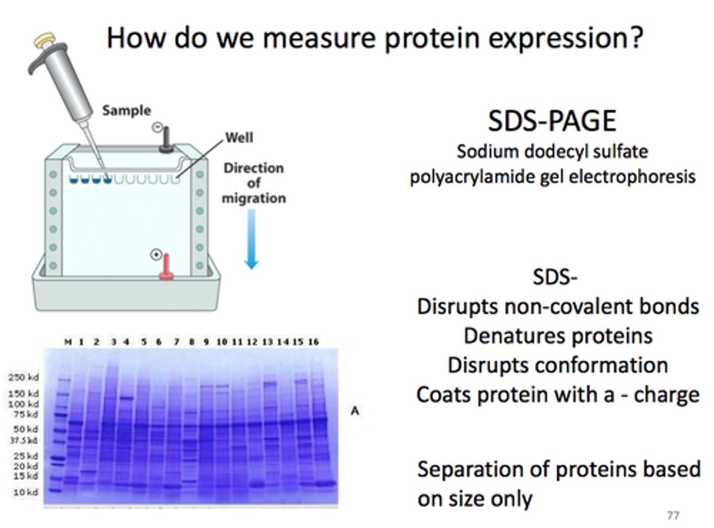
(- on top, + on bottom) Puts proteins in a gel made of polyacrylamide. Run electric charge, and proteins will separate based on charge and size down
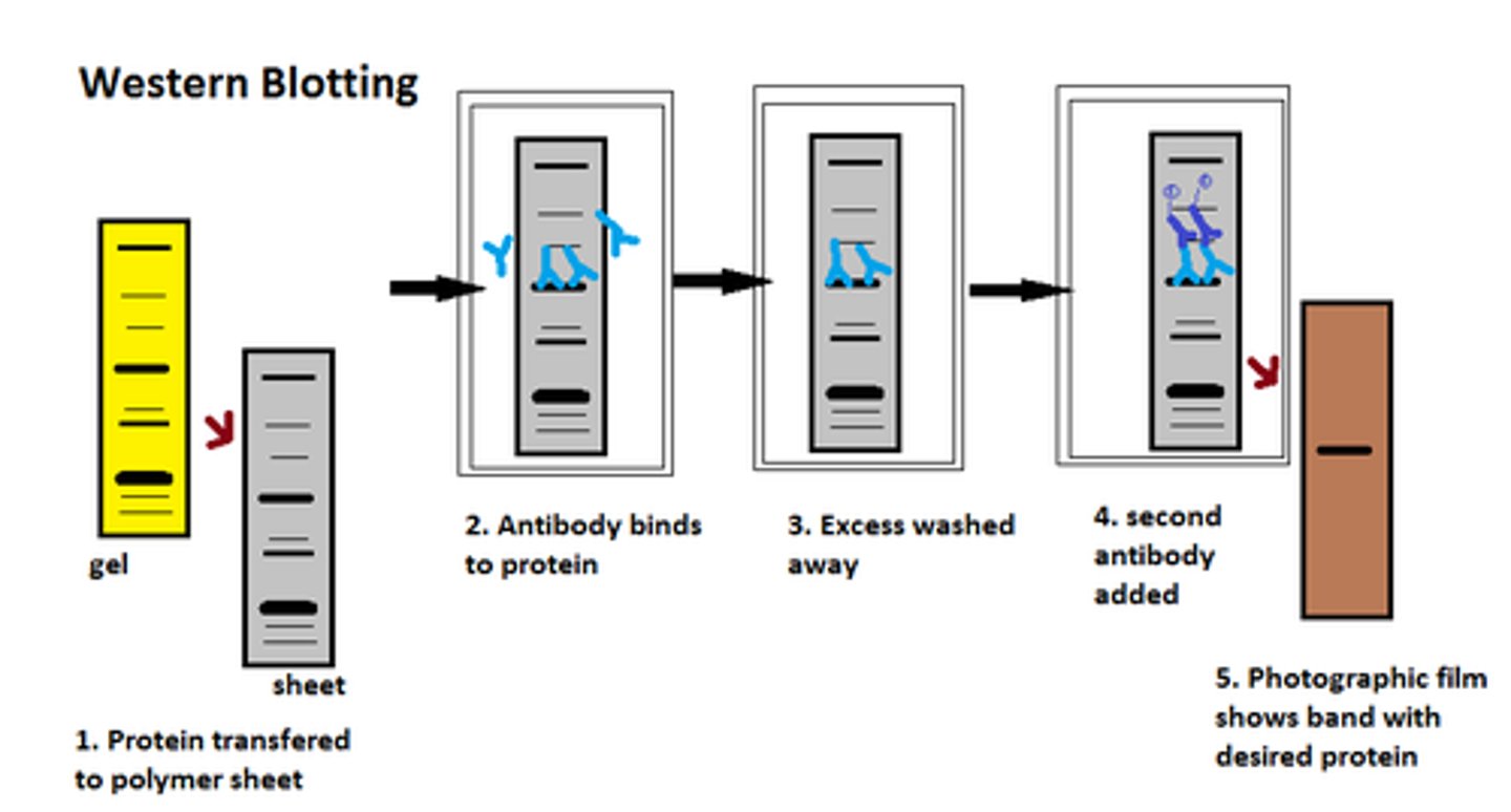
Western Blot
Get proteins after putting them through SDS page. Add primary antibody for protein, then secondary antibody that binds to primary with fluorescent part. Can be used to detect oncogenic fusion proteins, like SS18-SSX
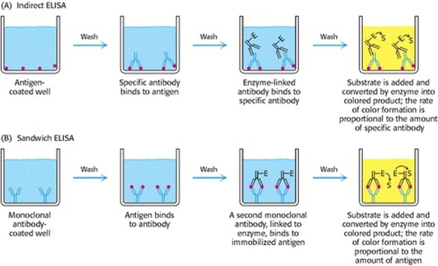
Enzyme-Linked Immunosorbent Assay (ELISA) 2 TYPES
1) Indirect Elisa - have ANTIGEN coated to wall of test tube. Antibody 1 binds to anitgen. Antibody 2 with enzyme binds to anitbody 1. Substrate is added that makes enzyme change color. Measures antibody concentration.
2) Sandwich ELISA - have ANTIBODY coated to wall of test tube. Antigen binds to antibody. Antibody 2 with enzyme binds to anitgen bound to antibody. Substrate is added that makes enzyme change color. Measures antigen concentration.
Difference between western blot and ELISA
Western blotting requires the proteins to first be separated by size using gel electrophoresis before they are transferred to a membrane for detection. In contrast, ELISA detects and quantifies the target protein directly from a liquid sample within a coated microtiter plate well without needing prior size separation.
Mass spec
1) Converts peptides into gas and reads m/z.
2) consist of three components: an ion source, a mass analyzer, and a detector.
3) Peptide mass fingerprinting can identify individual proteins within complex mixtures
Gel-electrophoresis SDS-PAGE
Uses SDS to denature proteins and bind to them. Makes all the proteins the same shape and charge, allowing them to be separated only based on weight