Part D: Surgery STATE
 Call Kai
 Learn
Learn Practice Test
 Spaced Repetition
 Match
Match Flashcards
 Knowt Play
1/45

There's no tags or description
Looks like no tags are added yet.
46 Terms
1. Diseases of the eyelids, conjunctiva and third eyelid
Conditions:
Eyelid → entropion, ectropion, laceration, eyelid neoplasia
conjunctival → conjunctivitis, dermoid/choristoma
Third eyelid → prolapse/cherry eye, third eyelid flap
1) Eyelid Disorders
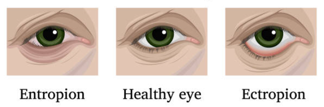
Entropion (Inward Rolling of Eyelid Margin)
eyelid margin rolls inward → hair/lashes to rub directly on cornea. Common in wrinkled puppies, like shar-peis, pugs, or in cats after chronic viral herpes/chlamydia conjunctivitis.. Can be sec. to ocular pain/ulcers or by scarring..
CS: Epiphora (excessive tearing), corneal ulcers, vision loss, blinking, discharge
Medical tx: Topical antibiotics (terramycin, erythromycin), procaine penicillin, or try topical antiviral drops in cats.
Surgical Techniques:
Puppies under 20w - dont do permanent surgery, just do temporary tacking with vertical lembert sutures (roll margin out)
Adults: Hotz-celsus procedure (permanent) - cutting out a strip of skin of lower eyelid, then closed w/simple interr.
Alternative: Permanent lateral tarsorrhaphy - remove v-shape part at lateral canthus (corner) to make the eye opening smaller or do arrowhead (removing arrowhead shape in corner, then close (pulls sideways)
Ectropion (Outward Drooping of Lower Eyelid)
→ Congenital in giant/hound breeds (Bloodhound, St. Bernard, Great Dane). → affects tear drainage, leads to excessive tearing, chronic keratitis. Fixed via a lateral wedge excision, V-Y plasty or modified kuhnt procedure..
Surgical Techniques:
Lateral Eyelid Wedge Excision: removing a full-thickness V-triangle out of the lid margin near the corner (lateral canthus), then close in 2 layers (simple continuous deep, simple interrupted skin), w/knots buried to prevent corneal irritation.
V-Y Plasty: Make a V-shaped incision 1 mm below the eyelid margin, excise underlying scar tissue, elevate skin flap, and close in a Y-shape.
Modified Kuhnt: eyelid is split like a sandwich into an outer skin layer and inner pink layer, we cut a V wedge only out of the inside layer (conjunctiva/tarsus) to tighten the structural wall. The skin on the outside is left intact, just stretched and sutured.
Laceration:due to →Trauma (bite wounds, sharp objects) → “laceration” - needs to be fixed immediately to protect cornea and maintain normal blinking. Sec. healing → fibrosis and corneal scarring (fibrosis ruins eye tear film!)
Surgery:
Direct Reapposition/closure (<1/3 margin loss): Align eyelid margin w/figure-8 suture. Close skin w/simple interrupted if over 1/3 margin loss = use flaps/grafts.
Temporary Tarsorrhaphy: if eyelid function/blinking is damaged post-op. → complete closure of lids to protect eye.
Post-Op: Artificial tears + topical and systemic antibiotics.
Eyelid Neoplasia
eyelid tumors are usually benign meibomian gland (the oil gl. lining eyelids) adenomas in dogs, but cats get mostly malignant SCC.
Dg: History, signalment, fine needle aspiration (use eyelid plate), chest X-rays (to check for pulmonary metastasis). Treat secondary infections prior to surgery.
Surgical removal:
if mass takes up less than 1/3 of the eylid length, a simple wedge excision and figure-8 suture, close skin with simple interrp.
If it takes up more than 1/3 on lower lid, we can do H-plasty pedicle advancement flap - removing square segment, then trimming little tissue triangles, then advance flap to margin..
If it at the lateral corner (canthus), we use a semi-circular flap cut twice the length of the defect. remove mass, make incision, triagnular exciion - advance skin flap, align w/figure-8.
Post-Op: Always submit tissue for histopathology.
Medical Tx options include chemo, radiation, or cryotherapy.
Eyelash & Other Margin Anomalies
Ectopic Cilia: hair growing straight through inside conjunctiva from meibomian gland, poking the cornea directly. Tx: wedge excision of cilia with conjunctiva, electro-epilation, or cryotherapy.
Distichiasis: Extra row of hair directly from Meibomian gland openings along lid margin. Tx: Cryosurgery or electro-epilation.
Trichiasis: normal hair pointing the wrong way. Tx: Surgical excision of facial/nasal folds or lid re-alignment.
Blepharitis: eyelid inflammation (bacterial, fungal, parasitic, or allergic).
Hordeolum: Localized suppurative gland infection (External = Zeis/Moll glands; Internal = Meibomian gland). Tx: Warm compresses - for relief, topical antibiotics/steroids (maxitrol)
Ankyloblepharon: eyelids does not open within 15 days post-birth. Tx: Manual separation and warm flushing.
2. Conjunctival Disorders
Condition | Etiology & Features | Surgical / Medical Treatment |
Dermoid (Choristoma) | Congenital skin tumor (normal skin tissue containing hair/glands located on conjunctiva/cornea). | Superficial conjunctivectomy or keratectomy. To surgically remove mass, a temporary stay suture is set through mass, lifting mass up for easier excision. |
Chalazion | Non-infectious, chronic granulomatous blockage of Meibomian/tarsal gland → Bump on eyelid. | Surgical incision and drain it. |
Neoplasia of conjunctiva | Dogs: Hemangioma, limbal melanoma. Cats: Lymphosarcoma (LSA), SCC. | Localized surgical excision (benign/small under local block; large require general anesthesia). |
Symblepharon | Pathological adhesion of conjunctiva to conjunctiva or cornea. Common in cats (post-FHV-1) | Surgical breakdown (indicated only if vision, drainage, or eyelid motility is impaired). can be keratectomy (surgical dissection, preventing re-adhesion → membrane graft/contact lens). |
3. Third Eyelid Disorders
a) Cherry Eye (Third Eyelid Gland Prolapse)
= when tear gland tucked inside a dog`s third eyelid pops out of place → red cherry-like mass in the inner corner of eye. Occurs when the anchor of the gland to the periorbita is weak/loose (laxity of CT). Common in Bulldogs, shi tzus.
Pre-Op: Topical antibiotic-corticosteroid combination (if cornea is intact) to reduce inflammation.
Treatment: Replacement, NOT excision (removal leads to Keratoconjunctivitis Sicca aka dry eye)
Morgan Pocket Procedure: tucks the popped-out gland back into the pocket - by 2 conjunctival incisions on opposite sides of gland → suture opposing margins together w/simple continuous to enclose gland within a pocket → make sure to tie starting and finishing knot on the side facing away from the eye to prevent irritation.

b) Plasma Cell Infiltration (Plasmoma / Pannus of 3rd Eyelid)= Immune-mediated inflammation common in German Shepherds; causes depigmentation, thickening, and irregular surface of the third eyelid margin. Tx: Lifetime topical immunosuppression (corticosteroids, cyclosporine, or tacrolimus).
Cartilage Deformity (Everted / Inverted Cartilage)= the free border of 3rd eyelid rolls outwards. Common in large breeds. Tx: Surgical excision of the deformed/bent portion of the cartilage stem.
Third Eyelid Flap (Procedure Note/surgical technique linked)
Indication: Temporary mechanical protective bandage for cornea after trauma like superficial non-healing/indolent ulcers or bullous keratopathy. Contraindicated in deep ulcers, descemetoceles, or corneal perforations (creates excessive pressure and blocks monitoring).
Technique: Suture passed from dorsolateral upper eyelid conjunctiva through outer surface of third eyelid (around cartilage, avoiding posterior surface), back out upper lid, tied over a stent. Remove after 14 days.
Enucleation: the complete removal of an eyeball from the eye socket. etio: malignant tumors, severe trauma (fight), blindness, glaucoma. Whole eye out, surgeon cuts lateral canthus, dissects out the globe, ligates the optic nerve, pulls out the entire third eyelid and gland → snips off the eyelid margins, stitches it up.
NOTES:
very briefly about the most common disorders of eyelids - wanted to know about entropion & ectropion + surgeries related to this.
another time → only wanted to know about entropion + ectropion - defintion &CS, disorders we have in eyelashes - but did not want to talk much about it. Then asked about infectious diseases of eye (chlamydia, calici, herpes), asked alot about chlamydia + tx, what drops to use in the eyes (treat w/systemic ATB (long) + also topical ATB drops).
2. Diseases of the cornea – Keratitis, ulceration, perforation
Anatomy of the Cornea
Corneal Layers: Epithelium → stroma → Descemet’s membrane → Endothelium.
Limbus: Junction between the cornea and the sclera.
1. Keratitis (Non-Ulcerative Inflammation)
= Inflammation of the cornea.
Etiology:
Trauma
Infectious: Feline Herpesvirus-1 (FHV-1), Chlamydia (cats), Fungi.
Immune-Mediated: Chronic Superficial Keratitis (Pannus) — common in middle-aged German Shepherds, or surgery.
Eosinophilic Keratitis: cats.
Clinical Signs: Redness, corneal opacity, blepharospasm, 3rd eyelid inflammation, white round raised spots (eosinophilic), or stromal abscessation (fungal).
Medical Treatment: Topical antibiotics, dexpanthenol gel (lubrication), topical corticosteroids (CS) or cyclosporine (only if no ulceration is present).
Surgical Interventions (for non-healing superficial defects):
Grid Keratotomy: Scratching grid lines into the stroma with a needle to promote cell adhesion/healing.
Diamond Burr Debridement: Mechanical burring/polishing to strip unattached epithelium and promote healing.
2. Corneal Ulceration
Most common in brachycephalic breeds.
Etiology:
Trauma & Eyelid Abnormalities: Entropion, trichiasis, ectopic cilia.
Ocular Surface / Systemic Disease: Dry eye (KCS), Diabetes mellitus, Cushing’s disease, Hypothyroidism.
Infectious: Secondary bacterial infection (Pseudomonas, Staphylococcus, Streptococcus).
Idiopathic: Boxer indolent ulcer (spontaneous superficial defect with non-adherent edges).
Ulcer Classification
Type | Tissue inv. + typical |
Simple Superficial | Loss of corneal epithelium only; heals rapidly without scarring. |
Indolent Ulcer | Superficial ulcer where epithelium fails to adhere to underlying stroma (e.g., Boxer ulcer). |
Stromal Ulcer | Loss of both epithelium and varying depth of corneal stroma; heals with scar formation. |
Descemetocele | Deep ulcer extending through full stroma down to Descemet’s membrane; high risk of rupture. |
Melting Ulcer | Rapid enzymatic liquefaction of stroma caused by bacterial proteases/collagenases (released by Pseudomonas, b-hemolytic Strep). |
Perforating Ulcer | Full-thickness rupture leading to corneal opacity (white), vascularization, and possible iris prolapse. |
Clinical Signs: Ocular pain, blepharospasm, photophobia, epiphora, discharge, corneal edema, opacity.
Diagnostics:
Fluorescein Stain: Positive uptake confirms epithelial loss (adheres to open stroma; Descemet's membrane stays negative).
Aerobic Bacterial Culture & Sensitivity: Essential for infected or deepening stromal ulcers.
Treatment:
Corticosteroids are strictly contraindicated as they increase matrix metalloproteinase activity (speeds up protease prod) → speeds up stromal melting.
Medical Protocol:
Topical Antibiotics: Gentamicin (G+focus), Chloramphenicol (G+&-)
Anti-Proteases: EDTA
Mydriatics: Topical atropine/tropicamide
Systemic Analgesia: NSAIDs
Debridement: Manual removal of loose epithelial lips.
Surgical Procedures (Conjunctival Flaps): make flap from bulbar conjunctiva and suture over affected area of cornea. Flap is removed after some time,. affected area will be permanently opaque.
Hood Graft: Indicated for small or peripheral defects. Simple interrupted sutures to suture flap over affected area, starting at the corners of flap.
360-Degree Graft: Indicated for large or central severe defects. Covers the entire cornea and is sutured to itself in a horizontal mattress pattern.
3. Stromal Abscess
= Focal accumulation of yellow-to-white inflammatory/infectious infiltrate trapped within the stromal layer.
Medical Tx: Topical chloramphenicol, natamycin (antifungal), atropine; systemic flunixin and trimethoprim.
Surgical Treatment: Surgical debridement combined with a pedicle conjunctival flap.
4. Corneal Perforation & Penetration
Etiology: Trauma (scratches, bite wounds, foreign bodies), mostly seen in brachycephalic dogs, or progression of an untreated deep ulcer.
Types:
Penetrating - full-thickness wound where object enters cornea but does not exit
Perforating - involves wound/foreign body to both enter and exit through cornea
Clinical Signs: Severe pain, opacity, purulent discharge, eye enlargement
Diagnostics: Seidel Test (Fluorescein stain applied to detect clear aqueous humor leakage streaming through the yellow stain).
Treatment:
Immediate foreign body removal (if present),
intensive topical antibiotics,
dexpanthenol gel lubrication,
surgery
conjunctival flap if penetrating, for protection to prevent endophthalmitis (tissue covering),
keratoplasty/corneal grafting (transplantation of corneal tissue),
superficial keratectomy (excision of a part of corneal epithelium and stroma).
3. Diseases of the iris and retina
Anatomy:
Uvea: Layer of tissue under the white of the eye (sclera). Consisting of 3 parts, iris (colored part), choroid (layer of bv + CT bw. sclera + retina), and ciliary body (secreting aqueous humor - transparent fluid into the eye). - basically iris is the most anterior part of uveal tract!
Retina: Innermost, light-sensitive layer of tissue located near the optic nerve. Its function is to get focused light from the lens → convert light into neural signals → then send these to the brain for visual recognition.
A) Diseases of the Iris
1) Congenital Diseases:Congenital abnormalities of uvea - relatively frequent, but usually have little to no adverse effect on vision.
Heterochromia: Difference in color between the two irises, or between parts of the same iris (hypopigmentation or hyperpigmentation).
Persistent Pupillary Membrane (PPM): Failure of regression of the pupillary membrane that closes the fetal pupil. Attached to the iris at one end, with the other end potentially floating in the anterior chamber. Heritable in the Basenji.
Anterior Uveal Cysts/Iris cysts: hollow, transilluminate.
Polycoria (more than 1 pupillary opening within iris), aniridia (partial/no iris), coloboma (tissue defect/notch in iris/choroid, retina), dyscoria (abnormally shaped pupil),
2) Uveitis
= Inflammation of the uveal tract/uvea (iris, ciliary body, and choroid).
Uveitis is anatomically classified as: Anterior, posterior, intermediate uveitis and panuveitis
Anterior Uveitis: Primary to the anterior segment. Includes iritis (inflammation of iris) and iridocyclitis (inflammation of both iris and ciliary body)
Etiology: Infectious, secondary to corneal/scleral/systemic disease, idiopathic, immune-mediated, toxic, or traumatic.
Viral - canine hepatitis,. herpes, distemper, FeLV, FIV, FIP, rhinotracheitis. Bacterial - rickettsia, lepto & bruce. Fungi, parasite - leishmania & toxoplasma.
Patho: Sensitized lymphocytes enter the uveal tract, participating in antibody formation or cell-mediated immunity.
Complications: recurrence, corneal scarring, corneal endothelial degen., cataracts & secondary glaucoma
Clinical Signs: Photophobia, blepharospasm, epiphora, discharge, impaired vision, conjunctival hyperemia, swollen iris, low intraocular pressure (IOP).
Diagnosis: Increased schirmer tear test, reduced pupillary reflex, visual exam w/light, reduced then increased IOP. Diff. dx: conjunctivitis, glaucoma, superficial keratitis.
Treatment: mydriatics (atropine drops - to dilate pupil), cycloplegics (paralyze the smooth mm to stop spasms),
NSAIDs or Corticosteroids - prednisolone (never combine systemic steroids & systemic NSAIDs due to risk of GI ulcer!),
antibiotics.
3) Hyphema
= Presence of blood in the anterior chamber; serves as a strong indicator of underlying uveitis.
Etiology: Trauma, clotting disorders, Collie eye anomaly, tumors, uveitis, glaucoma.
Treatment: Identify underlying cause, prevent recurrent bleeding via cage rest, and control uveitis.
4) Neoplasia (uveal tumors): primary - adenocarcinoma or melanoma, sec. - lymphosarcoma. CS - swollen eye, tx - partial iridectomy or laser surgery
5) Iris atrophy: irreversible degeneration/thinning of iris → reduced function (light reaction). Etio - inherited (cats), old age (chihuahua), secondary to glaucoma or unveitis.
B) Diseases of the Retina
1) Collie Eye Anomaly (CEA)
= inherited, recessive gene condition - in collies.
Defect: underdevelopment of the choroid (choroidal hypoplasia - pale area) and coloboma (holes in the optic disc/ocular tissue) → can lead to retinal detachment and early blindness. bleeding.
Diagnosis: Genetic testing (confirms disease presence or identifies carrier dogs), opthalmoscopy
Retinal DETACHMENT: retina separates from underlying tissue at the back of the eye. due to infection, truama, uveitis, catacts.. CS: Acute vision loss and fixed, dialted unrespons.pupils. Treat underlying cause and laser retinopexy (injecting gas pubble to push retina back and laser probe to seal tears):
Retinal Dysplasia
= abnormalilty in retinal anatomy - forming “folds” and “rosettes” instead of smooth layers, degeneration, white pupil, cataracts, nystagmus and blindness.
→ causes: Congenital or acquired in utero from viral infections like canine herpesvirus. Most often in labs/cavalier.
Diagnosis: Ophthalmoscopic exam at 4–8 weeks of age.
Progressive Retinal Atrophy (PRA)
= An inherited, progressive degeneration/Dysplasia of photoreceptors (rods and cones). Common in cocker spaniels, poodles, beagle..
Clinical: starts with night blindness (nyactalopia) - rods die first, so dog struggle to see in low light, then day blindness, where cones die next → total blindness
Dg: behavior signs, maze testing, opthalmoscopy - hypereflective tapetum (back of eye looks unusally shiny/bright due to retinal thinning), pupil dilation and shrunken retinal bv. Confirmed via electroretinography (ERG). Tx: none.
4. Diseases of the lacrimal glands and duct. Keratitis sicca
3 layers of the tear film:
outer oily (lipid) layer - made by the meibomian (tarsal - lower eyelid) gl. Prevents tear evaporation.
Middle aqueous (watery) layer: made by the main lacrimal gl. (upper eyelid) and gl. of third eyelid. Supplies O2, moisture, nutrients to cornea
Inner mucin layer: made by conjunctival goblet cells - helps watery tears stick evenly to the hydrophobic corneal surface.
Tear drainage pathway: tears wash across eye and drain into the lacrimal puncta (tiny holes near the medial corner) → through 2 canaliculi → into the nasolacrimal duct → emptying out at the nasal punctum inside the nose.

Diseases of lacrimal gl. & duct:
A) Excretory system: when tears cannot drain, they spill over the lower eyelid onto the phase (epiphora). Cause fluid backup/infection.
1. Dacryocystitis
= inflammation of the lacrimal sac/duct, usually by a trapped FB (grass, seeds, plant)
CS: Epiphora (excess tearing), painful swelling, secondary conjunctivitis, sometimes a draining fistula/abscess in lower eyelid (abnormal open tunnel, unatural discharge path - pus, fluid, drains onto face)
2. Imperforate Puncta
= Congenital or acquired (inflammatory/debris/FB) lack of an opening (lacrimal punctum is blocked) at start of the tear duct → overflow of tears. cs: tearing, mucopurulent discharge, conjunctivitis.
Diagnostics for drainage:
Fluorescein passage/staining - apply dye to eye, if no dye appears at the nose within 5 min, the duct is obstructed.
Dacrocystorhinography - contrast x-ray of tear duct to pinpoint exact site of blockage
Treatment: flushing the duct with saline, topical ATBs (ofloxacin, fluoroquinolones, gentamicin), anti-inflammatory drops. chloramphenicol.
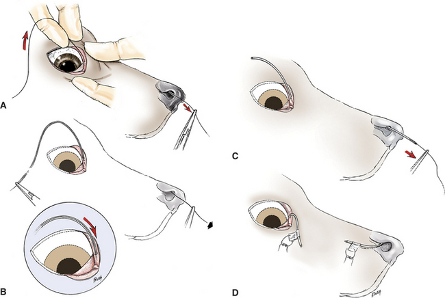
Imperforate puncta: surgical micro-incision to open up membrane over punctum. Can also do for dacryocystitis - temporary catheterisation - passed into punctum, through duct and out through nostril - during healing as a spacer..
Irreverisble obstruction - conjunctivorhinostomy - making a brand-new surgical bypass passage for tears directly into the nasal cavity.
B) Secretory system: problems here affect how tears are made or structured → eyes dries out/cornea looses its protective barrier.
1) Aqueous Deficiency - middle tear film
Aetiology: Deficiency in lacrimal and nictitating gland components of tear production → Dry Eye.
Developmental: Aplasia/hypoplasia of glands (predisp. Yorkshire Terrier, Pug)
Acquired: Immune-mediated, Distemper, surgical removal of nictitating gland, trauma, drugs (sulfonamides), hypothyroidism etc.
“underlying mechanism” - that causes classic KCS..
2) Mucin Abnormality - inner tear film
Aetiology: Evaporative dry eye associated with chronic conjunctivitis and Keratoconjunctivitis Sicca (KCS).
Clinical Signs: Features of KCS, often without ocular discharge; normal aqueous tear production.
Diagnosis: Clinical signs, Fluorescein Break-Up Time (FBUT), conjunctival biopsy (goblet cell quantification).
FBUT Values: Normal dog = ~20 seconds; Mucin deficiency = <5 seconds.
Treatment: Topical mucomimetics, mucin replacers, topical corticosteroids (CCS).
3) Meibomian Gland Dysfunction (Lipid Deficiency) - outer tear film
= lack of oil layer from glands → Increased evaporative tear loss, premature tear film break-up, dry eye.
Developmental: Eyelid defects affecting gland number and distribution.
Acquired: Marginal blepharitis (e.g., Staphylococcus, Streptococcus), meibomianitis.
Pathophysiology: Secretions thicken and obstruct ducts due to glands being inflamed/blocked etc. Gland rupture leads to a diffuse granulomatous response, secondary infection, and extensive eyelid swelling.
Keratoconjunctivitis Sicca (KCS / Dry Eye)
→ Deficiency in aqueous tear production and/or mucin production (gland of the 3rd eyelid/lacrimal gland) → corneal drying and irritation (dry inflammation of cornea & conjunctiva). Mostly in cavalier, bulldogs
Etio: Autoimmune (most common), drug-induced (atropine, sulfonamides, general anaesthetics), viral (Distemper), chronic blepharoconjunctivitis, trauma, metabolic disease (hypothyroidism), congenital, or iatrogenic (removal of prolapsed 3rd eyelid gland).
Pathogenesis
The precorneal tear film consists of three layers:
Outer Lipid Layer: Prevents evaporation (Meibomian glands).
Intermediate Aqueous Layer: Provides hydration and nutrients (Lacrimal & Nictitating glands).
Inner Mucin Layer: Anchors tear film to corneal epithelium (Goblet cells).
Deficiencies disrupt stability, triggering → chronic inflammation → secondary infection → corneal/conjunctival dehydration → hyperaemia → squamous metaplasia and hyperkeratinisation → corneal thickening.
Clinical Signs (Usually Bilateral)
Acute: Keratitis, conjunctivitis, ocular pain, blepharospasm, corneal ulceration, mucoid discharge, dry appearance.
Chronic: Mild discomfort, corneal keratinisation/pigmentation, mucoid/mucopurulent discharge, dull corneal surface.
Diagnostic Testing
Schirmer Tear Test (STT):
Normal: 15–25 mm/min
(Note: Some references classify diagnostic cutoffs below 10 mm/min or 8 mm/min; standard clinical diagnosis begins at below 15mm/min).
Rose Bengal Stain: Identifies dead epithelial cells - necrotic corneal/conjunctival epithelium (stains red)
Treatment Protocol:
Medical Management (Try Conservative Tx for at least 4 weeks first):
treat underlying cause, clean with NaCl or diluted betadine.
Lifelong cyclosporine (stimulates tear prod. + stops autoimmune destr.), preventative ATB, topical prednisolone (to decrease inflammation, CI if corneal ulcers are present).
methylcellulose, vit.A, hyaluronate - for hydration/lubricant
Surgical Options (If unresponsive to medical treatment):
3rd eyelid flap - to protect cornea
Conjunctival Flap
Parotid Duct Transposition (PDT): Reroutes the parotid salivary duct to the conjunctival sac to substitute saliva for tears.
5.Glaucoma
Etiology & Pathophysiology
Glaucoma is characterized by an abnormal increase in intraocular pressure (IOP > 25–30 mmHg) resulting from obstruction of aqueous humor outflow through the trabecular meshwork at the iridocorneal angle → accumulation of aqueous humor.

Patho:
Buphthalmos: Chronic elevation of IOP → global enlargement/stretching of the eye globe.
Corneal Changes: Stretching breaks Descemet’s membrane (producing linear corneal striae) and damages the endothelium, leading to corneal oedema.
Acute IOP spikes paralyze the iris sphincter and dilator muscles → fixed, dilated pupil.
All ocular structures are affected - pain, buphthalmia, epi-scleral congestion, mydriasis, lens luxation, ciliary body atrophy, phthisis bulbi (shruken, non-functional globe), retinal atrophy & blindness.
Classification
Primary Glaucoma (Genetic / Breed-related; Usually Bilateral):
POAG (primary open-angle glaucoma) ex. Beagle
PNAG / PACG (primary closed/narrow-angle) ex. american cocker spaniel
Congenital - goniodysgenesis ex. basset hound
Secondary Glaucoma (Consequence of existing disease; Usually Unilateral):
Causes: Uveitis (inflammatory cells/fibrin blocking outflow), lens luxation/subluxation, lens swelling (iris bombée), or intraocular neoplasia.
SOAG (secondary open angle) & SNAG (secondary narrow angle)
Clinical Presentation
Presentation | Clinical Signs |
Acute | pain, red eye (episcleral congestion), depression, anorexia, diffuse corneal oedema, fixed & dilated pupil (mydriasis), acute vision loss/blindness, corneal vascularisation. |
Chronic | Buphthalmos, lagophthalmos, secondary keratitis, lens luxation, corneal striae (Descemet's tears), retinal & optic nerve atrophy, eventual phthisis bulbi. |

Diagnosis
Tonometry (IOP Measurement): Head must be held vertical, + avoid pressure on neck. IOP > 25mmHg.
Gonioscopy: Special lens used to visualize the iridocorneal angle (drain opening) and anterior ciliary cleft (“drain pipe underneath”, where tears drain) → to see whether entry is open (open-angle/closed) and if it is formed incorrectly (diagnose goniodysgenesis)
Ophthalmoscopy (Direct & Indirect)
Treatment Protocol
A. Medical Management - to decrease production or increase outflow. Depending on cause and state of angle. Prevent glaucoma in the other eye.
Systemic drugs - reduce fluid production → acetozolamide, beta blockers (timolol, betaxolol)
Topical
reduce aqueous humor - CAIs, ex. dorzolamide
Increase outflow - epinephrine, pilocarpine, latanoprost
Class / Route | Mechanism of Action | Examples & Considerations |
Emergency Osmotic Diuretics | Rapid hyperosmotic fluid draw from the eye | Mannitol IV or Glycerol (Most effective in primary glaucoma & lens subluxation; use with caution in cardiovascular patients). |
Prostaglandin Analogues (Topical) | Potent increase in uveoscleral outflow; rapid miosis (needed when goal is to rapidly pull down the intraocular pressure!) | Latanoprost. - in case of severe acute glaucoma - 1st choice. (Contraindicated in anterior lens luxation or uveitis). |
Carbonic Anhydrase Inhibitors (CAIs) | Suppress aqueous humor production (for maintenance control, together with b-blocker) | Topical: Dorzolamide (Trusopt). Systemic: Methazolamide, Acetazolamide (Monitor serum potassium). |
Beta-Blockers (Topical) | Decrease aqueous production (reduces ciliary blood flow) | Timolol, Betaxolol. (Use with caution in cardiac or asthmatic/pulmonary patients - lowers heart rate/function). |
Miotics / Parasympathomimetics | Constrict pupil (miosis) to pull iris away from the angle and widen outflow space | Pilocarpine. (Can cause local ocular irritation and systemic side effects). |
Sympathomimetics | Increase outflow facility | Epinephrine. |
Analgesia | Systemic pain relief | NSAIDs or opioids as needed for intense ocular pain. |
B. Surgical Management
To Decrease Aqueous Production (Visual or Non-Visual Eyes):
Targets: partial or complete destruction of ciliary body
Cyclocryotherapy: Destruction of ciliary body epithelium (freezing probes) or Cyclophotocoagulation (using YAG laser = burns the ciliary body to stop production - NB! i notata det sto at det her increase outflow ??? men gir ikje heilt mening det??)
To Increase Outflow (Visual Eyes):
targets drainage path
Gonioimplants / shunts - passively drain aqueous humor to the subconjunctival space
Lens extraction - remove blockage
iridectomy (create bypass)
End-Stage / Non-Visual Painful Eyes:
Enucleation or Evisceration with Intraocular Prosthesis.
6.Diseases of the lens. Neoplasia of the eye
Diseases of the Lens
1. Cataract→ An opacity of the lens or its capsule resulting from structural changes in lens fibres or proteins (breaks down, clumps together).
Classification:
By Location: Anterior capsular, anterior subcapsular, cortical, equatorial, nuclear, posterior subcapsular, posterior capsular.
By Age of Onset: Congenital, developmental, juvenile, senile.
By Etiology:
Primary: Inherited.
Secondary: Uveitis, trauma, infection, diabetes mellitus, radiation, toxic, nutritional deficiency, senility.
By Consistency: Fluid, soft, hard.
Classification acc. to Stage of Development
Stage | Pathological Characteristics | Vision Status |
Incipient | Early, focal opacity. | Sight unaffected. |
Immature | Extensive opacity involving most of the lens; tapetal reflection still partially visible. | Vision impaired, but animal still has sight. |
Mature | Complete lens opacity; no tapetal reflection visible on ophthalmoscopy. | Complete blindness in affected eye. |
Hypermature | Proteolysis causes lens liquefaction and resorption; capsule appears wrinkled; anterior chamber deepens. | Blind; high risk of secondary phacolytic uveitis. |
Clinical Signs
Increased lens opacity and altered lens volume.
Visual impairment progressing to complete blindness. Non-painful in itself.
Diagnosis: Opthalmological exam (opaque lens, increased size)
Treatment
Medical Management:
Mydriatics (Atropine, Tropicamide): Dilate pupils in early stages to permit vision around focal opacities.
Aldose Reductase Inhibitors: Slow progression in diabetic cataracts.
Antioxidants (Topical/Systemic): Vitamin E, zinc ascorbate, carnosine.
Surgery (Definitive Treatment):
Prerequisites: Functional retina, controlled ocular inflammation (pre-op topical steroids/NSAIDs), absence of active co-existing ocular disease, patient/client compliance.
Extracapsular Cataract Extraction (ECCE): Removal of the lens while leaving the capsule.
Intracapsular Cataract Extraction (ICCE): Complete removal of the entire lens along with its capsule.
Phacofragmentation: Ultrasonic disruption and irrigation/aspiration of the lens nucleus through a micro-incision.
2. Lens-Induced Uveitis (LIU)
→ Inflammation of uvea triggered by the exposure of lens proteins (Ag`s) to the aqueous humor. Hypersensitivity type IV reaction (Ag + Ab → AgAb complex → uveitis).
Phacolytic Uveitis: Degradation of lens proteins (cataract) - in hypermature cataracts (lens capsule cannot hold onto proteins) → leakage → humoral & cell-mediated immune response. Mostly a mild, self-limiting if managed. Common in older dogs. Tx: topical anti-inflammatory agents (CCS, NSAIDs), surgery not typical.
Phacoclastic Uveitis: Traumatic rupture of lens capsule → exposes massive amounts of intact lens Ag to aqueous humor (class II MHC T-cells + MF → severe cell-mediated immune reaction). Rapidly progressive, destructive inflammation. Tx (google): surgical removal of damaged lens + anti-inflammatory therapy.
Tx general for LIU → Topical anti-inflammatory agents (corticosteroids - 1st line), NSAIDs
3. Lens Luxation
→ Displacement of the lens from the hyaloid fossa due to tearing of the ciliary zonules. (lens zonules = tiny thread-like fibers holding the lens of eye firmly in place/suspensory ligaments).
Etiology/classification:
Primary (Hereditary): Inherent weakness/abnormal insertion of lens zonules, leading to rupture early in life (<5yrs). predisp. in terrier breeds, miniature schnauzers.
Secondary (Acquired) to: blunt trauma, chronic glaucoma (buphthalmos stretching zonules), uveitis, tumors, cataracts
Clinical Manifestation by Position
Subluxation: Partial zonular rupture. Presentation includes an aphakic crescent (half-moon edge of lens in pupil), instability of iris and lens.
Anterior Luxation: Lens displaces into the anterior chamber. Presents as an acute emergency: glaucoma, corneal edema.
Posterior Luxation: Lens drops into the vitreous cavity. May be clinically silent or induce ocular inflammation / glaucoma.

Diagnostic Findings: Iridodonesis (iris vibration), lens movement, vitreous strands/fibers floating throught he pupil into anterior chamber - Opthalmological exam.
CS: Pain, whitening of eye, squinting, epiphora (excessive tearing), edema, glaucoma, uveitis.
Treatment Protocol
Medical Stabilization: Reduce IOP using hyperosmotics (Mannitol IV) or carbonic anhydrase inhibitors; manage secondary anterior uveitis with topical anti-inflammatories. (Note: Avoid miotics like Pilocarpine in anterior luxation as they trap the lens in the anterior chamber).
Surgical Extraction: for anterior luxation lens - in visual or potentially visual eyes via intracapsular extraction (ICCE) or phacoemulsification (w/USG energy).
Enucleation: Indicated if the eye is permanently blind, hyper-pressurized of unmanaged high IOP, and painful.
Neoplasia of the Eye
Tx: radiation, Laser ablation, cryosurgery, chemotherapy, enucleation depending..
1) Intraocular & Uveal Neoplasia
Iris Melanoma / Anterior Uveal Melanoma:
Most common primary intraocular tumor in dogs.
Manifests as dark hyperpigmentation of the iris, progressive mass growth, dyscoria (abnormal pupil shape), and secondary glaucoma due to angle occlusion.
Metastatic Potential: Slow-growing; potential to metastasize to lungs, liver, and spleen.
Treatment: Diode laser photocoagulation (for small/localized lesions) or enucleation.
Ciliary Body Neoplasia (Adenoma / Adenocarcinoma):
Arises behind the iris.
Manifests as a pink/red mass visible through the pupil, secondary retinal detachment → secondary glaucoma. swollen conjunctiva/prominent 3rd eyelid, mydriasis.
Treatment: Surgical excision or enucleation.
Uveal Lymphoma (Lymphosarcoma):
Secondary metastatic manifestation of systemic lymphoma. Bilateral pinkish-white infiltrates/mass in the anterior uvea, anterior uveitis, hyphaema, and secondary glaucoma.
Treatment: Systemic chemotherapy.
2. Other:
Eyelid Neoplasia:
Dogs: Predominantly benign (Meibomian/Sebaceous gland adenoma, papilloma, melanoma).
Cats: Predominantly malignant (Squamous cell carcinoma [SCC], especially in white-pigmented eyelids).
Treatment: surgical resection, creyo, chemo, laser ablation.
Conjunctival & Corneal Neoplasia:
Conjunctival: Commonly malignant (SCC, hemangiosarcoma, melanoma).
Corneal: Rare; primary malignant melanoma.
Orbital Neoplasia: Almost exclusively malignant (Osteosarcoma, Fibrosarcoma, Adenosarcoma).
(Quick Reference)
                            [ Main points  ]
                                     │
      ┌──────────────────────────────┼──────────────────────────────┐
      ▼                              ▼                              ▼
 [ CATARACT SURGERY ]       [ LENS LUXATION ]              [ NEOPLASIA ]
 ├─ Extracapsular (ECCE)    ├─ Anterior (Emergency!        ├─ Eyelid (Dog=Benign,
 ├─ Intracapsular (ICCE)    │   Glaucoma + Edema)          │   Cat=SCC/Malignant)
 └─ Phacoemulsification     └─ Posterior (Vitreous/Silent)  └─ Orb
NOTES:
just started with different classifications of cataract, then they just asked about treatment. Only wanted the names of different procedures (ex. phacofragmentation, intra and extracapsular extraction). Nothing on etiology or complications.
For lens lux - only said difference between posterior and anterior before they cut me off
Neoplasia - just listed what structures are most prone, types of tumor, usual treatment (ex. cryo).
7. Proptosis of the eyeball. Blindness.
1) Proptosis = Protrusion of the eyeball (anterior displacement of eyeball from orbit). Exophthalmus means the same thing, and this term is usually used when describing proptosis.
Aetiology
Congenital (Rare): mucocele (cyst of salivary gland). Brachycephalic breeds. Arteriovenous fistula.
Acquired: TRAUMA (primary cause), abscess, neoplasia, Orbital hemorrhage and swelling, acute eosinophilic myositis, periorbital cellulitis (foreign body, infection), vascular disorders.
Pathogenesis
Traumatic proptosis may follow blunt trauma (e.g., being hit by a car, fight with another animal) → during trauma, the globe is luxated from the orbit, and eyelid spasms prevent its retraction → secondary orbital hemorrhage and swelling displace the globe further from the orbit.
Complications: Corneal ulceration, enophthalmia, optic nerve degeneration, keratoconjunctivitis sicca, and medial rectus muscle injury.
Clinical Signs
Exophthalmos (protrusion of the globe from the orbit).
Fever, reddened discoloured swelling posterior to last upper molar, pain on jaw manipulation.
Treatment
Conservative: Systemic antibiotics, moistening of eye.
Surgical: Replace eye — lateral canthotomy and complete temporary tarsorrhaphy (for 2w). with usually two or three interrupted horizontal mattress sutures (use sutures on the skin and eyelid margins, be careful to not penetrate the conjunctiva).
                    [ CLIENT CALL / INITIAL ADVICE ]
                                   │
               Keep eye MOIST (Sterile Saline / Lube)
               DO NOT touch or rub ──► IMMED. CLINIC VISIT
                                   │
                                   ▼
                       [ CLINICAL EVALUATION ]
                                   │
         ┌─────────────────────────┴─────────────────────────┐
         ▼                                                   ▼
[ GLOBE SAVABLE? ]                                  [ GLOBE UNSAVABLE? ]
├─ Direct/Consensual PLR present                   ├─ >2 Extraocular muscles torn
├─ Intact globe & cornea                           ├─ Complete optic nerve avulsion
└─ Intraocular structures intact                   ├─ Ruptured globe / Hyphema
         │                                         └─ Severe posterior segment damage
         ▼                                                   ▼
   REPOSITION SURGERY                                   ENUCLEATION
PLR = pupillary light reflex test.
2) BLINDNESS= state of being totally sightless in both eyes. Blindness can be temporary or permanent, complete or partial.
Visual deficits may result from lesions at any level of the central visual pathways from the optic nerve to the occipital lobe of the cerebral cortex (optic nerves, optic chiasma, optic tracts, and optic radiation).
Causes
Disease affecting structure of the eye: Cornea, anterior/posterior chambers, iris, lens, retina, or optic disc.
Disease affecting neurological pathway and CNS: Cranial nerves and CNS.
Diagnosis
Evaluation includes thorough ophthalmic and general physical examinations + neuro exam because many systemic diseases may cause blindness.
Subjective/Visual Tests: Menace test, dazzle reflex, maze test in both light and dark illumination, electroretinography, and visual evoked potentials.
Other: Cerebrospinal fluid analysis (culture, serology, cytology), CT, MRI, US, and X-ray, blood test.
Types of Vision Loss
Central/cortical blindness: total or partial loss of vision caused by damage to the brain`s occipital cortex.
Can be acquired, congenital or transient. Typical: ischemic stroke, cardiac surgery, encephalitis, meningitis.
Acute vision loss: sudden onset of vision loss, bilateral loss is more common. Caused by trauma, glaucoma, intraocular hemorrhages, retinal detachment.
Progressive vision loss: slow, developing loss of vision by chronic lesions - glaucoma, keratitis, uveitis, cataract etc.
Congenital vision loss: present from birth due to structural developmental defects/inherited issues - gross ocular malformation (anophthalmos/microphthalmos), collie eye anomaly, retinal dysplasia.
NOTE:
The queen of eyes wanted to know → what is proptosis, main cause (trauma), if client called you and said “my babys eye is hanging out what do i do” → you say keep it moist and come to the clinic ASAP. After you need to decide if you enucleate or put it back in and if you put it back in how to do it → put it in and suture it in place, monitor for next 2weeks.
For blindness, all she wanted to know was about glaucoma, and problems with the retina that can cause blindness.
8. Evaluation and therapeutic surgical methods for eye diseases, diagnosis and therapy.
PHYSICAL EXAMINATION
Clinical Signs: Discharge (mucous = irritation, green = infection), red irritated eye, cloudiness, difference in pupil size, aversion to light, squinting, exophthalmos.
Observation: Should assess symmetry, conformation, and gross lesions.
Palpation: around eye and head, exclude skin and dental problems as the origin of eye issue.
OPHTHALMIC EXAMINATION
A standard ophthalmic exam is a comprehensive series of tests that checks vision and the health of the animal's eyes. When examining the eye, a dark room with red light is recommended.
Examination / Category | Tests & Descriptions |
|---|---|
a) Behavioural and Vision Examination Tests the animal's ability to navigate and orientate itself. | • Tracking: Drop cotton ball and check if dog follows. • Maze Test: Placement of obstacles around the room and assess the dog's ability to navigate around objects (in darkness and light), stairways. • Placing: See if they place their feet on a surface when they can see it. |
b) Neuro-ophthalmic and Vision Tests Localizes a defect to the nervous system or the eye. | • Pupillary Light Reflex (PLR): Shine a bright light into the lateral aspect of each eye and evaluate pupil constriction. (Tests CN II and III) • Menace Response: Make a threatening movement towards the eye. Normal response is to blink with aversion of head. (Tests CN II, III, VI, and VII) • Dazzle Reflex: Shine a strong light into each eye in turn. Normal is to blink/avoid. • Palpebral Reflex: Touch of medial and lateral canthus usually initiates a normal blink response. (Tests CN V, VI, and VII) |
c) Tear Film Products Tests (Quantitative) | • Schirmer Tear Test: Measure aqueous layer of tear film for 60 seconds. - Normal: >15 mm/min; <8–10 mm/min indicates risk of Keratoconjunctivitis sicca (dry eye syndrome). • Phenol Red Thread Test: Fast, accurate method (15 sec); recommended for small mammals. - Normal (cats): 18.4–27.7 mm/15 sec. |
d) Fluorescein Test (Qualitative / Structural) | • Corneal Staining / Ulcer Evaluation: Applies dye to evaluate corneal surface integrity or drainage. Outer epithelium is lipid-selective (repels water-based dye); damaged stroma absorbs dye, revealing green spots at ulcer sites. (Use strips instead of drops to prevent Pseudomonas infection.) • Tear Breakup Time: Evaluates tear film quality by measuring how long a fluorescein drop remains on the eye surface. - Normal: 20 sec. • Jones Test: Evaluates nasolacrimal duct passage by placing dye on the cornea and observing passage to the nose/mouth. If negative, try flushing. also have seidel test - to check for corneal perforation, aqueous humor will break up fluorescein. |
e) Rose Bengal Test | • Stains dead corneal and conjunctival epithelial cells red, - shows defects in cornea/conjunctival epithelium. • example: Useful for feline herpesvirus keratitis diagnosis. |
f) Intraocular Pressure (IOP) Asess this first during examination before handling (to avoid raising pressure) | • Target Range: Normal IOP is 15–25 mmHg (Low = uveitis; High = glaucoma). • Tonovet / Tonopen: Preferred options; probe touches cornea 5x (Tonovet). • Schiøtz Tonometer: Older apparatus; not suitable for cats. • Retropulsion: Palpate eyeball firmness through eyelids (avoid jugular pressure as it artificially raises IOP). |
g) Ophthalmoscope | • Direct Ophthalmoscope: Higher magnification; examines fundus and anterior segment using built-in filters. • Indirect Ophthalmoscope: Lower magnification; produces an inverted/upside-down image. • Fundus Camera: Specialized imaging (e.g., smartphone adapter lens). • Optical Coherence Tomography (OCT): High-resolution cross-sectional imaging of retina layers, cornea, and iridocorneal angle. |
h) Gonioscope | • Uses a specialized goniolens to inspect the drainage angle. • Identifies goniodysgenesis to assess predisposition to primary angle-closure glaucoma. |
i) Ultrasound | • Evaluates lens luxation, retinal detachment, retrobulbar lesions, and neural structures. • Applied via gel directly to the cornea or closed eyelid following topical anesthesia. Done when there is opacity and difficult to see inside eye. |
j) Electroretinography (ERG) | • Uses electrodes placed near the retina to measure electrical wave responses and evaluate function of sensory cells in retina. |
k) Corneal Pachymetry | • Measures exact corneal thickness; primarily utilized in clinical research contexts. |
SURGICAL TREATMENTS OF EYE AND EYELID
Eyelid laceration repair - by margin aligned w/figure-8-suture & simple interrupted for skin.
Eyelid surgery:
Entropion (inward rolling) - temporary (lembert tacking sutures) or permanent (hotz-celsus / Y-U plasty)
Ectroption (outward drooping) - V-Y plasty or modified Kuhnt
Eyelid mass excision - Can do V-excision, H-plasty, or semicircular flap.
Enucleation (removal of eye)
Transpalpebral - eyelid margins sutured closed first, entire globe and lid margins removed
Transconjunctival - eye removed directly through conjunctiva, 3rd eyelid and eyelid margins are excised secondarily.
Evisceration (removal of internal content only - replace by implant), exenteration (complete removal of eye + tissues), tarsorrhaphy
GENERAL/MEDICINAL TREATMENT
Antibiotics: Gentamicin and neomycin (G+), cephalexin and chloramphenicol (G-).
Anti-fungals: Itraconazole.
NSAIDs: Flunixin.
Corticosteroids: Dexamethasone, prednisolone (not if broken epithelium).
Parasympathomimetics: Carbachol, pilocarpine.
ACE-inhibitors: Demecarium, neostigmine.
Others: Glaucoma (carbonic anhydrase inhibitors, beta blockers, diuretics, mannitol).
9. Surgical diseases of the pinna and external ear canal. Methods of the treatment. Lateral resection of the external ear canal. Partial and total ablation of the EEC. Osteotomy of the bulla tympanici.
Anatomy:
External ear: pinna (cartilage), external auditory meatus, hair follicles, sebaceous & ceruminous glands
Middle ear (within temporal bone): tympanic membrane, bulla tympanica, malleus, incus, stapes (auditory ossicles - middle ear bones)
Auditory tube: connects nasopharynx & tympanic cavity - opens when yawning/swallowing
Inner ear: bony labyrinth (semicircular canal, cochlea, vestibule), membranous labyrinth (sacs and tubules filled w/endolymph)

Diseases:
I. Aural Hematoma
Bleeding bw. cartilage and skin of pinna (swellings - fluid-filled) - cartilage separated from the skin, due to broken blood vessel from bleeding occurring inside tissue. Eardrum can rupture if not treated.
etiology: Otitis, allergy, trauma/scratching, ear mite/head shaking, blood disorders, cushings and systemic diseases. Predisp. in dogs with long pinnae (cocker spaniels). Rare in cats.
Treatment:
Conservative: punction and aspiration of content (can reoccur). giving glucocorticoids (cause vasoconstriction).
Trans-pinnal suturing: Make curved/S-shape incision, remove hemorrhages. Suture skin and cartilage together w/simple interrupted. Not too tight, can use buttons, leave the incision open. Apply compressive bandage.
II. Wounds of the Pinnae
Treatment: Depends on the severity of the wound:
a) Superficial: local tx with chlorhexidine.
b) Deep: flush w/chlorhexidine + suture
c) Penetrating: sutures on lateral and medial pinnae, drainage at outer edge of wound. Cryotherapy to reduce oedema if present.
III. Otitis Externa (see internal q.7/infectious q.1)
et: allergy, autoimmune (eg, pemphigus), endocrine, bacterial, fungal (aspergillosis), parasites, viral (canine distemper), foreign bodies.
IV. Other - of auricle & external ear
Tumours: papillomas, SCC, hemangiomas..
Otitis ossificans: ear canal wall = ossificated, visible on x-ray
Otitis media/interna
Frost bite of the pinna
V. Surgery of the Ear Canal - methods:
Lateral Resection (Zepp’s Method)
Flap opened in lateral wall of vertical canal → improves airflow & drainage for otitis externa (that has not responded to tx.), hyperplasia of ear canal or to remove small neoplastic lesions. CI: in stenosis/otitis media.
Procedure: 2 incisions along the lateral wall of the vertical canal → stop when at the level of horizontal canal → cut of excess skin/cartilage and suture w/simple interrupted.

Partial Ablation (Vertical Canal Ablation)
Removed only the vertical canal, while keeping the horizontal canal. For severe disease/tumors in vertical canal while horizontal is still healthy.
Procedure: T-shaped incision on skin, remove vertical canal and damaged tissue. Fix the opening of the horizontal canal to the skin in the bottom of the T-incision → close in a T-shape.

Total Ablation (TECA)
Removing the entire ear canal (vertical & horizontal)
In case of end-stage otitis (chronic - unresponsive to tx.), inflamed bulla, ossification, stenosis, epithelial hyperplasia, neoplasia.
Osteotomy of Bulla Tympanica
For removing bony canal + any infected parts of middle ear. Often in combo w/total ear canal ablation.
Indicated for - otitis media/damage to middle ear + non-responsive otitis (confirm w/x-ray), polyps and tumors
Lateral bulla osteotomy → improve drainage and relieve chronic inflammation & pain.
Ventral → for inflammatory nasopharyngeal polyps in cats that come from bulla tympanica and grow through the ear canal/auditory tube. (opens bulla via ventral neck incision).
10. Surgical diseases of the salivary glands and ducts. Symptoms, diagnosis and therapy.
Important Salivary Glands Include: Parotid, mandibular, sub-lingual and zygomatic gland.
General Diagnosis: CS, palpation, sialography (x-ray + iodine contrast), USG. FNA.
I. Anatomy of the Salivary Glands & Ducts
Salivary glands = exocrine glands, producing saliva through a system of ducts. There are 4 important glands in small animals (+ some small):
Gland | Location | Duct Path & Opening |
Parotid | at base of auricular cartilage | Opens at labial mucosa (mouth) level of 4th upper premolar |
Mandibular | below parotid (junction of maxillary&linguofacial v.) | Opens near rostral border of frenulum (under tunga) |
Sublingual | Direct under oral mucosa, lateral to tongue (under base of tongue) | Opens near frenulum (closely assoc. with mandibular) |
Zygomatic | Ventral to zygomatic arch | Opens lateral to last upper molar |

Salivary glands are most often removed to treat salivary mucoceles or neoplasia.
mandibular and sublingual salivary glands are most often removed to treat cervical, sublingual and pharyngeal salivary mucoceles.
Neoplasms (usually adeno-carcinomas or carcinomas) occur most freq. in the parotid, and the mandibular salivary gl.
II. Salivary Mucoceles / Sialocele
Accumulation of saliva within the salivary duct or gland capsule, mostly in sublingual and mandibular glands. They have different location acc. to which gland/area affected.
Sublingual mucocele = Ranula (swelling under tongue)
Etio: Trauma/inflammation blocking duct/capsule or genetic
CS: Swelling, pain, dysphagia, enlarged mass. Depend on the site of saliva accumulation. For ex. a pharyngeal mucocele can obstruct airways → resp. distress.
Dg: Palpation, sialography (radiographic imaging)
Tx: Periodic drainage or surgical removal:
Extirpation (Removal of salivary gland): Incision ventral to ear canal, over the mandibular salivary gl., and divide the platysma mm. Open gland capsule, pull the gland caudolaterally, sharp dissect until the lingual nn is identified, and dissect the digastric mm. Ligate vessels, place drain, and close the mm and skin w/absorbable sutures. (main goal is complete removal).
Marsupialization (Ranula): Drain the ranula, open the duct (to bypass the blockage), and suture the skin to make a new opening for saliva to drain out. (main goal is decompression by slitting cyst open and suturing the cut edges to adjacent skin)
Other:
Sialadenitis: Inflammation/abscess of the salivary gland. May abscess and form salivary fistula that rupture through skin. Rarely a clinical problem in cats/dogs. Et: Trauma/infection. Tx: drain, ATB, removal may be needed in recurrent infections.
Sialoliths:Ca phosphate stones in salivary gland from ascending foreign body (grass).
Neoplasia: Carcinoma, adenocarcinoma - metastasis common. More in cats > 10yrs old. Generally low occurrence in dog/cat.
Fistula: abnormal connection between two hollow spaces. Salivary fistula is an uncommon problem.
Ptyalism / Hypersalivation: Excessive salivation - by either hypersecretion of saliva or inability to swallow normal saliva production. Et: Dysphagia, toxins, rabies, nausea, fever, fear. Tx: glycopyrrolate.
Hypoptyalism: Decreased saliva production. - dry mouth (xerostomia). Et: Atropine (drug), dehydration, fever, anesthesia. Tx: pilocarpine.
EXAM NOTE:
Only mentioned stones and common salivary glands affected, can be checked with USG. That was all Trbolova wanted to hear (maybe due to lack of time + she wasn’t really listening).
Dec 2021: Trbolova told me to start with explaining all the salivary glands, their location and where the ducts go and open. She wanted me to show where they are located on my throat. I didn't know the location of all the ducts so I tried to start talking about mucoceles and sialoceles but she didn't want to hear it and just went back to the location of the damn ducts. I also tried to explain about other surgical diseases like sialoliths, neoplasia but she didn't appear to be interested in those either. She wanted me to describe extirpation and marsupialization. I think that knowing these procedures is what saved me from failing. So, learn the anatomy of all the ducts if you get this question as this apparently is what she wants here.
11.Diseases of the lips. Mouth diseases. Gingivitis. Tartar. Neoplasia of the soft and hard tissue in mouth. Cleft palate. Oronasal fistula. Surgical diseases of the nose.
A) Diseases of the Lips
1) Lip Fold Dermatitis
Chronic skin inflammation of drooping lips and lip folds due to accumulated moisture - bacteria, poor oral hygiene. Predisp. in spaniels and Saint bernards.
CS: Redness, inflammation, bad smell, swelling, pain on palpation.
Treatment: clean w/ chlorhexidine, keep dry; surgical correction of droopy lips.
2) Feline Eosinophilic Complex
Forms: Eosinophilic plaque, eosinophilic granuloma, and indolent ulcers. (“hypersensitivity reaction”)
CS: Lesions around mouth (also inside), redness, pain, swelling (bleeding possible), not wanting to eat, halitosis (chronic bad breath).
Dg: history, physical exam, FNA, biopsy. Tx: fluids, nutrition, ATBs (amoxicillin), CCS (prednisone), cyclosporine
Other: Tight lip syndrome (shar pei), primary cleft palate (hare lip) - defect of lip and incisive bone.
B) Diseases of the Mouth
General: Redness, bleeding gums, not wanting to eat, bad breath, dysphagia.
1) Stomatitis:Inflammation of oral mm, in response to dental plaque / immune system dysfunction.
CS: Red, ulcerative "kissing ulcers" on mucosal surfaces in contact w/teeth (most in upper lip, next to 4th premolars/canines).
Treatment: Plaque control, ATBs, topical anti-inflammatories/NSAIDs, dental extraction of contact teeth with plaque buildup.
2) Gingivitis:Inflammation of gums, by poor diet + lack of hygiene (food, bacteria build up bw. gums and teeth → forms plaque, calcifies into tartar, can progress to periodontitis).
Treatment: Scaling (plaque/tartar removal), antibacterial gel (Stomodine gel).
3) Periodontal Disease & Tartar
Infection/inflammation of periodontium (gingiva, periodontal ligament, alveolar bone).
Plaque: yellow/clear, soft film of bacteria, can calcify in 2w if not removed → hard calculus (tartar).
Gingivitis → periodontitis (subginigival plaque, loss of CT, deep pockets) → advanced (tartar mineralization, pus, bone loss, loose teeth). Periodontal pockets can lead to systemic spread (ex. pneumonia).
DG: CS, dental radiography, periodontal probing.
Tx/prevention: Mechanical scaling, gingivectomy, gingival flap procedures, extractions, splinting, bone grafting, ATB, NSAIDs, stomodine gel, home care: daily brushing, chew toys.
C) Oral Neoplasia
Benign: Epulis (local mass from ginigiva or periodontal lig/stroma), peripheral odontogenic fibromas (most common firm gum mass from periodontal lig.) & canine peripheral ameloblastomas
Malignant: melanoma, SCC, Fibrosarcoma, osteosarcoma (tongue neoplasia less common).
Tx: surgical excision (incl. bone via mandibulectomy or maxillectomy), radiotherapy
D) Congenital Oronasal Fistula (Cleft Palate)
Abnormal mouth-nasal cavity communication due to failure of palatine halves fusion in utero. (Primary = cleft lip, secondary = hard/soft cleft palate).
Et: Genetic, teratogenic, or nutritional. Brachycephalic breeds predisp.
Leads to high neonatal mortality, survivors show poor growth, nasal regurgitation of food/milk, resp. signs, aspiration pneumonia.
Treatment:
Conservative: Orogastric tube feeding, custom palate guard until surgery.
Surgical: Post-12 weeks of age, using overlapping or sliding bipedicle flaps to make an airtight nasal floor
E) Diseases of the nose:
1) Oronasal fistula: abnormal opening bw. nose and mouth.
Et: trauma, canine tooth extraction, severe periodontal disease
Treatment: Surgical correction, flap technique if needed (require air tight, tension free, closure). Tube feeding until surgery, ATB for rhinitis, treat nasal infection prior to repair.
Brachycephalic syndrome (BOAS) - includes:
Stenotic nares: malformed, abnormally narrow, or inward collapsing nostrils during inhalation.
→ dyspnea, open-mouth breathing, laryngeal prolapse
→ Surgical corrections:
Alar Wing Amputation: Ventral alar wing removal without sutures.
Wedge/Punch Resection: Removal of an identical tissue wedge from each nasal wing.
Alapexy: removing small triangles from lateral side, and suturing the alar wings back.
Elongated soft palate: extends too far back into pharynx, going past epiglottis and physically obstructing the laryngeal opening.
Tx: shorten it with scalper or laser (post-op swelling can be issue)
narrowing of trachea/hypoplastic
Eversion of laryngeal saccules + laryngeal collapse (sec. abnormnalities, acquired over time)

Exam Focus
Briefly about some diseases, basic about lip fold dermatitis and treatment. Wanted to know mostly about tartar, how it develops and treatment. Talked about brachycephalic breeds + basic surgeries. Did not mention neoplasms or other diseases.
Asked to focus just on brachycephalic - nares, elongated soft palate. Asked about the trachea. Whats the main problem caused by soft palate surgery - swelling.
12. Diseases of the teeth – oligodontia, polyodontia, brachygnatia, pulpitis
Dental Anatomy & Formulas
Tooth Anatomy: Composed of Enamel (outer calcium phosphate layer), Dentin (tubular hard tissue), Pulp (inner neurovascular tissue), Cementum (root-binding tissue), and Periodontal Ligament (anchors tooth to jaw).
Tooth Types: Incisors (cutting/scraping), Canines (holding/tearing), Premolars (holding/breaking), and Molars (crushing/grinding).
Dental Formulas:
Cat: 2X (Incisior 3/3, C1/1, PM3/2) - 26 milk teeth
Adult: 2x (incisior 3/3, C1/1, PM3/2, M 1/1 - 30 adult teeth
Dog: 2x (incisor 3/3, C1/1, PM3/3) - 28milk teeth
Adult: 2x (incisor 3/3, C1/1, PM3/3, M:2/3) = 42 adult teeth
Triadan System: Uses pig permanent dentition as a baseline; 1st digit = quadrant (1=upper right, 2=upper left, 3=lower left, 4=lower right), 2nd & 3rd digits = tooth position (I = incisor, C=canine, P=premolar, M=molar).
Developmental Anomalies
Number:
Anodontia: Total absence of teeth.
Oligodontia: Missing > 4 teeth
Often P 1-2 or incisors. Often in brachycephalic
Et: genetic, trauma. Tx: place implants, no other tx.
Polyodontia: Excess teeth
(usually 2nd/3rd premolars, incisors). Et: retention of milk teeth.
causing crowding and malocclusion; extracted only if problematic.
Hypodontia: 1–2 teeth.
Pseudohypodontia: Retained un-erupted teeth (confirmed via X-ray).
Eruption Barriers: Impacted (position/trauma - cannot erupt due to path being blocked) vs. Embedded (lack of eruptive force).
Size & Shape: Microdontia/Macrodontia (size); Germination (two crowns, one root), Fusion (completely fused teeth), supernumerary roots, and dens invaginatus.
Enamel Defects: Hypoplasia or hypocalcification from trauma, infections, or nutritional deficits.
Jaw Alignment (Brachygnathia)
Prognathia (Maxillary Brachygnathia): Short maxilla (underbite) leading to rotated, crowded teeth; common in brachycephalic breeds.
Mandibular Brachygnathia: Short mandible (overbite).
→ Malocclusion, dysphagia, pain, and oronasal fistulas; treated with bite plates or surgery.
Pulpitis & FORL
Pulpitis: Inflammation of the dental pulp within its rigid chamber, causing pressure buildup, restricted blood flow, hemorrhage, tooth discoloration, and necrosis. Diagnosed via X-ray/transillumination and treated with extraction. Et: bacteria, degeneration, truama, periodontitis.
FORL (Feline Odontoclastic Resorptive Lesion): Idiopathic tooth resorption by odontoclasts in older cats, causing pain, salivation, and bleeding; diagnosed via X-ray and treated with extraction (pulp can get infected → abscess, infection can spread to heart).
13. Fracture of the teeth. Extraction of the teeth. Endodontia, exodontia. Local analgesia of the head in the dog.
Tooth Fractures
Most common in canine teeth and upper 4th premolars
caused by trauma or hard objects
Types:
Classified by location (enamel, crown, root, or combination)
& whether the pulp is exposed (complicated) or not exposed (uncomplicated).
Patho: Pulp exposure → allow bacteria to infect root canal → tooth necrosis & root tip bone destruction → systemic bacterial spread.
Can cause carnassial tooth root abscess (upper 4th premoval fracture, commonly causes a draining facial skin wound below the eye).
Dg: Dental exam, x-ray
Treatment: Extraction (if pulp is affected), root canal therapy (endodontics), or conservative antibiotics.
Surgical treatment of teeth:
1) Tooth Extraction (Exodontia)
Indications: Fractures, mobile teeth, retained baby teeth, root abscesses, pulpitis, FORL, crowding.
Procedure Steps:
Do x-ray pre-op.
Gingival incision, in multi-root teeth - use drill to divide into single root segments and remove each.
Breakdown of periodontal ligament by using elevator (apply continuous pressure for 20-60sec)
Extraction w/forceps and check that it is whole.
Clear debris from alveolus and lavage, compression - place collagen sponge, suture.
Complication: Bleeding, damage of vessels and unerrupted teeth, damage to alveolar bone, gingival laceration, oronasal fistula, infection, damage to mandibula
2) Endodontia - root canal.
Treatment of dental pulp to preserve the tooth.
Indications: Fractures w/exposed pulp, pulpitis, necrosis, pulp abscess/cyst/granuloma or dev. abnormalities.
Procedure: Use endodontic burr to gain access to canal → plase a small file (pin) - and x-ray to assess length of canal → clean w/file, disinfect and dry canal → place sealer (resin/zinc-oxide based) → fill (obturate) w/gutta-percha to seal it. Restore the access site with composite, harden w/UV light.
3) Local Analgesia/anesthesia of the Head
Drugs: Lidocaine 2% (up to 3 hrs) and Bupivacaine (NEVER give IV due to severe cardiotoxicity).
Nerve Blocks:
→ top floor = Upper jaw (maxilla)
→ Bottom floor = Lower jaw (Mandible)
For both, we have 2 choices, 1. putting needle in hole near the FRONT → numbs only the front teeth on that side, or 2. Put needle in hole at the BACK → numbs the ENTIRE side (front, middle and back teeth)
For upper jaw/Top teeth:
Infraorbital block (maxillary n. block): Injected at infraorbital foramen; blocks upper lip and front half (of mucosa) (does NOT block teeth).
Maxillary Block: Injected behind the last upper molar; blocks all maxillary teeth, lip, mucosa, and soft tissue on that side.
For LOWER jaw / lower teeth:
Mental: Injected near 2nd lower premolar - to inject at mental foramen; blocks lower incisors, soft tissue, and lower lip rostral to mental foramen.
Mandibular (Inferior Alveolar n.+mandibularis n.): Injected medial to mandibular notch/angular process; blocks all mandibular teeth )(lower teeth+lip)

14. Surgical diseases of the esophagus. Hiatal hernia. Esophageal feeding tubes
Anatomy:
Esophagus → Muscular tube connecting pharynx to stomach, divided into cervical and thoracic parts.
Histo Layers: Mucosa, submucosa, muscularis (skeletal in dog, skeletal and smooth in cat), and adventitia.
Lacks a serosal layer (which normally produces pluripotent cells), resulting in prolonged healing times.
Signs of esophageal problems: dysphagia and regurgiation - effortless, without warning signs, food that hasnt reached the stomach. From obstruction or muscular weakness. Can also be vomiting, reflux, hypersalivation, anorexia, resp. distress, esophagitis..
Diagnosis by: X-ray with contrast, endoscopy, CT, Fluoroscopy (real-time x-ray movie - shows dynamic movement of food/liquid as it passes through), palpate/history
Surgical Diseases of the Esophagus (See q. in internal)
Obstruction
Etiology:
Persistent right aortic arch/vascular ring anomaly where the right aortic arch fails to regress. Esophagus is trapped and encircled by the arch (right), lig. arteriousum (left/dorsal) and heart base (ventral)
Foreign bodies (predisp. in narrow areas - thoracic inlet, base of heart, hiatus of diaphragm).
Stenosis / stricture (luminal narrowing) due to functional (spastic, esophagitis), mechanical (FB), or compressive (enlarged Lnn, hematoma).
Tx: depends on cause → Endoscopic retrieval, surgical removal - esophagotomy, hydration, balloon-catheter luminal dilation, partial esophagectomy (severe). Note: do not close w/simple continuous suture.
Diverticulum→ Localized pouch-like sac/dilation of the esophagus (commonly above a stenotic area).
Et: Muscle rupture, hernia, compression, or traction (incorrect suture technique). Excise if large.
Perforation →caused by: Ingestion of sharp objects, trauma.
Dg. by using iodine contrast x-ray ONLY (barium is contraindicated if suspected perforation). Tx: surgical repair / esophagectomy.
Megaesophagus
→ Esophageal dilation → to food/liquid accumulation.
Can be congenital or acquired. By vascular ring anomalies, myasthenia gravis, cushing, severe esophagitis, achalasia, idiopathic etc..
Tx: Upright feeding positioning (prevents regurgitation/aspiration) and prokinetics (controls acid reflux).
Tracheoesophageal fistula → dont use barium.
Hiatal Hernia
→ Protrusion of the abdominal esophagus, gastroesophageal junction, and sometimes gastric fundus through the esophageal hiatus into the caudal mediastinum. (“Means that the hole (hiatus) is too wide/loose, so parts of stomach gets squeezed/pushed through the hole, out of abdomen and into back of chest cavity (as diaphragm separates chest from abdomen”).
Etiology: Congenital abnormalities of hiatus, trauma.
Types:
Type I (Sliding Hernia): Most common in small animals; intermittent displacement of lower esophageal sphincter and gastric fundus into the thorax (slides back and forth).
Type II: Less common; displacement of the gastric fundus only.
Treatment:
Midline abdominal celiotomy - cut from xiphoid process to pubis, inspect and replace herniated contents, break down adhesions w/blunt dissection, close hernia w/single later simple continuous. Evacuate air from chest before closing up.
Diaphragmatic hiatal reduction, Gastropexy (suturing stomach to abd.wall/diaphragm), diaphragmatic plication (flattening of diaphragm).
Surgical TREATMENT of esophagus:
Esophagostomy Tube Placement
Indication: Long-term enteral feeding for anorexic patients with oral/pharyngeal disorders and functional distal GIT.
Contraindications: Esophageal dysfunction (megaesophagus, strictures, esophagitis).
Placement & Anatomical Landmarks: Left mid-cervical region; tube length measured from placement site to the 7th–8th intercostal space.
Placement Technique:
Insert forceps through the mouth into proximal cervical esophagus.
Turn tip outward to tent skin caudal to the mandibular ramus (avoid jugular vein).
Make a small skin incision over the tented tissue and puncture the esophagus.
Grasp feeding tube with forceps, pull through esophagus and out mouth.
Redirect tube down the esophagus toward the stomach.
Secure with a Chinese finger-trap suture and light bandage.
Surgical Esophagostomy (Ventral Surgical Approach)
Technique: Ventral midline incision; dissect cervical musculature while avoiding the jugular vein, carotid artery, and thyroid gland. Locate esophagus on the left side of the trachea, place stay sutures, make incision, perform procedure (e.g., foreign body removal), and close using apposition sutures.
Procedure | Core Concepts & Surgical Steps |
Esophagotomy | • Indication: Incision into the esophagus to remove a foreign body (FB). • Approach: Ventral midline cervical, lateral thoracotomy, or ventral midline celiotomy (depends on FB location). • Surgical Steps: Move trachea ventrally, cut sternothyroid muscle, retract paired sternohyoid and sternocephalicus muscles. Then make incision into lumen. Stay-sutures can be used. • Caution: Avoid thyroid gland and carotid sheath (carotid artery, vagus nerve, jugular vein). • Close in 2layers: 1. Inner: Mucosa + Submucosa (inverted intraluminal knots) 2. Outer: Adventitia + Muscularis + Submucosa (simple continuous w/extraluminal knots) For closing - we want monofilament, synthetic, absorbable suture. |
Esophagectomy | • Indication: Partial resection - to remove diseased esophageal segments. • Surgical Steps: Clamp lumen on both sides, dissect diseased section, place stay sutures for alignment, and close using the same 2-layer closure as esophagotomy. • Post-Op Care: Nothing by mouth for 24–48h (healing starts at 24h), then feeding in small portions. • Complication Risk: Resecting a segment 2x longer than the diameter risks sutures breaking apart - might need relacement. |
Esophageal Feeding Tubes (rubber tube placed into esophagus, on left side of neck (mid-cervical region). | • Indication: Long-term feeding for anorexic patients with oral/pharyngeal disorders (requires a functional GIT distal to the esophagus). • Contraindications: Esophageal dysfunction (megaesophagus, strictures, esophagitis). • Care: Always flush with water after feeding. |
Esophagostomy (Procedure for making new opening in esophagus for placing feeding tube) | • Surgical Steps: 1. Measure and mark tube length (to 7th–8th ICS). 2. Pass forceps through mouth into esophagus, direct tip against left neck skin, and incise over the bulge (bw. the jaws of forcelps). 3. Pull the tube into the incision, into the mouth, then fold back to redirect down the esophagus. 4. Verify placement: Blow air into tube (listen at mouth) or confirm via X-ray. • Securing: Purse-string suture, Chinese finger-trap suture, and light neck bandage. |
NOTES:
December 21: She only wanted to hear about esophagostomy (indication and describe the procedure). • midline incision (ventral approach), dissection of which muscles, avoid vena jugularis and carotid artery and thyroid gland. Locate esophagus on left side of trachea, place stay sutures and incise esophagus. Do what you came for (remove obstruction for example) and stitch back up. Which suture and material. Apposition suture (means bringing tissue edges directly edge to tedge).
15. Acute abdomen. Types. Etiology. Symptoms, diagnosis and treatment. Traumatic, hypovolemic and septic shock. Emergency and critical care
I. Acute Abdomen
→ Sudden, severe abdominal pain. In many cases a medical emergency.
Patho: Pain originates via stimulation of Type C and A nerve fibers located in the capsules of abdominal organs, parietal peritoneum, and mesentery. Terminal nerve ends are triggered by mechanical stretching or irritation from inflammatory mediators (substance P, histamine, serotonin).
Types
Conditions needing immediate surgical intervention (acute): acute obstruction (GIT - FB/intussusception, Thrombus ex.), rupture (bladder, bv, cysts), perforation (intestinal wall)
These conditions often make an exudate, cloudy/yellow/opaque.
not requiring immediate surgical intervention (less acute): Pancreatitis, gastritis, enteritis, colitis, acute hepatitis, acute prostatitis, peritonitis
these conditions often made a clear/turbid transudate
CS: Abdominal distension, inappetence, lethargy, fever, vomiting, diarrhea, ileus, various stages of shock, dyspnea.
Causes of Distension (6 F's): Fat, Food, Fluid, Flatus (gas), Feces, Fetus (or Tumor).
Diagnostics:
Physical Exam & Triage: Evaluate perfusion (mm, CRT, pulse quality, temp), posture (arched back / "praying posture"), palpation (superficial, deep, ballottement), stool evaluation (melena).
Diagnostics: abdominal radiographs (plain/contrast), USG, abdominocentesis & fluid analysis, exploratory laparotomy. hematology/biochem.
Treatment:
Stabilize First: Airway/oxygenation, intubation if needed, IV catheter placement, fluids, blood transfusion.
Medical Therapy: Treat according to dg., ATB, gastric protectants, analgesia, shock tx etc.
II. Shock
→ drop of blood flow through the body & cellular hypoxia - state of inadequate tissue perfusion leading to decreased oxygen delivery - impaired cellular metabolism.
CS: pale mm, slow CRT (>2,5s), tachycardia + weak pulse, tachypnea, hypotension, cool peripheries, hypothermia, lethargy, collapse.
Types of Shock
1. Hypovolemic Shock (Most Common)
From inadequate blood flow due to a decrease in blood volume. Can progress to MODS (multiple organ dysfunction syndrome). subtypes:
Hemorrhagic: Acute severe blood loss from trauma or internal bleeding.
Fluid loss shock: GIT, renal or surgical causes of severe dehydration
Kidney: Fall in MAP → RAA and ADH systems triggered → severe renal vasoconstriction, decreased glomerular filtration, electrolyte imbalance, and prerenal azotemia.
Lung: Fluid retention and capillary permeability changes lead to pulmonary fluid congestion, impaired CO2 clearance, respiratory acidosis, and acute respiratory insufficiency.
2. Traumatic shock
From severe damage to muscles and bone, the pain causes a depression of the vasomotor centre and peripheral vasodilation which leads to a decrease in blood pressure → ischemia.
3. Distributive / Vasogenic Shock
Caused by excessive, widespread vasodilation expanding vascular capacity without actual blood loss, causing an inability of normal blood volume to fill the enlarged vascular bed → low bp, fall in blood hydrostatic pressure, less venous return to the heart, less cardiac output, ischemia, accumulation of endotoxins.
Subtypes:
Septic Shock: Driven by Systemic Inflammatory Response Syndrome (SIRS) secondary to severe sepsis, pancreatitis, or trauma. Not reactive to fluid tx, or vasopressors. can cause DIC.
Anaphylactic Shock: Hypersensitivity response to drugs, toxins, insect bites, or allergens. Tx: adrenaline, CCS, antihistamines.
Emergency & Critical Care Protocol
ABCD Rule:
A – Airway: Secure patent airway (intubate if severely depressed).
B – Breathing: Oxygen, bronchodilators if indicated.
C – Circulation: Hemostasis, immediate IV fluid resuscitation:
Moderate to Severe Shock: Lactated Ringer's or NaCl at 20–40 ml/kg over 15–30 min (repeat as needed).
Very Severe Shock (Full Shock Bolus): 60–90 ml/kg in dogs , 40–60 ml/kg in cats .
D – Drugs:
Vasopressors: Dopamine / Vasopressin to maintain blood pressure via vasoconstriction.
Antimicrobials: Ampicillin (or broad-spectrum coverage) for sepsis.
Anaphylaxis Care: Adrenaline (epinephrine), corticosteroids, antihistamines.
Contraindicated Drugs: Avoid vasodilators, alpha-2-agonists (eg, dexmedetomidine, xylazine), and inhalant anesthetics, as they worsen cardiovascular collapse.
16. Surgical diseases of the stomach. Foreign body, dilation and volvulus. Etiology, symptoms, diagnosis and therapy. Pericardioperitoneal hernia, neoplasia of the stomach
Anatomy of stomach: Located in the cranial abdomen behind the diaphragm and liver, behind the last rib. Divided into cardia (connects to esophagus), fundus, body, greater curvature, lesser curvature, and pylorus (connects to duodenum).
I. Foreign Bodies
Ingestion of non-digestible material (stones, metal objects, thread/linear bodies, toys) → can obstruct / perforate the gastric wall / intestine → peritonitis.
CS: Signs of acute abdomen, vomiting, non-productive retching/regurgitation, abdominal distension, anorexia, and lethargy.
Diagnosis: Plain/contrast x-ray, USG, Endoscopy
Treatment:
removal by endoscopy
Surgical removal via gastrotomy (or enterotomy if the FB migrated into the intestine).
II. Gastric Dilatation and Volvulus (GDV)
→ dilation & twisting of stomach, acute and life threatening. Mainly affecting deep-chested, large or giant dog breeds.
Et: Air swallowing, overfeeding, decayed food, and exercise too close to eating.
Patho: Stage 1 (Dilatation) - stomach fills with gas/fluid (“Simple bloat”) - Stage 2 (Volvulus) - bloated stomach rotates along its axis, blocks cardia and pylorus
Clinical Signs: Bloated, dyspnea (pressure on diaphragm), hypovolemix shock (compression of caudal v. cava and portal v.), salivation, arched back posture, pale MM (first hyperemic then cyanotic), abdominal tympany.
Diagnosis: Abdominal x-ray (right lateral view showing the classic "double-bubble" or "boxing glove" appearance separating gas compartments)
Therapy: Stabilize first! Orogastric intubation (mouth to last rib), methadone for pain, gastrocentesis (careful of splenic puncture). Surgical:
premeds (alfaxolone, benzodiazepines), induction (propofol), maintenance (isoflurane),
cranial ventral midline laporotomy, remove damaged tissue (sometimes spleen), decompress and untwist stomach, gastropexy
Give lidocaine, opioids and fluids peri-operatively. Lidocaine bolus 1mg/kg followed by infusion of 50ug/kg/min helps to reduce cardiac arrhythmias and reduce the risk of acue kidney injury.
III. Gastric Neoplasia
Benign: Adenoma (polyps), leiomyoma (polyps).
Malignant: Adenocarcinoma (most common wall neoplasm), lymphoma, leiomyosarcoma, carcinoma.
CS: GIT signs, weight loss
Diagnosis: Abdominal x-ray (contrast), gastroscopy, + biopsy.
Treatment: Surgical resection (partial gastrectomy) - cutting out a part of stomach wall w/tumor, if negative for gross metastasis
IV. Pericardioperitoneal Diaphragmatic Hernia (PPDH)
→ A congenital defect resulting in an abnormal developmental connection between the peritoneal cavity and the pericardial sac, allowing abdominal organs (like stomach or liver) to herniate into the pericardium. → dyspnea, tachycardia, GIT signs. Tx: Midline celiotomy (close to diaphragm), imp. to not close the pericardium and do a pericardiocentesis before closing the abd. incision.
Also gastric ulcers - common in dogs, tx. by partial gastectomy.
Surgical Procedures of the Stomach
1. Laparotomy / Celiotomies
Surgical opening of the abdominal cavity.
Surgical Approaches:
Pre-umbilical / Ventral Midline (Linea Alba): Standard approach.
Paramedian (off center) / Paracostal (parallel to last rib)
Cut through external oblique, internal oblique, rectus abdominis, transverse muscles and peritoneum
2. Gastrotomy
Incision into the gastric lumen. (into stomach)
Indications: Foreign body removal, full-thickness biopsy, neoplasia resection.
Surgical Technique:
Incision at ventral midline - place stay sutures on stomach, secure w/laparotomy sponges
exteriorize stomach, make incision on avascular part (avoid the curvatures - avoid right and left gastric artery and left gastroepiploic artery), remove FB. Change sponges, gloves and surgical set to avoid contamination.
Close stomach in 2 layers, w/cushing or lambert - layer 1 (inner) - mucosa and submucosa w/inverting continuous pattert (lembert, connel/cushing), layer 2 - muscularis and serosa w/inverting pattern (lembert, cushing). Abdominal wall closure - 3 layer closure, linea alba, subcutan. tissue and skin.
3. Gastropexy
fixation of stomach to another area to prevent movmeent. Used for GDV as tx or prevention.
Surgical Techniques:
Incisional Gastropexy: (Standard technique) Incising the seromuscular layer of the antrum and suturing it directly to an incision in the transverse abdominis muscle.
Circumcostal - loop a seromuscular stomach flap around 11th or 12th rib and reconnect
Muscular flap - connect a flap from the stomach muscle to the abdominal muscles
Belt loop gastropexy (make a flap of stomach, put through muscle and reconnect to stomach - similar to circumcostal but around muscle instead of rib)
4. Gastrostomy
Making an artificial opening into the gastric lumen for a feeding tube direclty into stomach.
closure of stomach - 1st cushing or simple continuous - serosa + muscular + submucosa. 2nd - lambert/cushing serosa and muscular layer.
Can also mention here: pyloroplasty, bilroth 1 and 2 (anastomosis)
17. Diaphragmatic hernia. Pericardioperitoneal hernia. Trauma of the abdominal wall and abdominal organs. Umbilical hernia. Traumatic hernia of the abdominal wall. Umbilical hernia
Definition of Hernia: An abnormal protrusion of an organ or tissue through normal or abnormal body openings.
Complications:
Obstruction of blood flow → necrosis or,
Entrapment of hernial sac contents → causes pain, acute abdomen.
1. Diaphragmatic hernia
Protrusion of the abdominal content into the chest cavity, through the esophageal hiatus, into the caudal mediastinum.
Et: Congenital, truamatic (blow to the abdomen → increased abdominal pressure and cause lungs to deflate - the pressure difference causes diaphragm to tear)
Clinical Signs: Respiratory distress, Pale mm, Tachypnea, Tachycardia, Cardiac arrhythmia, Regurgitation, Vomiting, hypersalivation, anorexia
Diagnosis: history, CS, x-ray
Treatment: Midline abdominal celiotomy (from xiphoid to pubis)
Inspect and replace herniated content → break down adhestions with blunt dissection, close diaphragm w/simple layer of simple continuous sutures w/synthetic absorbable material, aspirate pleural fluid and vacuate air from the chest before closing
2. Pericardioperitoneal Diaphragmatic Hernias (PPDH) - q.16
Definition: A congenital communication between the abdomen and the pericardial sac. Usually occurs together with cardiac abnormalities and sternal deformities.
Clinical Signs:
Asymptomatic.
Variable GIT, cardiac, or respiratory signs (vomiting, anorexia, muffled heart sounds, tachypnoea, dyspnea).
Imaging findings: Enlarged heart, gas-filled structures inside the pericardial sac.
Diagnosis: History, clinical signs, X-ray, USG.
Treatment:
Oxygen (conservative).
Surgery: Replace organs into abdomen, close diaphragmatic hole with sutures.
3. Traumatic Hernia of the Abdominal Wall (Abdominal hernia)
Classification & Definition:
Abdominal hernias are "false hernias" because they do not contain a hernia sac.
External Abdominal Hernias: Defects in the external wall of the abdomen that allow protrusion of abdominal contents.
Internal Abdominal Hernias: Occur through a ring of tissue confined within the abdomen or thorax (e.g., diaphragmatic hernia).
Etiology: Secondary to trauma (bite wounds, hit by car), congenital.
Clinical Signs:
Swelling or protrusion at the location of the hernia.
Entrapment of hernia may cause tissue strangulation and necrosis → fever, vomiting, sepsis, secondary peritonitis.,
Diagnosis: Palpation, history, clinical signs, radiography, ultrasound.
Treatment: Surgical closure. (open peritoneum, locate hernea and close w/simple interrupted/continuous, close abdominal wall).
Trauma of abdominal organs: splenic rupture, liver rupture, kidney rupture, internal bleeding (damage to major vessels - portal vein), ureter rupture, bladder rupture (broken pelvis).
4. Umbilical Hernia
Definition: A hernia through the umbilical ring, seen in young animals.
Lined by a peritoneal sac = true hernia.
Etiology:
Usually congenital (flawed embryogenesis).
May be acquired (e.g., too short clipped or chewed umbilical cord after birth).
Complications:
Small hernia: Not a problem.
Large hernia: May allow strangulation of intestine.
Diagnosis: Palpation, radiography, USG.
Treatment:
If not spontaneously closed by 6 months of age: Surgical treatment.
Suture muscle edges together (usually done when the animal is neutered).
extra on traumatic ? → Traumatic Diaphragmatic Hernias (Pleuroperitoneal)
Occurs when continuity of the diaphragm is disrupted such that abdominal organs can migrate into the thoracic cavity. Caused by congenital / trauma. It is by abrupt increase in intra-abd. pressure (that accompanies forceful blows to abd. wall) → lungs will rapidly deflate → large pleuroperitoneal pressure gradient → cause muscular part of diaphragm to tear. CS - asymptomatic (congenital), resp. distress, pale MM, tachypnea/cardia, cardiac arrhtyhmia, signs related to herniated organ (liver, GIT ex.). Dg by history, cs, x-ray (displacement of lungs, abdominal organs present in thoracic cavity, loss of diaphragmatic line).
Treatment: Surgical – reposition organs, suture diaphragmatic hole.
18. Surgical diseases of the pylorus and spleen. Etiology, diagnosis and therapy. Pyloroplasty, splenectomy
Pylorus: Transition from stomach to intestine. Layers: Serosa - muscle - submucosa - mucosa.
Spleen: Spleen – left side of abdomen, connected with lig. gastrosplenicum, lig. splenorenale, lig. splenocolicum.
Diseases of the pylorus
1. Pyloric Stenosis
Muscle thickening of the sphincter obstructing the outflow. Et: Congenital (brachycephalic) or acquired (infection/tumour).
CS: Delayed gastric emptying, vomiting (few h after food), hypersalivation, dehydration.
Dg: Contrast X-ray, endoscopy, biopsy (exclude neoplasia).
Tx: Pyloroplasty or Billroth I.
2. Obstruction of Pylorus
Etiology & Patho: From foreign body or hypertrophy of mm, can happen post parvovirus infection. Can cause ileus, gas accumulation and thinner stomach wall.
Clinical Signs: Vomiting, electrolyte and acid-base imbalance, dehydration, anorexia.
Diagnosis: X-ray w contrast, endoscopy.
Treatment: Surgical removal of FB.
3. Neoplasia -Uncommon. Tend to be malignant.
Surgical Procedures of the Pylorus:
Pyloroplasty
Goal: Aimed to widen the pyloric opening.
Methods:
Fredet-Ramstedt pyloroplasty: Cut only through muscle layer, allow mm to bulge out.
Y-U shaped pyloroplasty: Full thickness Y incision through pyloric lumen, pull tissue from stomach to form U. Make a flap and suture with simple interrupted.
Heineke-Mikulicz pyloroplasty: 1–1.5 cm full thickness incision parallel to axis, then suture the edges in opposite direction.
Gastric Resection Procedures
Billroth I (Gastroduodenostomy): Remove pylorus, reattach duodenum where pylorus was cut. For neoplasia or outflow obstructions where pyloroplasty is not enough.
Billroth II: Remove pylorus and part of duodenum and attach jejunum to stomach by side-to-side anastomosis. Used when extent of lesion prevents end to end anastomosis of pyloric antrum to duodenum.
Diseases of the spleen:
1. Torsion - common with GDV, prevents blood drainage → congestion and enlarged. CS of acute abdomen, collapse, vomit, abdominal distension and weight loss. Dg. by x-ray & USG. Treat by splenectomy, prophylactic gastropexy w/partial splenectomy.
2. Rupture - from trauma or neoplasia (hemangiosarcoma / hemangioma). Immediate hemorrhage into peritoneum - hemorrhagic shock. CS of distended abdomen, pale MM, vomit, hypotension and acute abdomen. Dg by abdominocentesis, x-ray, USG, ex-lap. Treat by partial/total splenectomy, shock tx, if small injury - compression of abd. cavity.
3. Neoplasia
Hemangiosarcoma, hemangioma – vascular endothelial origin, metastasis on mesentery. Same cs as others.. + icterus, pale mm. Dg by x-ray, USG - fluid filled cavities inside spleen. Treat by total/partial splenectomy + chemotherapy.
Splenectomy - surgical procedure
Standard Surgical Approach & Ligation Steps:
Ventral midline incision.
Elevate spleen.
Cluster ligate the 4 short gastric vessels that supply cranial spleen.
Ligate major splenic artery and vein that supply middle part.
Cluster ligate the 4 small omental tributaries that supply the caudal part.
Surgical Types:
Partial Splenectomy: Clamp both healthy side and affected side, place simple interrupted suture between the two and then continuous suture of the end of the remaining part.
Total Splenectomy: Double ligate vessels, puncture gastro-splenic ligament, place a hemostat on the big vessels, ligate and then cut spleen away.
19. Surgical diseases of the small intestine and cecum. Aetiology, symptoms, diagnosis and therapy. Enterotomy and enterectomy
Anatomical:
Duodenum: 25cm, least mobile, common bile duct and pancreatic duct open at papilla duodeni and accessory pancreatic duct opens caudal to this at the minor duodenal papilla.
Jejunum: Most mobile, longest part. Begins left of the mesenteric root at duodenojejunal flexure
Ileum: passes from left to the right side, joins ascending colon at the ileocolic orifice. Has antimesenteric vessels. Root of the mesentery attaches jejunum and ileum to the dorsal wall.
Celiac and cranial mesenteric arteries supply the SI, Lnn lie along the vessels

Surgical Diseases / Conditions of SI & Cecum:
1. Obstruction
Types: Extra or intraluminal, or intramural (inside intestinal wall), partial or complete degree of obstruction.
Et: FB, tumours, intussusception, water metabolism imbalance.
Patho: Leads to distention with gas and fluids, malabsorption, acid-base imbalance and cardiovascular disturbances. Can cause necrosis and leakage of GIT bacteria to tissues (E. coli, Clostridium).
CS: Vomitus, tenderness, constipation (depends on location and extent).
DG: USG (quick), X-ray (compare intestinal parameters to the last rib), gas in the intestine, & Barium study - slow passing if decreased peristalsis, faster if enteritis.
Tx: Fluids, laparotomy, enterotomy, enterectomy if necrosis, cut within 12h of diagnosis.
2. Linear Foreign Body - More likely to cause partial obstruction, mucosal damage and necrosis because they resist peristaltic movement and the intestines gather around it causing plication (drawstring effect), can cut through mucosa - most in cats.
Never ever pull the end of a linear FB!!
CS: Acute abdomen.
Dg: X-ray better than USG, palpation.
Tx: Enterectomy/enterotomy.
3. Incarceration and Strangulation
Definition / Aetiology: Incarceration (ingesta can’t pass - intestine becomes trapped) or strangulation (obstruction of blood flow) usually from entrapment from traumatic/congenital hernia, duodenal ligament rupture or compression of mesenteric veins.
CS: Vomiting, anorexia, pain, arched back, dehydration, depression - hypovolemic shock.
Dg: USG.
Tx: Stabilisation, surgery.
4. Intussusception
Definition / Aetiology: Invagination of intestine caused by exaggerated peristalsis, neoplasia, enteritis, linear FB. Usually in young animals.
Most commonly at duodenojejunal or jejunoileal junction, or ileocecocolic junction.
CS: Vomiting, regurgitation (if cranially), pain, diarrhoea.
Dg: Palpable mass in cranial abdomen, Increased radiopacity on x-ray and gas behind it (double thickness), USG: ring inside another ring (target) in transverse view.
Tx: Exploratory laparotomy, manual reposition. Enterectomy if necrotic.
5. Infarction of SI Wall / Acute Mesenteric Ischemia
Blocked blood flow from embolism, thrombosis (HCM - hypertrophic cardiomyopathy) → necrosis. Treat by enterotomy/enterectomy.
6. Neoplasia
Types: Lymphoma (very common in SI of cats), adenocarcinoma (LI), mast cell tumour, leiomyosarcoma, leiomyoma, stromal tumours, polyps (duodenum in cats, colon/rectum in dog).
Diagnosis (Dg): Palpation, x-ray, USG, endoscopy, biopsy.
Surgical Procedures
Enterotomy - opening the intestine
Procedure: Access abdominal cavity through linea alba (“Ventral midline celiotomy”), exteriorise segment and keep it moist.
Incision: Incise antimesenteric side slightly cranial to FB (to suture only healthy tissue), make sure no luminal content spills.
Hygiene: Change gloves and everything after removing FB.
Closure: Close with simple interrupted, simple continuous or Cushing - extraluminal knots. Can inject w saline to check if suture is tight. Flush peritoneum before closing.
Enterectomy - removing a segment
Indication: Removal of a segment and anastomosis. For ischemia, necrosis, neoplasia, irreducible intussusception and ulcer. (connects healthy segments w/anatostomosis).
Procedure:
Isolate desired part, evaluate vitality.
Place 2 non-crushing forceps at each end, make sure resection edges are close to a vessel (better healing).
Cut segment away.
Lumen Discrepancy Adjustments:
For segment w larger lumen: resect a small wedge to make it smaller.
For segment w smaller lumen: transect at an oblique angle.
Anastomosis Technique:
Suture Order: 1st suture on mesenteric border, 2nd on antimesenteric border - then close between.
Hand Sutured Methods: End to end, side to end, side to side.
Single-Layer Rule: If you close in 1 layer - only the Gambee suture.
Staple Anastomosis: Triangulating end to end, inverting end to end, side to end or functional end to end. Staple closure more expensive but quicker to use.
Surgery of the Cecum - Removal of Cecum: Partial or complete typhlectomy.
Surgical Complications & Precautions
Complications of SI Surgery: Peritonitis, leakage, inflammation.
Precaution: Important to be careful of gallbladder when operating on duodenum.
EXAM NOTES:
which different diseases we can have - briefly spoke (foreign bodies, intussusception, ileus, strangulation…).
How do you determine if you need to do enterotomy vs. enterectomy? → classical CS of abdominal disease, full examination, x-ray (native and contrast) and ultrasound - wanted this as USG is faster than x-ray!
When would you do enterectomy over enterotomy - do if there is necrosis and a section needs to be removed! (wanted nothing on the procedures itself)
what cs for SI problems generally, diagnosis + what could we see on each - x-ray for foreign body, USG for intussusception.
common complications for SI surgery = peritonitis, leakage, inflammation..
Tx of peritonitis - lavage, then not close completely (open peritoneal drainage - used for severe generalized septic peritonitis in dogs and cats when initial source of infection has been adressed but severe contamination and inflammation remains) vs. primary closure and closed suction drains - can be good to keep abdomen sealed against envir. bacteria using special negative-pressure (vacuum) active bulb drains, but may clog, may need planned second-look surgery if fluid cytology worsens (GOOGLE).
DId not want any details on diseases, no list.. no descriptions of procedures either..
20.Surgical Diseases of the Large intestine. Prolapse ani and recti. Colonpexy.
Anatomy - LI, also known as the large bowel = last part of GIT, consisting of cecum, colon, rectum and anal canal. Water is absorbed here + remaining waste materinal is stored as feces. All conditions affecting SI can affect the LI as well (obstruction, strangulation, infarction, neoplasia, intussusception etc.)
Diagnosis: x-ray with or without contrast, USG, Colonoscopy, abdominal palpation.
Surgical diseases of LI:
1) Prolaps Ani and Recti (Prolapse of Anus and Rectum)
Definition: Protrusion of the mucosa (complete-all layers, incomplete-mucosa), common in young animals with severe diarrhoea and tenesmus.
Causes: Severe enteritis, parasites, disorders of rectum (FB, laceration), neoplasia, urethral obstruction, cystitis, dystocia, colitis and prostatic disease → anything that makes animal strain
CS: Elongated, cylindrical mass protruding from anus
Tx: Decrease edema with hypertonic solution, manual repositioning with lubrication. Can do perianal purse string suture, colonopexy or rectal resection(necrotic)
2) Neoplasia of LI
Types: Polyps, adenocarcinoma, lymphosarcoma, leiomyoma, more comonin dogs.
CS: Blood/mucus in faeces, constipation, prolapse, tenesmus, vomitus, anorexia
Dg: Imaging, colonoscopy, cytology, biopsy
Tx: Surgical removal - recurence
3) Constipation and Obstipation
Description: Obstipation is a higher degree of constipation.
Causes: Caused by dehydration, hernias, distended abdomen. neuro/endocrine disease or behavioural in cats .
Surgery: Colostomy, partial colectomy
4) Faecal Incontinence
Definition: Inability to pass faeces voluntarily. Congenital or neurological
Causes: Cauda equina, myasthenia gravis, trauma (surgical), rectal prolapse, tumour, age, obesity
Tx: Opioids, surgery: repair sphincter and anal muscles (can use implant)
Surgical Procedures
Colonopexy → Fixation of colon with surface of abdominal wall to prevent caudal movement (prolapse) . Done for recurrent rectal prolapse to create permanent adhesions between serosal surface of colon and abdominal wal.
Technique: Make seromuscular incision on colon and another incision on body wall, suture the two together to fixate colon to abdominal muscle wall 2-3 cm lateral to the linea alba through the peritoneum and the underlying muscle of the left abdominal wal.
Colotomy
Incision into the colon - biopsy, FB, impaction.
Closure: Close 1. simple continuous 2. cushings/ conell
Colectomy
Removal of a section- megacolon, chronic intussusception, neoplasia.
Technique: Same procedure as for SI
Typhlectomy
Resection of a part of cecum - impaction, perforation, inversion, severe inflamation, neoplasia.
Colostomy
Creation of an opening between the colon and body surface (rarely done in pets)
EXAM NOTE:
ONLY wanted to hear about the SURGERIES of the LI, nothing on megacolon etc. Just mentioned about rectal prolapse and howt o treat it, and what tumors were most common and that you could remove if possible. And also how to do colonopexy and she did not ask about anything else after saying this. Same for me - only surgeries + how generally they are done.
21.Perineal Hernia. Diseases of the perianal glands and perineal neoplasia. Diagnosis and treatment.
Perineal Hernia
= Weakening of pelvic diaphragm (weakening of muscles forming it) → resulting in displacement of rectum, prostate, bladder or fat in hernia sac.
Perineal hernia is a lateral protrusion of a peritoneally lined hernial sac between the levator ani and either the external anal sphincter muscle or the coccygeus muscle + obturator muscle.
The hernia can be unilateral or bilateral, and is more common in old male dogs of the breeds dachshund, Welsh corgi, boxers, collies, and Pekingese.
Types: Caudal (bw. levator ani, sphincter + obturator - most common), sciatic, dorsal or ventral.
Etiology: hormonal imbalance (testosterone), prostatic disease, chronic constipation, and weakness of the pelvic diaphragm due to chronic straining. Tenesmus (feeling of needing to defecate, then followed by involuntary painful + ineffective straining), megacolon, weakness of sphincter
Clinical Signs: Constipation, tenesmus, swelling ventrolateral to anus - soft mass, can reposition.
Diagnosis: Digital rectal palpation, X-ray, USG. Laboratory findings usually show azotaemia, hyperkalaemia, hyperphosphatemia (blood).
Treatment:
to relieve of pain/by stool evacuation: Enema, manually, laxatives. (only temporary, surgery is indicated), relieve urinary obstruction by catheterization or cystocentesis.
Surgery:
Corrective perineal herniorrhaphy - Internal obturator transposition or superficial gluteal muscle transposition. - Elevate specified muscle from the ischium to cover the opening - basically can take m. obturatorius and use this to cover the opening (transpositioning).
Other surgical tx: colonpexy, cystopexy, castrate male dogs (might me testo mediated).
Perianal Fistula / Furunculosis
= Septic inflammation of perianal skin with deep ulceration, abscess and infection.
etiology: Anal gland inflammation → can form abscess, create abnormal connection (anal gland burst from the pressure).
Clinical Signs: Moist, foul-smelling perianal lesion, painful defecation, pain on palpation.
Diagnosis: Clinical examination, Biopsy to exclude neoplasia/histology.
Treatment:
Prednisolone, cyclosporine (antibiotics)
Surgery: Aim is to remove necrotic, damaged tissue. Removal of anal sacs (Sacculectomy). Skin flap or skin graft.
Perineal Neoplasia - perineal adenoma/adenocarcinoma. Et: stimulated by testosterone - usually in older intact males. cs - rectal bleeding, tenesmus, licking.
Dg by biopsy, x-ray (metastasis). Treat by surgical excision and castration. Sliding advancement or pedicle advancement flaps.
Perianal glands
Anal sac - orificium, ductus and sac. Located bw. internal and external sphinter mm.
Sacculitis - Infection of anal glands. More common in small breeds
etiology: clogged opening, bacterial overgrowth, failure of emptying during defecation (diet/diarrhea), poor muscle tone (obesity), excessive secretion.
Clinical Signs: scooting, licking, straining to defecate, pain.
Diagnosis: palpation – enlarged anal sacs - purulent or bloody discharged when emptied.
Treatment
Emptying and flushing of sacs. ATB, anti-inflammatory.
Surgery: Anal Sacculectomy.
SURGERY - Anal Sacculectomy
→ the removal of the anal sacs, + its structures. Most often indicated for definitive tx of chronic anal sacculitis + also for neoplasia. We have closed and open method.
Be careful of the caudorectal nerve when dissecting the sac free - stay as close to anal sac as possible.
1) Open Method: direct incision into duct and anal sac - gives better visualization but more contamination.
Insert scissors into anal sac → incise the skin, subcutis, external sphincter, duct and sac. Separate (dissect) the sac from the external sphincter. Suture the sphincter, subcutis and skin.
2) Closed Method: preferred over open because it does not transect the sphincter or open the gland.
Attempts to keep anal sac completely intact, does not transect sphincter mm. Dissection is directed from the base of gland towards the duct.
Empty the rectum, flush and disinfectanal sac. It is possible to fill the sac wtih resin for better control and distention.
Make a 3-4cm long incision over each sac (4 and 8 oclock), locate the sac with blunt dissection. Grasp the sac and dissect from base toward the duct, until it is only attached to the duct. Ligate the duct close to the anal wall and transect. suture the subcutis and skin.

insert penrose drain if needed
Post op - systemic analgesia, stool softeners.
EXAM NOTE: kept going back to perianal hernias, less about anal glands. Treatment 2 types of surgery, diagnosis and etiology, 2 types anal glands surgery and how to.
22.Hernias - General information. Inguino-scrotal hernias. Symptoms, diagnosis and therapy
Hernia is: A protrusion of an organ or a part of it (usually abdominal contents) through a defect in the wall of the anatomical cavity in which it resides (usually abdominal wall, diaphragm or perineum).
Classification
Type | Definition / Examples |
True hernia | Enclosed in a peritoneal sac (hernial sac) → congenital umbilical hernia |
False hernia | Not in a sac, usually from trauma/surgery → traumatic abd hernia (gunshot) |
Reducible | Can be manipulated back into cavity |
Irreducible | Adhesions formed, hernia is fixed and can’t be replaced |
External abdominal | Defect in external abdominal wall → scrotal/umbilical |
Internal abdominal | Through a ring of tissue within body wall → diaphragmatic hernia |
Consequences of Herniated Tissue
Strangulation: Stop of blood flow → necrosis
Obstipation: Accumulation of intestinal fluid
Incarceration: Worse than obstipation, retained fluid causes inflammation
Diagnosis: Physical exam, palpation, x-ray, USG.
Principles of Hernia Repair: Contents must be viable and returned into normal location. Hernia sac must be removed, make sure you only suture healthy tissue and with minimal tension.
Specific Types of Hernias
1) Inguinal Hernia
Uni- or bilateral protrusion of organs/tissue through the inguinal canal along with the vaginal process (not inside it), most often on left side.
Etiology: Congenital (late ring closure from late testis descent) or traumatic.
Clinical Signs: Painless, soft swelling. If incarcerated (meaning that the tissue/organ that is herniated is trapped in inguinal canal and cannot be manually pushed back/reduced into abd. cavity) – vomiting, lethargy, depression.
Differential Diagnosis: Mammary tumours, cysts, abscess, haematoma.
Treatment: Surgery – caudal abdominal midline laparotomy.
2) Scrotal Hernia
Abdominal contents within the vaginal process/scrotum. Less common than inguinal but more risk of strangulation. Might be associated with testicular tumours.
Clinical Signs: Pain, lameness.
Treatment: Surgery w/wo castration.
3) Femoral Hernia
Rare. Abdominal contents or fat protrudes through femoral canal. May be mistaken for inguinal hernias.
Etiology: From trauma or from subtotal pectineal myectomy (surgery for HD).
Clinical Signs: Swelling on the medial area of thigh, may extend into the inguinal area.
Additional Hernias: Paracostal hernia, ventral/subxiphoid hernia, cranial pubic ligament rupture.
Surgical Approach & Technique
Incision (make according to location of hernia):
Caudal abdominal midline (inguinal)
Over inguinal ring (scrotal)
Parallel to the inguinal ligament (femoral)
Retract skin and bluntly dissect to reach hernia sac.
Reduce contents, amputate the base of the hernia sac and close the opening (horizontal mattress, simple continuous or mattress).
Close inguinal ring/femoral canal by simple interrupted – mesh or tissue flap if needed.
23.Surgical diseases of the trachea. Collapse of the trachea. Diagnosis and therapy.
Surgical Diseases of trachea:
1. Tracheal Collapse
→ Progressive weakening of the cartilage → decreased lumen → obstruction of airflow.
Types
Cervical: Inspirum
Thoracic: Expirum
Grading
Grade | Reduction in Lumen Diameter |
|---|---|
Grade 1 | 25% reduction |
Grade 2 | 50% reduction |
Grade 3 | 75% reduction |
Grade 4 | Non-functional lumen |
Etiology:
Failure of ring development, laryngeal paralysis, megaesophagus, Spirocerca lupi granuloma, enlarged lymph nodes.
More in middle-aged small breeds (Yorkies, Pomeranians).
Predisposed in obese animals.
Clinical Signs: Chronic dyspnea, harsh goose-honking cough (worse in excitement or hot weather).
Diagnosis: CS, X-ray (LL, DV), endoscopy, palpation (cough when touching trachea).
Treatment:
Medical / Supportive: O2, butorphanol (sedative and anti-tussive), methadone, anti-inflammatories, chondroitin sulphate and glycosaminogen supplement, bronchodilators (theophylline), CCS, weight loss.
Surgical:
G1 + 2: Dorsal tracheal plication – horizontal mattress suture
G3 + 4: External or internal prosthetic rings (stent) - this means by using artificial structural support as grade 3 and 4 = severe, flattened/non-functional lumen.
2. Foreign Body
rare, caused by aspiration of rocks, teeth, bone or food. Leads to dyspnea, cough, resp. obstruction if large.
Dg: x-ray, endoscopy. Treatment by surgical removal - Tracheobronchoscopy (place a foley catheter beyond the FB, inflate and pull back with FB), or tracheotomy
3. Tracheal Rupture
Uncommon, Caused by bite wounds, choke chains, ET tubes (direct tear in cats, in dogs overinflation → necrosis → rupture). Leads to dyspnea, cough, stridor, SC emphysema (cats) and pneumonthorax.
Dg by tracheobronchoscopy. Treat by cage rest, O2, suture the rupture if it dones heal with rest.
4. Tumors
Intra- or extratracheal.
Types: Lymphosarcoma, SCC, or adenocarcinoma.
CS: Dyspnea, weight loss, palpable mass.
Diagnosis: Palpation (mass, coughing, enlarged lymph nodes), X-ray, tracheobronchoscopy (best).
Treatment: Tracheal resection and end-to-end anastomosis, combined with radiation.
5. Tracheostomy
→ Creating an opening into the trachea.
for: Manage critically ill patients, upper airway obstruction.
Types: Transverse flap (horizontal or vertical), inverted ventral wall flap.
EXAM NOTE:
start - only wanted degrees of tracheal collapse & treatment. mention the 4 degrees, 1-2 is treated by suture procedure (tighen the muscle layer) while degree 3-4 is by external prosthetic rings or internal fixation.
Diagnosis - x-ray / endoscopy
24.Trauma of the chest wall and lungs. Neoplasia of the lungs. Pneumothorax. Diagnosis and therapy. Lobectomy, thoracic drainage, thoracocentesis.
Lung Lobes
Right: Cranial, middle, caudal, accessory
Left: Cranial (cranial and caudal part), caudal lobes
Trauma to the Chest Wall & Lungs
Et: Hit by car, bite wounds, gun-shots, falling etc.
CS: Respiratory distress, hypoventilation, emphysema.
Dg: CS, pneumothorax, haemothorax seen on x-ray, USG (fluid or air).
Tx: Stabilise (fluids, 02, control bleeding), thoracocentesis, bandage, anesthesia etc.
Pneumothorax
→ Air in pleural space causing partial or complete lung collapse – emergency! Spontaneous (disease, pneumonia, infection, cancer), traumatic (open/closed) or iatrogenic (condition from complication of medical tx/diagnostics)
Types
Open: Penetrating wound into thorax.
Closed: Hole in the lungs allows air to escape.
Tension pneumothorax: Injured soft tissue on the thoracic wall or in the lung creates a one-way valve that sucks in air when animal breathes and traps the air in pleural space.
Aspect | Findings / Procedures |
CS | Respiratory distress, shallow, rapid breathing. |
Dx | Dyspnea + dull/absent lung sounds, x-ray (heart apex elevated from sternum (lateral view), air space around lungs making them compressed). |
Tx | Thoracocentesis, bandage/surgically close penetrating wounds, thoracostomy tube. |
Neoplasia of the Lungs
Primary (Rare): Adenoma, bronchoalveolar adenocarcinoma, hemangioma, hemangiosarcoma, SCC.
Secondary: Mammary carcinoma, osteosarcoma, lymphosarcoma, hemangiosarcoma…
→ Non-productive cough, dyspnea, dysphagia. Diagnose by imaging, biopsy, cytology. Treat w/thoractomy → lobectomy (partial/total), remove affected part of thoracic wall.
Diff. dx:
Surgical Procedures & Interventions
Thoracocentesis:clip and scrub 6-7 ICS on both sides (animal standing or in sternal recumbency) → insert needle to cranial portion of rib (upper dorsal third for pneumothorax, lower 2 thirds if fluid), 45 degree angle downwards parallel to body wall → have air in the syringe, when in pleural space the air empties - confirms location → aspirate air or fluids (stopcock allows removal of fluid without air getting in) → stitch skin closed or cover w/antibiotic cream and gauze.
Thoracic Drainage / Thoracostomy Tube
For permanent drainage. Nerve block → incision on dorsal third of lateral thoracic wall in 10–12 ICS → make a tunnel under the skin to 8th ICS cranio-ventrally → enter the cranioventral pleural space → put in a drain with purse string suture and secure with chinese finger trap.
Thoracotomy:Access chest cavity (lung tumour, heart surgery).
Entry Options:
Intercostal thoracotomy: Incision between ribs 3–4. Used for one sided lesion.
Median thoracotomy/sternotomy: Incision along sternum. Visibility of both sides.
Transcostal: Incision perpendicular to last rib.
Procedure: Incision in skin → cut latissimus dorsi, serratus, external oblique, pectoralis, scalenus & pneura → elongate thoracic cavity, flush w/saline and drain → close w/simple interrupted and circumcostal sutures. Evacuate air from thorax after closing muscle (before closing subcutis and skin) by thoracocentesis or a pre-placed tube.
Lobectomy:Complete or partial removal of lung lobe – via intercostal or medial thoracotomy.
Partial (removing part of lobe, ex. mass/injury on edge): seals and cuts through the lung tissue
Complete (severe tumor): cuts off blood vessels first, then seals, clamp at main bronchus into lobe, then cut and remove the whole lobe, close in 2layers - layer 1 . sew stump of bronchus, then layer 2 fold over and oversew cut edge of bronchus.
Note after surgery on lungs: Check bronchus for air leaks by inflating the lungs! Always remove all fluid or air from the thorax before closing subcutis and skin!
EXAM NOTE:
Chest wall trauma
thoracocentesis - placement & procedure! - procedure to do when not opening the thorax, easy tx for pneumothorax.
Air (pneumothorax): 7th-9th ICS, dorsal/upper 1/3 of the thorax (or middle-to-high)
Fluid (hydro/hemothorax): 7th-9th ICS, ventral/lower 1/3 of thorax
Needle technique: insert at cranial border of rib to avoid the neurovascular bundle (IC artery, vein + nerve running along caudal border)
Thoracotomy: final step after closing a thoracotomy, approach/indications, closure layers - 1. rib circumcostal sutures/muscle approximation around ribs, 2. IC muscle/deep thoracic wall muscle layers (serratus ventralis, latissimus dorsi..), 3. subcutaneous tissue layer, 4. skin
Anatomy of thoracic wall muscles!
Intercostales externi & interni, serratus ventralis thoracis, scalenus, pectorales (profundus&superficialis), rectus abdominis (cranial insertion), rhomboideus
Latissimus dorsi, trapezius

chest wall/thoracic trauma = very painful, needs analgesia (opioids).
X-ray in pneumothorax
Also - maybe: ABCD - how to stabilize patient if wound in chest, dont focus on pulmonary neoplasia, keep brief - ex. primary vs. secondary, diff.dx like abscess/granuloma etc.
25. Surgical therapy of the kidneys and surgical diseases of the ureters. Ectopic ureter
Kidney: capsule - cortex - medulla - renal pyramids - pelvis - hilus - renal artery & vein - ureter
Surgical Procedures of the Kidney
Nephrotomy:Incision into the kidney - to remove calculi or neoplasia.
→ Cut capsule - cut the equator to access pelvis - remove stones and flush - simple continuous
Nephrectomy:Partial or complete removal of a kidney. For trauma, neoplasia, pyelonephritis, hydronephrosis
→ Ventral midline abdominal incision - expose kidney by elevating duodenum - free the kidney from sublumbar attachment - double ligate arteries, ligate vein, ligate ureter - cut kidney free
Nephrostomy:Cannulation of kidney by creating a fistula from the kidney to the skin. For urinary obstruction (calculi) or leaks.
Pyelolithotomy:Opening renal pelvis for removal of calculi - less trauma to parenchyma than for nephrotomy. Only if renal pelvis is sufficiently dilated
Surgical Diseases of the Kidney
Trauma
Rupture of parenchyma, arteries or veins→ hypovolemic shock, hemoperitoneum
Tx: stabilise, laparotomy - nephrectomy
Nephrolithiasis (Kidney Stones)
Caused by high Ca in urine and blood, alkaline pH of urine and recurrent UTI
CS: hematuria, vomiting, recurrent UTI, dysuria, polyuria
Dg: USG, urinalysis, x-ray
Tx: acidifying urine (diet or medication), increase water consumption, nephrotomy or pyelolithotomy if the pelvis and proximal ureter is dilated enough to remove stones.
Hydronephrosis
Dilation of renal pelvis as a result of outflow obstruction or backflow of urine (UTI, urolithiasis, obstruction).
Dg: Diagnosed with ultrasound
Tx: get rid of blockage with diet/drugs/surgery
Urinary Obstruction
Causes: hypo-contractility of bladder muscle, urolithiasis, neoplasia, urethrospasm, sacral lesions
Leads to accumulation of metabolic waste → post-renal azotemia
CS: distended bladder, pollakiuria, dysuria, abdominal distention, pain, stranguria
Dg: urethrography w iodine, USG, biochemistry (blood), neuro exam & Myelography (only for functional neurogenic urinary obstruction - neuro lesion affecting bladder).
Tx: bethanechol or metoclopramide for detrusor muscle atony, diazepam or acepromazine for functional obstruction, ureterotomy to remove calculi if mechanical obstruction.
Ectopic Ureter:Urethral opening that should be at the trigone of bladder is in the wrong place. Congenital, most in females, labrador, golden or huskies predisposed. Ureters most commonly open into the urethra, can also open into uterus or vagina. Can cause hydronephrosis and hypoplastic bladder (because it doesn’t fill properly)
Types:
Intramural: ureter goes into bladder at trigone but continues down and opens in urethra (attaches to bladder but does not go into it)
Extramural: ureter does not go into bladder but straight to urethra or vagina
CS: dribbling urine, urinary retention, UTI, vaginitis, vulvitis
Dg: CS, urography and urethrography w iodine
Tx: alpha-adrenergic agonist (phenylpropanolamine) to increase sphincter tone.
Surgical Procedures for Ectopic Ureter
Intramural: ureterostomy, neoureterostomy, laser ablation
Neoureterostomy:For intramural ectopic ureter where ureter attaches to bladder but does not go into it. Do a cystotomy, make an incision through the bladder into the ureteral lumen, suture ureteral mucosa to the bladder (simple interrupted), place a short catheter into the distal ureter and fix it in place.
Cystoscopically Guided Laser Ablation:laser cuts new ureter opening
Extramural: Ureteroneocystostomy- re-implanting ureters into bladder. Isolate ectopic ureter, ligate and transect it and place a stay suture (so you dont lose it). Incise the bladder mucosa and make a short, oblique submucosal tunnel in the bladder wall. Pull ureter through the opening and suture it to the bladder mucosa.
Unilateral Ureteronephrectomy- removal of ureter and kidney if significantly damaged/abnormal
EXAM NOTE:
Lead with ectopic ureter first
Define anatomic basics - urethral opening is diplaced outside the normal bladder trigone (trigonium). Predisp. - congenital, commonly in young female dogs (labradors, retriever, husky). Cardinal CS: urinary incontinence / continuous drippling of urine since birth/weaning.
2 types & match their surgical repairs - intra vs. extramural.
explain 1 sentence on each surgery
anatomy of kidney surgery - fibrous capsule, cortex, medulla, renal pelvis, hilus, renal artery/vein, ureter. Take care around the adjacent adrenal glands to avoid causing endocrine disruption or severe vascular bleeding. Incison: ventral midline laparatomy (preumbilical).
Closing nephrotomy - closing renal parenchyma w/mattress sutures (or continuous patterns)
Simplify procedures! Only mention SURGICAL diseases, diagnose an ectop ureter or urinary tract obstruction → CONTRAST Radiography (excretory urography / IV pyelography - IVP) or contrast CT /cystoscopy.
26. Surgical diseases of the urinary bladder and urethra. Cystotomy, cystectomy, urethrotomy and urethrostomy in dogs and tomcats
Bladder: serosa - muscular layer - submucosa - mucosa
Trigone of bladder = dorsally. Ligaments = median + 2 lateral ligaments (note during cystotomy, because - injury to lateral lig → contain distal ureters, umbilical arteries, fat..)
Urolithiasis (NB: this is q.44 in internal, just know how to do surgical removal: Cystotomy/Urethrotomy - hydropulsion - pushing stones from urethra to bladder).
Neoplasia of Urinary Bladder
Transitional cell carcinoma in trigonum, lymphoma, adenocarcinoma, scc, leiomyoma..
CS: hematuria (blood in urine), pollakiuria (frequent urination), incontinence (leakage urine), PUPD (Polyuria/polydipsia)
Dg: USG, x-ray, biopsy
Tx: surgical excision (cystectomy) if possible, NSAIDs, chemotherapy
Also: Cystitis(inflammation of urinary bladder = wall is thickened), feline lower urinary tract disease (non-obstructive or obstructive), incontinence, urine abdomen (bladder rupture), urethral prolapse (testosterone mediated)
Surgical Procedures of the Bladder
1) Cystotomy:Incision into the bladder to remove uroliths, fix ectopic ureter or do a biopsy. Incision ventrally of bladder.
Procedure: Catheter is placed pre-surgery to push stones/debris from urethra to bladder → abdominal incision close to pelvis - incise linea alba - enter peritoneum, gently lift/pull bladder aside, pack with laparotomy sponges wet with sterile saline (careful of ureters and the two lateral lig. that support bladder) → place stay sutures on bladder, open it in the middle to avoid vessels → remove stones, place catheter from inside bladder out through urethra to flush while first catheter is removed. Only suture submucosa and muscles, not the mucosa itself - 1 layer. (but depends on each situation).
we can remove ish 40-70% of bladder
2) Cystostomy:Surgical creation of an opening into the urinary bladder and catheterisation with foley catheter. Midline approach, muscle splitting inguinal approach, laparoscopy or percutaneous approach.
Used for urinary obstruction, traumatised/surgically repaired urethras or for bladder atony.
3) Cystectomy:Surgical removal of a part of the bladder (neoplasia) or a total cystectomy (usually combined with urethrectomy).
Total cystectomy: Ligate ureters and urethra and disconnect from the bladder, ligate and resect arteries and veins, insert 2 catheters into the vagina and connect end of each ureter to one catheter by end to end anastomosis.
we need to preserve trigonum!
Anatomy of the Urethra !!:
In male cats: urethra leaves the bladder caudally → under the prostate, along colon → turns below bulbourethral gland → opens caudally at the tip of penis (pointing bakcwards).
Urethra is directed upwards if you are entering from penis with a catheter, so aim up and then it curves and levels (up then in).
In male dogs: it leaves the bladder caudally, goes under the prostate and then curves at the level of the testes and starts going cranially into the penis though the os penis and past bulbus glandis. Dog urethra is most narrow when it passes through os penis (at the base of the penis)
Note: Narrow part is where the problem usually is (base of the penis).
Surgical Procedures of the Urethra
1) Urethrotomy: Opening the urethra to remove stones that cannot be hydropulsed. Prescrotal, scrotal and perineal.
2) Urethrostomy!: making opening in the urethra, in the case of chronic/recurrent urethral blockages (calculi, urethral stricture, neoplasia) that cannot be managed medically or for neoplasia needing penile amputation. (WE AMPUTATE DISTAL PART - part that goes into penis! - then reroute the urethra - making new opening/stoma where urine exits)
Procedure: Place a urinary catheter and secure with a chinese finger trap.
Incision of skin:
Prescrotal (dogs - base of penis, narrow urethra) - incision prescrotal and dissect down to urethra
Scrotal (dog) - incision around scrotum, remove testicles (if intact), dissect to urethra
Perineal (most for cat) - incision around scrotum and prepuce in cats, between scrotum and anus in dogs
Prepubic (both) - ventral midline from umbilicus to pubis, dissect down to urethra.
Subpubic (Cats) - ventral midline from umbilicus to pubis, perform pubic osteotomy (remove pubic rami), dissect down to urethra - exteriorise urethra
In cat - amputate penis “Perineal urethrostomy - standard”: Place urinary catheter w/finger trap suture → incise subcut. tissue until penis is isolated, dissect around, the paired m.ischiourethralis is isolated, elevated from bone, locate bulbourethral glands, dissect retractor penis muscle, remove to expose urethra on dorsal surce of penis, incise urethra until seeing catheter, incise along urethra up (2-3cm).
Penis amputated - due to narrow penile urethra, short.
Place sutures attaching urethral lining to the skin (full thickness bites), beginning at the apex of urethrostomy, then complete by placing interrupted or continuous sutures around, creating a drain border of urethral mucosa. Leave a urinary catheter in for 2 days (2-5 in dog)
Dogs - scrotal urethrostomy: penis is preserved - dog can still void through prepuce, urethra is incised at scrotal level and anastomosed to the scrotal skin. while perineal (salvage only) - when this is done, penis is amputated (same as in cat) - last resort option!
LITT ekstra notes:
Bladder Closure: To close bladder - Monofilament, synthetic absorbable suture.
Non-inflamed: appositional - tryggest å si dette i guess (single layer simple continuous) or inverting 2 layer (1 layer simple continuous, 2nd cushing/lembert) (IFØLGE PROF SÅ APPEARENTLY INVERTING RIP BLADDER BUT WHAT??)
Inflamed: single layer simple continous or simple interrupted. Never invert an inflamed bladder (inverting it cause tension on stitch line - sutures can tear as an inflamed wall is thick and stiff)
(NEVER everted, as it expose internal submucosa/mucosa to the outside, delaying primary healing, risk of urine leakage! - GOOGLE + research papers seie detta, men altså null peiling at this point… )
Intrapelvic Urethral Anastomosis
If urethra is torn (pelvic fracture, catheterisation, surgery)
Cystocentesis
USG guided, ventral or lateral approach. Must be aseptic.
Which crystals are not visible in USG? Urate and cysteine
27. Head and spinal trauma. Emergency and critical care. Diseases of the peripheral nerves on the fore and hind limbs.
Head trauma
Direct tissue damage, free radical production, brain herniation, oedema, haemorrhages etc.
Concussion - impact trauma
Contusion - bleeding on the brain
Coup-Contrecoup - contusion at the site of impact and on the opposite side on the inside from the brain hitting the inside of the skull
Diffuse axonal - strong shaking or rotation motion, tearing of nervous tissues
CS: Lethargy, confusion, disorientation, bleeding from nose or ears, pupil dilation, stumbling, seizure, shock, paralysis, semicomatose or comatose (cs depends on area damaged)
Dg: Hx, CS, x-ray (bone trauma), MRI/CT (brain)
CSF tap contraindicated (due to risk of brain herniation from a suddend rop in ICP)
check corneal, pupillary light and palpebral reflex (BUT imp. to stabilize patient before any exam done!)
Tx: ABCD, fluids, mannitol 0,5-1 ml/kg IV (brain oedema, increased ICP), keep head elevated, control seizures, maintain normal glucose levels, temp and BP. keep the spine still (sedation), neck brace
Surgery: craniotomy - open a piece of the skull to relieve pressure from brain swelling (rarely done - poor prognosis).
Spinal trauma
Can be fractures / luxation of vertebrae,. Intervertebral disc herniation, wobbler..
Fractures - most often of the cervical vertebrae, from hyperextension, hyperflexion or compression.
Injuries to spinal cord are divided into primary injury and secondary injury, also:
Ventral: vertebral body, intervertebral disc, ligament on ventral side
Dorsal: lamina, pedicles, dorsal spinal process, articular process, ligaments on dorsal side
CS: sensation loss, loss of bladder control, exaggerated reflexes/spasms, back pain, difficulty breathing, weakness, incoordination, paralysis
Dg: Hx of accident, CS, neuro reflex examination, radiography/myelography, MRI
Tx: same as for head trauma. Strict rest for 2-4 weeks. Best to avoid surgery.
Surgery: decompression of spinal cord with laminectomy and removal of the compressive disc material, stabilisation of unstable vertebrae
Diseases of the peripheral nerves
Caused by such as → (Mnemonic = VITAMIN D)
V - vascular (thrombosis, ischemia, hemorrhage),
I - inflammatory (neuritis),
T - traumatic/toxins (brachial plexus trauma, tick paralysis),
A - autoimmune - myasthenia gravis),
M - metabolic (hypothyroid neuropathy, uremic/hepatic neuropathies),
I - idiopathic (facial nerve paralysis),
N - neoplastic / nutritional (thiamine def., nerve sheath tumors (PNST) etc.
Classification:
Neuropraxia - mildest, temporary loss, reversible
Axonotmesis - damage of axons, loss of function depends on how many axons involved
Neurotmesis - nerve is severed - sensory and motor function is lost
Dg: Hx, CS, neuro exam, x-ray, palpation, electromyography
Tx: suture of nerves is possible (depends on degree of lesion)
Immediate repair - 8-12h, clean, sharp wound
Early delayed repair - 2-6w - suture after contamination and inflammation is less
Diseases of the peripheral thoracic limb nerves
→ Suprascapular, subscapular, musculocutaneous, axillary, radial and median nerve
CS: paresis, paralysis, weakness, atrophy, inability to walk or stand
Loss of sensation of palm surface → paralysis of median nerve
Leg remains flexed, dragging of paw → paralysis of radial nerve
Atrophy of biceps brachialis muscle → musculocutaneous nerve paralysis
Atrophy of supra and infraspinatus muscles → paralysis of supra scapular nerve
Diseases of the peripheral pelvic limb nerves
→ Femoral, cranial gluteal nerve, ischiadic, peroneus communis, tibial and pudendal nerve
CS: hind limb paralysis/dysfunction, loss of sensitivity, muscle atrophy, pain
28. Diseases of the cervical vertebrae and spinal cord. AAI and Wobblers syndrome. Etiology, symptoms and therapy. Myelography
NOTE: Wobbler - definition, breed predisp., cs, name of cs where dog drags the paw and get bruises (knuckling), tx, dx. Basics, surgery - do surgery to decompress spinal cord.
C7 - T13 - L7 - S3 - C6-23
Share overlapping cs - neck pain, ataxia, lowered head, proprioceptive and motor deficits, weakness and UMN signs of limbs, paresis, knuckling of paws.
diagnostics: physical/neuro exam, x-ray (LL - normal and stressed x-ray), MRI, CT, myelography
Tx - neck casts, rest, NSAIDs (CCS in wobbler)
Atlanto-Axial Instability (AAI) - (“C1-C2 compression”)
Ligaments between atlas & axis: dorsal atlantoaxial, transverse and alar ligaments
→ Instability or excessive movement of the atlanto-axial joint. More in small breeds. Causes vertebral subluxation and cord compression. Usually diagnosed at around 1 year.
Aetiology:
Trauma: rupture of dorsal atlantoaxial ligament, fracture of dens or cranial body of axis
Congenital: hypoplasia/aplasia of dens, nonfusion with the apical growth plate, absence of ligament, dorsal deviation of dens
Dg: x-ray - LL and stress radiographs - flexion shows increased distance between dorsal arch of atlas and spinous process of axis
Surgery: ventral or dorsal fusion of the joint by a wire or medullary pins
Wobbler Syndrome (“C5-C7 compression”)
(Caudal Cervical Spondylomyelopathy of C5-6/C6-7)
Definition: Disorders of the caudal cervical vertebrae and intervertebral disc (spondylopathy) resulting in compression of the spinal cord (myelopathy) in large dogs. Doberman, Great Dane, and Rottweilers predisposed. Causes a wobbly gate of the front legs.
Aetiology (Congenital, traumatic, inflammatory, neoplastic or nutritional):
Degeneration of intervertebral disc or disc protrusion
Congenital osseous malformation of vertebral bodies
Vertebral tipping - dorsal malposition of vertebrae body
Hourglass compression - canal stenosis compressing the spine from all sides
Hypertrophy of ligamentum flavum/vertebral arch malformation - dorsal compression
X-ray: caudal cervical malformation, malalignment of adjacent vertebrae, narrowing of canal, malformation of articular process, disc collapse
Surgery: decompression and stabilisation by ventral/dorsal pins, ventral slot or dorsal laminectomy, disc replacement
Cervical Disc Disease (IVDD in Cervical Part of Vertebra)
→ One of the discs between the cervical vertebrae (mostly C2-3) becomes damaged and ruptures. More in poodle, beagle, dachs. Can be Hansen I or Hansen II. Grades: 1.ataxia, 2.pain, 3.paresis, 4.paralysis and 5.loss of deep pain. Diagnosis same as other, but also - checking bladder function, extruded disc fragments in spinal canal on imaging.
Tx: grade 1: rest for 3-4w with short harness walk, NSAIDs. Surgery if grade 2-5: Ventral slot, mid/caudal cervical hemi-laminectomy and facetectomy, dorsal cervical laminectomy
Myelography
→ special x-ray to look at spinal cord, after inecting a special dye (iodine-based contrast) into the subarachnoid space. → lights up on x-ray - outlines cord so we can see its shape and size.
Cervical myelography - cisternal puncture:
contrast is here injected at base of the skull/top of neck (into cisterna magna at atlanto-occipital space). Dose of 0.3ml/kg to look at one section, or 0.45ml for whole spine.
place animal on its side, or stomach with head bent down (ventral flexion). Needle goes into a landmark triangle formed by the back of the skull and the wings of atlas.
Lumbar myelography - lumbar puncture: Contrast injected at L5-6 or L4-5
Classification of myelographic lesions:
Extradural (disc protrusion), intradural (ex. tumor sitting inside fluid space), intramedullary swelling (shows problem is inside cord itself - inflammation/edema), intramedullary opacification (shows tissue damage/tearing)
Spinal Surgery - Decompression of Spinal Cord
(Remove parts of bones to relief pressure)
Ventral slot: a small window is drilled on the central ventral aspect of the disc and any herniated fragments of the disc are removed (ventral laminectomy)
Facetectomy: removal of facet joints
Foraminotomy: removal of the roof of intervertebral foramen
Fenestration: creation of a window in the lateral/ventral annulus fibrosis to remove nucleus pulposus from IVD
Laminectomy/hemi-laminectomy:
Dorsal laminectomy: expose dorsal aspect of spinal cord. (or ventral slot)
Hemi-laminectomy: removal of right or left half of bone and lig. flavum
Additional Notes
Can also mention: Schiff sherrington syndrome. Lesion on T3-L3
29. Diseases of the intervertebral disc. Diseases of the thoraco- lumbar vertebrae. Discospondylitis Spondylosis deformans. DISH. Fractures, luxation and neoplasia. Etiology, symptoms, diagnosis and therapy.
NOTE: spondylosis deformans - only wanted this. Etio, predisp., cs, dx (no tx needed). difference with DISH. How and why does spondylosis form on the spine.
Anatomy of discs: Outer fibrocartilaginous annulus fibrosus, inner gelatinous nucleus pulposus. The discs = Shock absorber, bw. two vertebrae.
Spondylosis Deformans
A non-inflammatory degenerative aging process, where the bone spurs (osteophytes) form along the ventral and lateral edges of the vertebrae (T9-T10, L7-S1 in dogs; thoracic spine in cats): WHy? → osteophytes form as a compensatory reaction to re-establish spinal stability across an unstable or degenerating disc space.
Typical: Bony spurs or bridges bw. vertebrae on x-rays, often an incidental finding, non-painful!
can be secondary to IVDD, or other problems
predisposed in boxers, german shepherd, flatcoat retriever, welsh corgi (large and giant breeds higher at risk - more load on spine).
CS: asymptomatic or stiff back. Dg: x-ray, MRI, CT
Tx: analgesia, surgery to remove spurs.
Diffuse Idiopathic Skeletal Hyperostosis - DISH
A form of spinal hyperestosis (Excessive bone growth) involving calcification / ossification of the soft tissues (tendons and ligaments) along the spine. (Arthritis form)
Typical: Continuous, flowing ossification along the vertebrae (resembling melted candle wax along the spine). Classic in middle aged to older boxers.!
CS: asymptomatic/or less flexible / less agility, painful nerve compression (rare). Dg: x-ray, MRI, CT
Tx: NSAIDs, ccs, surgery if severe to reduce compression.
Conditions:
1) IVDD - intervertebral disc disease (common in L7-S1 / T9-T13)
→ Progressive degeneration & dehydration of the nucleus pulposus → leads to displacement (herniation) of disc material into the spinal canal. This displaced material compresses the spinal cord, spinal nerves, nerve roots → back pain, weakness or paralysis.
The 3 types of displacement (herniation):
Bulging: inner nucleus pulposus stretching the outer annulus fibrosus → bulge into the spinal canal, without breaking through
Protrusion: discal mass bulges into the vertebral canal
extrusion: Inner nucleus ruptures completely through the outer annulus into the epidural space.
Hansen type:
Type I (Extrusion) - degeneration occurs via chondroid metaplasia (gel like nucleus dehydrates → mineralized into gritty material). Outer ring fails, and inner core extrude (bursts) directly into the spinal canal → acute, severe pain and rapid neuro sign (paralysis). TYPICAL in chondrodystrophic breeds (dachs, corgi)
Type II (Protrusion) - degeneration occurs by fibrous metaplasia. Outer ring slowly thickens and protrudes (bulges) into canal over time WITHOUT completely rupturing → Slow, chronic stiffness, pain, mild neuro signs.
Diagnosis: Neuro exam, history (breed), MRI, X-RAY, myelography (extradural lesion)
Tx: rest, pain relief (not NSAID), muscle relaxers, rehabilitation
Surgery - fenestration, laminectomy, ventral slot.
2) Discospondylitis
→ Bacterial infection (most common by S.aureus / S.pseudointermedius) of the intervertebral disc and endplates.
→ Basically bacteria travel through the blood from another site of infection like urinary tract or prostate infection → settles in disc space. Common in older, larger dogs (rottweiler)
cS: Progressive back or neck pain, reluctance to walk, stilted pelvic limb gait.
Dg: X-ray (PROGRESSIVE bone lysis, proliferation, sclerosis, narrow IV space), MRI, CT, FNA + culture.
Tx: Long course antibiotics, painkillers (codeine), cage rest, physiotherapy, surgery
2) Fractures and Luxations:
Disurption of the structural integrity (fracture of the vertebral body) or alignment (luxation/sublux) of spine, usually sec. to severe blunt trauma (like hit by car). Typical: abnormal alignment, compressed/radiopaque vertebral bodies or narrowed disc spaces on x-rays.
3) Neoplasia: Primary (osteosarcoma, meningioma) or secondary / metastatic. SPinal nephroblastoma - specific primary intradural spinal tumor affecting young german shepherds.
30. Syndrome caudae equina. Transitional lumbosacral vertebrae. Etiology, symptoms, diagnosis and therapy.
NOTE: Dogs prone to cauda equina, lameness linked, urinary incontinence as symptom, electromyography use, etio - compression of vertebrae, surgery tx approaches, differentiate head or spinal trauma on electromyography. Dx - MRI is nbest. Tx - NSAIDs, surgery. What is caudae equina ? - nerve groups from cord, ends at L6 or L7. 3 stk sacral bones.
Compression of the Cauda equina spinal nerves - Neurological deficits:
Motoric activities L6-S1
Lower urinary tract and anal sphincter S2-S3
Movement of the tail Ca1-Ca5
Cauda Equina Syndrome
→ Stenosis and compression of the lumbosacral nerve roots (cauda equina - L6-7, S1-3, Ca1-5). Affects giant and large dog breeds, old age (Doberman, Rottweiler). Compression from:
Prolapse of the intervertebral disc degeneration, stenosis of lumbosacral canal
Osteochondrosis of the os sacrum
Lumbosacral transitional vertebrae, hypertrophy of lig. longitudinale dorsale
Subluxation of the lumbosacral joint - trauma, discospondylitis or congenital
CS
Pain in lower back, reluctance to jump or rise, knuckling/dragging rear paws
Dysfunction of sphincters - hypotony - urinary and faecal incontinence, prolapse of the penis and preputium. Urinary retention (can’t feel that it has to pee)
Decreased sensitivity and motoric function, decreased proprioception, normal patellar reflex, uncoordinated tail movement, incoordination and weakness of hind limbs, atrophy of muscles in hind limbs, hind limb lameness (they wanna know what type of lameness)
Dg & Tx
Dg: Hx, neuro signs, x-ray, caudal myelography, CT, MRI, electromyography
Tx: ccs, NSAIDs, analgesia
Surgery: dorsal laminectomy (L7-S1) to relieve pressure, excision of herniated disc, foraminotomy
Transitional Lumbosacral Vertebrae
→ A congenital, abnormally formed vertebra which has anatomical characteristics of another vertebral region. Uni or bilateral. Most in German shepherds. Types:
Lumbosacral transitional segments - vertebrae with characteristics of both lumbar and sacral vertebrae L7-S1
Sacralisation - lumbar vertebra has a sacral wing instead of a transverse process
CS: signs of lumbosacral stenosis - back pain, uni/bilateral hind limb lameness, scoliosis, lumbosacral disc degeneration, signs of hip dysplasia, cauda equina syndrome
Dg and Tx: same as for cauda equina
Electromyography
Test to measure electrical activity of muscles and nerves. Measures nerve signals to muscles - if there is compression in the spine, the signals will be abnormal.
Can use to differentiate head or spinal trauma and localise lesion on spine
31. General diagnostics of the spinal cord diseases. Spinal reflexes. Imagine diagnostics. Radiology, CT and MRI
NOTE: neuro exam - what to see on animal, reflexes, cranial nerve exams, spinal injuries that we can dx with CT, MRI and normal x-ray. Just diagnosis of spinal diseases in general
Physical Examination
→ Hx, CS, clinical exam - to rule out other disorders that may look similar to a neuro disease
Neurological Exam - 7 Steps
To confirm the presence of a neurological problem and to localise the lesion
Mentation
Observation of behaviour and consciousness - normal, excitation, depression (apathy, stupor), coma. Alteration indicates intracranial disease.
Posture & Gait
Kyphosis (dorsal curve of spine), lordosis (ventral curve), scoliosis
Torticollis (head tilt - vestibular system or brain stem), opisthotonus
Wide-based stance - vestibular or cerebellum. Schiff-sherrington posture - increased tone in forelimbs, paralysis in hindlimbs. T3-L3 lesion.
Gait: ataxia, paralysis, spasms, tremor, forced movements
Examination of Cranial Nerves
Testing reflexes and responses of cranial nerves to detect abnormality - helps to localise lesion to a specific level of the brain or brain stem.
Ability to see and smell, pupillary light reflex, response to threatening movements towards face, dazzle reflex, palpebral and corneal reflex, swallowing reflex, nystagmus
Postural Reactions
Knuckling correction - to test proprioception, should correct in 1-2 seconds
Placing response - tactile and visual - moving towards a table, animal puts feet on table
Hopping reaction (lift front part) or wheel barrowing (lift hind part) - should hop/walk
Hemi-walking - lift one front leg and one back leg, should balance
Spinal Reflexes - exaggerated = UMN, less = LMN
Biceps reflex (C6-T1) - knock biceps tendon - contraction of muscle
Triceps reflex (C7-T1) - knock triceps tendon - contraction of muscle
Patellar reflex (L4-L6) - knock patellar tendon - kick with foot
Gastrocnemius reflex (L6-L7) - knock Achilles tendon - contraction of semimembranosus and semitendinosus muscle
Withdrawal reflex - squeeze digit - withdrawal of limb
Perineal reflex (S1-S3) - touch perianal area - contraction of sphincter
Panniculus reflex (C7-T2) - pinch skin at level of tuber coxae along the vertebrae - lateral thoracic nerves
Palpation
swelling, atrophy, pain
Nociception
Presence, decreased or absent of superficial (cutaneous - pinch skin) or deep (periosteal - increased pressure on a digit) pain. Loss of deep pain = poor prognosis.
Imaging
X-ray - spinal fractures, osteophytes, bone loss, subluxation, intervertebral space deviation
CT - accurate for spinal fractures and bone problems, less accurate for soft tissue damage
MRI - better for soft tissue damage affecting the spinal cord, IV discs, ligaments, nerve roots etc
Myelography - contrast medium (iodine into spinal fluid) and x-ray or CT. Spinal canal stenosis
32. Pediatric long bone diseases. Neoplasia of the bones. Etiology, symptoms and therapy
NOTE: 5 pediatric long bone diseases - panosteitis, osteochondrosis, HOD, legg-calve perthes diseases (predisp. in small breeds) - deifinition, etio, predisposed, cs, dg and tx. Neoplasia - main one is osteosarcoma and chondrosarcoma, legs affected, x-ray, treat medically or surgically. what to do when a customer come in with young dog with bone bain - dg probably growing pains. Is there any innovative tx for osteochondrosis - stem cells.
Panosteitis Juvenilis - Growing Pain
→ Typical in young, under 2 years old. German shepherds. Large breeds.
Self-limiting, inflammation and remodeling of bone
Typical with pain on palpation, shifting lameness.
Dg: <2y, CS, x-ray - areas of increased opacity in the medullary cavity
Tx: rest, analgesia, NSAIDs, self limiting.
Hypertrophic Osteodystrophy (HOD)
developmental, auto-inflammatory disease of bones, typ. in 2-8 mo old, fast growing large breeds.
→ Disruption of metaphyseal trabeculae - radius, ulna or tibia
Causes: too much vitamins and minerals, hypovitaminosis C (old myth?), inflammation, bacterial infection, after getting distemper or just the distemper vaccine.
CS: abnormal gait, limping, pain on palpation, enlarged metaphysis (soft tissue swelling), inappetence, lethargy, diarrhoea, fever
Dg: CS, acute onset, x-ray: looks like double physis line (radiolucent line parallel to growth plate / physis), collar formation around metaphysis, opaque line around metaphysis, soft tissue swelling
Tx: supportive care, analgesics, NSAIDs, ccs, ATBs, vit C - animals become very sick. Should recover in 10 days with care but can have multiple relapses
Legg Calve-Perthes Disease (necrosis of femoral head)
Ischemia / loss of blood supply → necrosis and collapse of the femoral head and neck (degeneration, arthritis)
TOy breeds, at 3-8 mo. Can be genetic, mineral imbalance.
Dg: breed, age, non-weight bearing lameness (Dog comes in holding one hindleg up), limited range of motion.
X-ray: frog leg view -decreased bone opacity, loss of roundness of femoral head, wider joint space in hip, shallow acetabulum, subluxation, neck thickening
Tx: NSAIDs, physio, decrease body weight, FHO (femoral head ostectomy) or total hip replacement
Osteochondromatosis:
extra growth of bone (cartilage-capped bony outgrowths).
Inherited, most in great danes, st.bernards. Linked with FeLv in cat. asympto. or pain/lame. Tz: surgery if spinal cord is compressed.
Slipped Capital Femoral Epiphysis
displacement of the femoral epiphysis in young cats (maine coon/siamese). Inability to jump. Tx: internal fixation in early stages, total hip replcament in late stages.
Rickets - Very Rare - Maybe Not Mention?
Vitamin D deficiency - inadequate bone formation in growing animal. From deficient diet or prolonged steatorrhea (vit D fat soluble). Bones don't mineralise - weak soft bones with large epiphysis and joint (widened metaphysis).
CS: stiff gait, enlarged joints, abnormal bone curvature
Can also mention: hip and elbow dysplasia (but other questions on that)
Neoplasia of the Bones
Primary Bone Tumours
Osteosarcoma (85%), chondrosarcoma, fibrosarcoma, hemangiosarcoma, giant cell tumour, liposarcoma. Mostly affects distal radial metaphysis, proximal humerus, distal femur, proximal tibia. Large breeds predisposed - Doberman! Bone responds to tumour by lysis, proliferation or both.
Secondary Bone Tumours
Carcinoma from mammary gland, lung or prostate
CS: pain, hypercalcemia, lysis of bone, pathological fractures, soft tissue swelling, new bone formation, commonly at rib shaft, vertebral bodies or humeral or femoral metaphysis.
Dg: biopsy (for definite diagnosis), x-ray : swelling, cortical destruction, bone lysis, sunburst, codman’s triangle
Tx: local resection of tumour or amputation
Can also talk about osteochondrosis and osteochondritis dissecans in this question!
33. Osteochondritis dissecans. Osteoarthritis, osteoarthrosis (DJD). General principles of the Arthrotomy.
NOTE: basics of OCD (etio is unknown, predisp. in growing large dogs why - quick growth and nutrition - in shoulder elbow stifle etc), arthrotomy - surgical incison of joint to see inside + can use cameras to be less traumatic. what is a joint, Osteoarthritis vs osteoarthrosis - osteophyte formation.
Osteochondritis Dissecans - OCD
Developmental disorder of young medium-large, rapid growing dogs (4-9m).
Osteochondrosis: Abnormal endochondral ossification of epiphyseal cartilage (shoulder, elbow, stifle, hock joints) that leads to cartilage thickening
Cause is unknown (overnutrition, Ca), rapid growth, trauma, hereditary..
→ Basal cartilage thickens and weakens. Cartilage closest to the bone stops getting enough nutrition from synovial fluid and dies off → cartilage cracks forming fissures and flaps - OCD, that can break off after a minor trauma/normal pressure on joints → synovitis and arthritis.
CS: unilateral forelimb lameness (improve w rest, worse after exercise)
Problem is usually bilateral even though lameness is unilateral.
Dg: CS, x-ray, arthroscopy confirms diagnosis.
Tx: Short term - NSAIDs, weight loss. Long term - arthroscopy/arthrotomy (surgical removal of flap), stem cell transplant
Osteoarthritis, Osteoarthrosis/Degenerative Joint Disease - DJD
Osteoarthritis - inflammatory
Osteoarthrosis - non-inflammatory, degenerative joint disease
Progressive degeneration of cartilage in joints with a change in bone surface, osteophyte formation and deformation of joint
Abnormal joint motion - increased load on normal cartilage or normal load on abnormal cartilage - rough surface, fissures, cartilage fragments → inflammation.
Causes
Primary: cartilage degeneration with age - osteoarthrosis
Secondary: response to joint instability (CCL rupture), abnormal loading of articular cartilage (hip/elbow dysplasia, trauma) or other joint disease (infection, immune mediated inflammation, secondary more common!)
CS, Dg & Tx
CS: pain, lameness (acute, chronic, persistent, intermittent), exercise intolerance.
Dg: x-ray, arthroscopy (early detection), synovial fluid analysis (increased vol and WBC)
Tx: NSAIDs, weight loss, physio, chondroprotective (glucosamine), steroids, librela
Surgical: arthroplasty, FHO, joint replacement, arthrodesis (fuse joints, carpal/tarsal), amputation.
Arthrotomy
Surgical exposure of a joint. Used for example in CCL rupture to see meniscus. Very invasive, can have long term degeneration after this.
Incision into joint - drain purulent material and lavage the joint space - suture capsule w absorbable material (don't need to close completely, synovial layer forms quickly), non absorbable can cause irritation - connect muscles in individual layers - close subcutis and skin. Debridement of osteophytes has little effect. OA is progressive regardless of tx.
34. Diseases of the shoulder joint. Etiology, diagnosis and therapy.
NOTE: Start with OCD of caudal head: specific aetiology → trauma of the caudal glenoid and specific symptom → m. supra/infraspinatus + deltoideus. Continue with shoulder joint instability → congenital laxity +++ of lig. glenohumeralis medialis/lateralis OR acquired abnormal angulation of glenoid concavity. (know basic anatomy, OCR, synovial joint)
Joint type: synovial ball and socket joint (glenohumeral joint) - formed by glenoid cavity of scapula + head of humerus.
OCD - See Q33 - Failure of endochondral ossification of epiphyseal cartilage. (but appearently from epichondral ossification failure? Idk…)
→ OCD of caudal humeral head. When immature cartilage fails to ossify → thickens, necrosis + forming painful cartilage flap / loose fragments from cracks in cartilage - displacement froom articular cartilage of caput humeri into joint cavity → lead again to osteoarthrosis - within shoulder joint. - Cause forelimb lameness, pain on joint extension/flexion, muscle atrophy - specifically of m.supraspinatus/infraspinatus/deltoideus.
Dg: cs - test range of motion, x-ray (flattening/deficit of caudal humeral head), arthroscopy. Tx: NSAID, rest, or chondroprotectants or surgical flap removal via arthroscopy / arthrotomy.
Instability (Subluxation)
Instability of the shoulder allows abnormal movement of humeral head in the joint. Low- to high grade. Causes: tearing/stretching of supporting structures, chronic/acute trauma.
CS: pain, lameness
Dg: physical exam (sedation, increased angles of abduction), x-ray, MRI, arthroscopy
Tx: NSAIDs, physiotherapy, exercise modification. Surgery: lig reconstruction, joint capsular imbrication (overlapping) or shrinkage, prosthetic stabilisation, shoulder fusion, total shoulder replacement.
Luxation
Displacement of shoulder bone from joint, from ruptured ligaments/muscles that hold the bone in place. Congenital (medial luxation in small breeds) or traumatic (cranial + caudal luxation).
CS: lameness, hesitant to walk, pain, swelling, discomfort
Dg: palpation, x-ray
Tx: Manual reduction - difficult (anaesthesia), Velpau sling. Surgical reconstruction of glenohumeral lig. and joint capsule. Transposition of biceps brachii/supraspinatus tendon
Transfer of biceps tendon: Open joint capsule - do osteotomy of greater tubercle of humerus (make deep groove) and move the biceps brachii tendon into it - secure with lag screw/K-pins (screw goes through tendon and into humerus)
Biceps Tendon Disease - Biceps Tenosynovitis
Seen in middle aged M-L breeds (Lab, Rottweiler)
Inflammation of biceps brachii tendon. It attaches to the scapula, crosses over the shoulder joint and widens to become biceps muscle. This tendon stabilises the shoulder joint and muscle for flexion of elbow and extension of shoulder. Causes: acute/chronic trauma, degenerative conditions like OCD.
CS: pain, forelimb lameness, change in gait, muscle atrophy
Dg: Hx, physical exam, x-ray, USG (enlarged hypoechoic tendon w fibre disruption)
Tx: cage rest, NSAIDs, ccs (depo-medrol). Surgery: Bicipital tenotomy (cut, heals to humerus) or tenodesis (cut and reattach to humerus).
35. Diseases of the elbow joint. Elbow dysplasia. Screening ED. Short radius and short ulna. Etiology, symptoms and therapy.
NOTE: Elbow dysplasia - etio, predisposition in young, fast-growing large/giant breeds, core primary lesions (FCP, UAP, OCD, Elbow incongruity/short radius/short ulna syndrome), CS & physical exam - what/where on x-ray, dx, IEWG, tx - medical (nsaid, rest, chondroprotectives etc, control weight), surgical - arthrotomy.
Elbow Dysplasia
→Dysplasia = irregular development of bone/joint.
Elbow dysplasia is a group of diseases in young, growing dogs, mainly large and giant breeds (German shepherd, Rottweiler, Great dane, St Bernards).
Genetic and environmental factors, Ca/P disturbance, growth hormone, trauma, fast growing (too much weight on skeleton) plus the primary lesions: FCP, RUI, UAP, OCD, MCD (Medial Compartment Disease)
Fragmented Coronoid Process - FCP
fissure/cracking and separation of coronoid process, more commonly the medial one.
CS: forelimb lameness in young dogs 5-11m, pain on hyperextension of elbow, possible joint effusion, swelling and muscle atrophy
Dg: x-ray
Tx:
Arthroscopy: subtotal coronoidectomy, release of biceps brachii, tenotomy of pronator teres muscle via incision of medial collateral ligament.
Arthrotomy: osteotomy of medial condyle of humerus, remove coronoid fragment from ulna, lag screw insertion
Radio-Ulnar Incongruence - RUI
Non synchronised growth of radius and ulna, shallow trochlear notch → non uniform contact of articulating surfaces. CS start at 5-7 m, similar signs as for FCP
x-ray: measure incongruency in mm - >4mm is severe.
Tx: surgery: proximal/distal ulnar segmental osteotomy
Ununited Anconeal Process - UAP
Anconeal process does not unite with the proximal ulnar metaphysis, should fuse by 6 months. Can be genetic or caused by hormones, nutrition or trauma. CS start at 6-12 m
x-ray: radiolucent line on the anconeal process
Tx: anconeal process removal, ulnar osteotomy, lag screw fixation
Osteochondrosis Dissecans - OCD
See Q33 - thickened cartilage - flap. Kissing lesion - surface on other side affected also
CS: lameness, synovitis, joint effusion, swelling, pain - dogs <10m
Dg: dog <10m, x-ray: radiolucent zone along articular margin, sclerotic margin, cartilage flaps attached or free in joint space. Synovial fluid analysis - inflammation, arthroscopy
Tx: restrict exercise, NSAIDs, chondro-protective supplements. Surgical flap removal with cauterisation or curettage (sharp spoon) until bleeding - promotes growth
Arthroscopy: removal of loose cartilage with curettage or hand drill
Arthrotomy: caudal, medial or lateral approach, remove flaps.
Screening and Grading - International Elbow Working Group (IEWG)
0: normal, no osteoarthritis / primary lesion
1: mild ED with osteophytes <2mm, sclerosis present
2: moderate osteoarthritis with osteophytes 2-5mm
3: severe osteoarthritis, osteophytes 2-5mm, obvious primary lesion (UAP, FCP, OCD)
X-rays: neutral (110°) and flexed (45°) mediolateral view of each elbow. Has to be min 12m old
Short Ulna Syndrome
Ulnar growth plates close prematurely (trauma) but radius keeps growing and bends. Can happen from abnormal cartilage turnover and overnutrition in large breeds. Joint instability → arthritis
CS: lameness, pain, angular limb deformity.
Dg: x-ray
Tx: cutting ulnar bone in puppy, corrective osteotomy and stabilisation of adults
Elbow Luxation
→Traumatic
Lateral displacement of radius and ulna from trauma.
CS: acute non-weight bearing lameness, flexed elbow, elbow rotated outwards (paw in)
Tx: closed/open reduction, if not possible - olecranon osteotomy, eliminate pull of triceps
→Congenital
Incongruency of joint surface - abnormal articulation. More in small breeds
CS: usually not painful, puppies carry leg in flexion and rotate internally
Tx: closed or open reduction, might need corrective osteotomy
36. Diseases of the hip joint. Arthritis, arthrosis (DJD). Surgical and medical therapy.
NOTE: arthritis, arthrosis definition, hip joint dysplasia, cs and dx (x-ray and how to position) - morgan line and norbert angle - how to measure norbert. Legg-calve perthes disease - etio, cs, dx, xray flattening of the femoral head, tx, medical and osteotomy -procedure (mm to resect, ligaments to avoid/resect, how to remove femoral head from body, close up manage).
Arthritis
Inflammation of the joint. Commonly affects males of large breeds. Inflammation promotes formation of fibrin and entering of leukocytes into joint. Can lead to osteomyelitis.
Septic/bacterial - hematogenous (bacterial endocarditis) or direct - staph, strep, corynebacterium, e.coli
Aseptic - trauma, immune mediated, idiopathic, tumours. Erosive or non-erosive.
CS:
Acute: pyrexia, severe lameness, joint swelling, warm on palpation
Chronic: weight bearing lameness, stiffness after rest, joint swelling
Dg: Hx, cytology and culture of synovial fluid, ECG (murmur from bacterial endocarditis), x-ray (first joint effusion and soft tissue swelling, later bone lysis, periosteal reaction, joint surface irregularities and subchondral sclerosis)
Tx: ATBs - enrofloxacin, doxycycline, NSAIDs, glucosamine, lavage and remove fibrin/purulent material by arthroscopy or arthrotomy
Arthrosis (DJD)
Non-inflammatory degeneration of cartilage in a joint - changes and deformation, osteophytes.
Primary (ageing and cartilage degeneration), acquired (trauma, instability) or secondary to HD/ED, patellar luxation, ruptured CrCL, OCD, infection, immune mediated.
CS: pain, lameness
Dg: x-ray: synovial effusion, perichondral osteophytes, subluxation of femoral head
Tx: NSAIDs, weight reduction, physiotherapy, chondroprotective, FHO, hip replacement
Legg Calve Perth Disease
A local ischemia causing necrosis of femoral head and neck → degeneration of femoral head, collapse of hip joint and arthritis. Uncommon, affects toy breeds (terriers). CS start at 3-8m (before closure of capital femoral physis). Genetic or from mineral imbalance, usually unilateral. Femoral head becomes flattened cranially, neck collapses and thickens, subluxation
Dg: breed, age, non-weight bearing lameness, limited range of motion.
X-ray: frog leg - decreased bone opacity, loss of roundness of femoral head, wider joint space in hip, shallow acetabulum, subluxation, neck thickening
Tx: NSAIDs, physio, decrease body weight, FHO or total hip replacement
Femoral Capital Physeal Fracture
In dogs <1y and cats <2y. Most common is trauma resulting in salter harris fracture.
Tx: surgical reduction and stabilisation with multiple pins
Luxation of Femoral Head
Femoral head is outside the acetabulum. From trauma (car accident) or hip dysplasia. A luxated femur creates a risk of rupturing surrounding structures and damage to femoral head.
CS: variable. Lameness, abduction
Dg: shortened or prolonged limb, place dog in dorsal recumbency and pull the legs up. Check for crepitus, pain, decreased motion etc. Hip x-rays
Tx: closed reduction if trauma is new and no fracture, or open reduction - capsule and joint reconstruction (pins), translocation of greater trochanter distally and caudally
Femoral Head Osteotomy (FHO)
Restores mobility to the hip, the muscles hold the femur in place and over time scar tissue forms between the acetabulum and femur (a false joint)
Craniolateral skin incision centred over the hip joint
Retract the biceps femoris caudally and tensor fasciae latae cranially.
Incise vastus lateralis and put it ventrally
Do a (partial or complete) tendonotomy of deep gluteal muscle, leaving a stump to to make suturing easier later
Incise the joint capsule and luxate the joint by externally rotating the limb, if lig.teres (round ligament) is intact, incise it
Identify the line of ostectomy at the junction of the femoral neck and femoral metaphysis and use an oscillating saw to cut - remove head and neck
Palpate the cut surface for irregularities, remove anything that isn’t smooth.
Suture the joint capsule - very important! To prevent the stump rubbing against acetabulum
Suture vastus lateralis and deep gluteal muscles, suture tensor fascia lata, subcutis and skin.
EKSTRA - https://www.imaios.com/en/vet-anatomy/anatomical-structures/norberg-angle-11111646516#
Positioning for x-ray:
HD score - dorsal recumbency in a V-trough. Pelvic symmetry, femoral alignment - femurs kept parallel to each other + to spine, pelvic limbs extended caudally. Femurs must be inwardly rotated so patellae are centered directly over the trochlear groove / distal femoral condyles.
Morgan Line - caudolateral curvilinear osteophyte - focal radiopaque line of osteophytes along caudal aspect of femoral neck → early, sign of hip joint instability + early sec. DJD.
Norberg angle (NA) - index measuring femoral head subluxation+ degree of dorsal/cranial acetabular coverage over the femoral head.
normal / dysplasia free hip - grade A over or like 105 degrees
Borderline - grade B - 100-104 degrees
dysplastic / subluxated - grade, C,D,E - over less than 100 or less than 90 in severe.
37. Hip dysplasia. Etiology, symptoms, diagnosis and therapy. Screening program. Grading system of the hip dysplasia.
NOTE: know grading system, tx (name of surgeries) - medical (chondroitin sulfate), severe grades - removal or prosthesis. dx by x-ray (hyperextended VD and standard L/L).
Dysplasia → Most common non-traumatic ortho disease in M-giant breeds, rare in cats Developmental disease → laxity of joints and secondary OA.
Subluxation: femoral head moves laterally → reduced Norberg angle (<105°). Femoral head is normally covered >50% by acetabulum
Secondary OA: first seen as Morgan line (osteophytes on femoral neck), later causes remodelling of head and acetabulum
Et: Inherited, multifactorial – environmental, genetic, nutrition, hormones, BW, trauma
CS: Pain, lameness, later “bunny-hopping” gait. Reluctance to exercise, muscle atrophy, asymmetry of lumbar area, difficult standing up
Dig: Physical exam (gait, range of motion, pain on palpation of hip joint)
Ortolani test: one hand on knee with pressure on femur, other hands thumb on greater trochanter → move bone from adduction to abduction (move laterally). In HD → clicking/popping, thumb “sinks”.
X-ray: thickening of femoral neck, secondary OA
Norberg angle: degree of subluxation and depth of acetabulum. Mark center of each femoral head and draw a line between them, draw a line from the center to the cranial rim of acetabulum. <105° is abnormal – shallow acetabulum/subluxation.
VD hyperextended (include 2 lumbars, sacrum, pelvis, femur, stifle joints): hindlimbs are extended and rotated inwards – patella centrally over the trochlear groove.
VD stress radiograph (PennHIP): accurate at young age (susceptible breeds 3-4 months). Legs held in neutral position → compressive (push into socket) and distraction (pull away from socket – use an object between knees) views.
VD frog leg position: hips and stifles are fully extended (show congruity of joints – acetabular filling)
Dorsal acetabular rim projection: DV/sternal recumbency, put legs cranially. >50% of acetabulum should be covered
Treatment:
NSAIDs, weight loss, joint care food, physio, chondroitin sulphate, glucosamine
Triple pelvic osteotomy (TPO) – young: cutting the pelvis to rotate the acetabulum part over the femoral head, then stabilizing pelvis with plates
Juvenile pubic symphysiodesis (JPS) – <5 months old: stopping development of pelvic floor by scarring the physis, but allow the rest of the pelvis to grow (rotates acetabulum externally, onto the femoral head → better coverage)
Femoral head + neck ostectomy (FHO): muscles will hold femur in place, scar tissue will form a false joint
Total hip replacement (THR): done as late as possible, when nothing else works
Screening for HD
Require minimum 2 views – LL and VD (extended). Pelvis must be symmetrical with no rotation Patient must be anesthetized, >12m old (large breeds >18 months).
Grading system depends on the country, Europe mostly uses FCI
FCI (federation cynologique international): grades A-E based on Norberg angle, joint congruency, femoral head center (FHC), and osteoarthritis (OA)
OFA (orthopedic foundation of animals) has 7 grades: excellent, good, fair, transitional, mild, moderate, severe
BVA (British veterinary association) measures 9 parameters and scores each side 0-58 points
A Normal
Norberg angle: >105°
Joint congruency: congruence between femoral head and acetabulum
FHC: medial to border of cranial rim of acetabulum
OA: none, narrow joint space, deep acetabulum, no Morgan line
B Near normal
Norberg angle: >105°
Joint congruency: mild incongruency
FHC: medial or on the border
OA: mild
C Mild HD
Norberg angle: 100-105°
Joint congruency: mild incongruency
FHC: on or lateral to the border
OA: mild
D Moderate HD
Norberg angle: 90-100°
Joint congruency: incongruent
FHC: lateral to the border
OA: present
E Severe HD
Norberg angle: <90°
Joint congruency: severe incongruency
FHC: very lateral to the border
OA: severe with joint deformation
38. Diseases of the stifle joint - patella luxation and fractures. Screening program and therapy. Radiological examination and assessment of the DJD in stifle joint.
NOTE: anatomy (ligaments + attach where), fractures, short etio, grades, surgeries to be done, tx.
Anatomy
Quadriceps becomes the quadriceps tendon which becomes patellar ligament (ligamentum patellae), 2 branches from patellar tendon on both sides: lateral and medial femoropatellar ligaments - attach to fibulas on both sides. Patella ligament attaches at the tibial tuberosity.
Medial and lateral collateral ligaments between femur and fibula
Patella should sit in the trochlear groove of femur and should not be possible to move
Patella Luxation
Displacement from the trochlear sulcus, one of the most common causes of lameness in the dog. Affects all sizes, may affect cats also. Medial, lateral or bidirectional luxation, medial luxation in small dogs being the most common.
Medial Patellar Luxation→ Most in middle-aged small breeds, gets worse with age. Congenital, developmental or traumatic.
Pathology: Malalignment of the quadriceps mechanism: quadriceps muscle group, patella, trochlear groove, patellar ligament, tibial tuberosity seems to have a key role, any abnormality can lead to anatomical changes and patellar instability.
Abnormalities: shallow trochlear groove, lateral rotation and bowing of distal femur, dysplasia of femoral epiphysis, poor alignment between distal femur and proximal tibia. Medial displacement of quadriceps
CS: skipping lameness - intermittent to continuous. Stiff gait, bow-legged stance, pain
Dg: palpable luxation while dog is standing, in lateral position, on flexion and extension
X-ray: patella absent from trochlear groove - lies superimposed on femoral condyles. Hypoplasia of medial condyle if medial displacement, shallow groove
Tx: NSAIDs, chondroprotectives, strict rest, physiotherapy. Surgery if grade 2-4
Lateral imbrication - tighten lateral soft tissues (ligaments) to prevent luxation
Trochlear sulcoplasty - excise a piece of bone and make the groove deeper
Trochlear wedge resection - resect a wedge from the cartilage, deepen sulcus, replace cartilage
Trochlear block resection - elevate an osteochondral block, remove the bone, replace cartilage
Tibial tuberosity transposition - cut a wedge of the tibial tuberosity, move it laterally to make it easier for the patella to fit into the groove, place pins or wires (around fabella) to keep it in the new position.
Grading
0: normal
1: Located in groove but can be pushed out, returns immediately. Asymptomatic, no surgery
2: Luxates with flexion, returns with extension, luxates when walking. Mildly bow-legged, intermittent non-weight bearing
3: permanently located medially, able to push back but returns medially. Moderate bow-legged, flexed stifles, frequently non-weight bearing.
4: permanently luxated, can’t push back. Severe bow legged and flexed stifles
→Lateral Patellar Luxation:Less common, more in large dogs from trauma. Usually associated with genu valgum deformity. (knees in)
Patellar Fracture
Uncommon. From trauma.
Tx: suture patellar ligament, Kirschner wire placement
Radiological Examination and Assessment of Stifle Joint
ML view with stifle at 90°, craniocaudal/caudocranial, lateral oblique views.
Check for swelling, joint effusion, mineralisation, osteophytes, evaluate size and shape of fat pad
39. Diseases of the stifle joint. Diseases of the ligaments in stifle joint- LCC and LCCa and collateral ligaments. Diagnosis and treatment.
Diseases of the Stifle Joint – Ligaments
Cranial Cruciate Ligament Rupture (CrCL/CCL)
CrCL limits internal rotation and cranial displacement of tibia – prevents hyperextension Can be partial or complete rupture.
It is most common in L breeds, but also seen in small. Often seen with patellar luxation
Et: Progressive degeneration (overload, obesity) or trauma (acute rupture)
CS: Lameness, effusion, OA, muscle atrophy, positive sit test, joint instability
Dig: History, CS (acute onset, non-weight bearing lameness or chronic intermittent lameness)
Tibial compression test: tibial tuberosity moves forwards when flexing metatarsal
Cranial drawer test: tibial tuberosity moves forward when moving tibia + femur oppositely
Sit test: sit with leg out (normal/non-rupture sit with stifle fully flexed under pelvis)
X-ray: joint effusion, smaller cranial fat pad, osteophytes on apex of patella
Tr: Hyaluronic acid injection, CCS, NSAIDs – not very successful. Surgeries: (aim to stabilise stifle joint)
Intra articular stabilization: replace CrCL with flap from fascia latae, medial tibia → middle tibial plateau → center of distal femur → lateral femur
Extra articular stabilization: sutures based technique, fabello-tibial stabilization (lateral fabellotibial suture) – wire through tibial tuberosity, around lateral fabella (mimics CrCL function)
Osteotomy of tibia: Modify the distribution forces:
TPLO - Tibial plateau levelling osteotomy: corrects slope of tibial plateau, preventing femur from sliding down plateau, tibial thrust eliminated
TTA - tibial tuberosity advancement: advance tibial tuberosity to make tibial plateau perpendicular to patellar tendon, tibial thrust eliminated
Caudal Cruciate Ligament Rupture (CaCL)
Less common
Et: Trauma
CS: Acute lameness (rarely causes problems)
Dig: X-ray, caudal drawer test (animal in dorsal/lateral recumbency, limb flexed, tibia parallel to ground → depression of proximal tibia)
Tr: Reduced exercise, NSAIDs - Suture stabilization, redirection of medial collateral ligament, popliteal tendon tenodesis
Rupture of the Collateral Ligaments
Ligaments responsible for stabilizing joint and prevent adduction/abduction
Et: Trauma
CS: Lameness
Dig: History, physical exam, palpation (extended joint) - Valgus/varus stress test: medial/lateral collateral ligament rupture
Tr: Reconstruction of lig. with suture (locking loop), prosthetic lig. placement
40. Fractures. Aetiology, symptoms. Biological, physical and clinical assessment of the fracture. Methods of therapy. Cerclage. Intramedullary pinning. Bone plate. External fixation of the bone fractures.
NOTE: types of pins, difference of wires, types of screws and where to use each, use pins where (anatomical names), etio of fractures, use of external fixators, give examples. (okay to just list for pins, wires, etc. Methods of therapy, why we use external fixation devices (for open fractures, easier access to site, if infection would occur for ex), size of pins. ok with fracture, cs, dx, tx, healing - depends on few things like fracture type - list them, client, what is callus - sec. bone healing.
Fractures
Continuity of bone is broken, discontinuity of the cortex with radiolucent lines.
Et: Trauma, metabolic bone disease (osteoporosis, rickets, osteogenesis imperfecta), or other bone diseases (bone cancer, collagen defects, breed predisposition, osteolysis)
CS: Obvious
Dig: X-ray: swelling, malalignment, radiolucent lines, fragments, callus
Tr: Based on the fractures assessment score
Incomplete fractures in young, healthy dogs – external splint/casts
More complicated – bone plates, screws, orthopedic wires, pins, bone grafts, etc.
Classification of Fractures
Simple: Only 2 fragments, does not break the skin
Compound: Breaks through the skin
Complex: Dislocation of bone
Comminuted: Multiple fractures – butterfly/wedge fractures of segmental (2 lines)
Complete: Involves the entire bone
Incomplete: Involves cortex only – greenstick, torus, fissure, folding
Avulsion: Chip fracture – separation of a small piece connected to a tendon near joint margin
Transverse: Fracture site is a straight line through the width of the bone
Oblique: Fracture line is oblique through the width of the bone
Impacted: Fracture fragment embedded in each other – compression (shortening of bone)
Salter Harris: Fracture of the physis
S = simple separation through the physis
A = above physis
L = below physis
T = through physis
ER = crush injury, erasure of growth plates
Also incomplete fracture with bridging of new periosteal bone over the physis
Fracture Assessment Score
Scored 1-10 (10 is the best).
An objective method that considers all fracture characteristics affecting the rate of healing.
Used to decide the strength and duration of fixation needed, and which way of fixation and management is better tolerated by pet and owner.
Biological factors: Predicts the duration or functional lifespan of the stabilization
Age – faster healing for younger animals
Soft tissue injury – compromise blood supply to the bone → decreasing healing
Region of bone affected – more distally, less tissue around → worse healing
Health
Mechanical factors: Predicts the ability of the limb for load bearing and how much weight it will have to bear
Fracture configuration – comminuted, non-reconstructable fractures → lower score
Patient size – larger dogs → lower score
Presence of single limb versus multiple limb injury
Clinical factors:
Post-op patient cooperation – active patient → worse score
Patient comfort – external fixator can inhibit normal use
Owner compliance – is owner willing to care for a external fixator? If not, internal fixation might be best
Methods of therapy - based on the fracture assesment score! 1)
Internal Fixation
1) Cerclage Wire – Orthopedic Wire
Hold fragments of bone together, good for spiral or oblique fractures with IM pin and fixator. Not strong enough to be used alone for diaphyseal long bone fractures. Can do inter-fragmentary wiring or cerclage wiring (wire around a bundle of fragments).
Full cerclage wiring: 360 wiring around the bone, for diaphysis of long bones
Hemi cerclage: goes through the main fragments and around the other fragments
Tension band wiring: specialized form of hemi cerclage (figure 8)
2) Intramedullary Pinning
Pins inserted into the medullary cavity, left for 6-8 weeks. Must fill 75% of the cavity.
They resist bending forces, but not rotation → combined with wires, external fixators, multiple pins, or plates and screws
Types: Steinmann pin, Kirschner wire, Rush pin, Kuntscher nail
3) Bone Plate / Screws
Plate is bent to fit the fractures bone and screwed into place. Titanium plates are better than steel plates (steel breaks more). It neutralizes all the forces. Can be used with IM pins for extra support. Most are left in for life, some are removed.
Types of plates:
Classic: screw plate onto the bone, head of screws are not threaded (they are not as stuck in place)
Locking: head of screws are threaded, so its locked inside plate to maintain stability (bridging)
Dynamic compression plate: slope of hole compress fragments (plate move as the screw is screwed into the bone), pushed 2 bone pieces together as you put screws all the way in
Function of plates: neutralize, compress, bridge, buttress plate (condylar fractures)
Types and sizes of screws:
Plate, position, or lag/compression screws
Self-tapping or non-self-tapping (non = threads must cut into the bone first)
Locking screw (threaded head) – used in combination with locking plates
Cortical screw – used in cortical bone
Cancellous screw – used in epiphysis and metaphysis (deeper threads)
External Fixation
→ Stabilizing a fracture externally by using transfixation pins placed percutaneously through the bone, then attached to connecting bars and gripping clamps.
Allows for stabilizing a fracture, while a wound in cared for. Significant swelling, bone loss, or soft tissue loss. Mostly used for open fractures or across joints. Used when fracture can be reduced to the point at which displacement is not more than half of the width of the diaphysis of the broken bone.
Axial rotation and alignment must be corrected and must be stable after reduction.
Half-pin splintage: Pins pass through skin on one side of the limb and the bone (both cortices) – bars and clamps are only on one side of the limb
Full pin splintage: pins pass through whole limb – clamps and bars on both sides.
Combination of half and full pin
External cast/splint: Mostly for metacarpal and metatarsal fractures (short bones). Must have enough swelling and hematoma to immobilize bone fragments
41. Primary and secondary bone healing. Complications of the fractures treatment and therapy. Arthrodesis
NOTE: primary vs secondary, how cells are working + steps in both. sometimes only needed complications, indications + method of arhtrodesis → last resort, which joints are better for it - have to remove all cartilage and use artificial bone graft + plates/pins.
Bone Healing
Fracture healing is influenced by several factors – fracture assessment score in Q40:
Biological: blood supply, type of fracture, location, cellular response, soft tissue injury, age
Mechanical: stabilization and fixation, patient size
Clinical: patient cooperation (minimal activity)
Primary Bone Healing – Direct Cortical
Requires rigid stabilization and compression to create contact healing, which suppresses callus formation. Primary healing is also seen in impacted fractures (spine, femoral head).
Contact healing: Fragments are in direct apposition and osteons are able to grow across. No callus of woven bone, just straight formation of lamellar bone.
Gap healing: 2 stages. First woven bone is formed by osteoblasts, then its replaced by lamellar bone. Then haversian remodeling happens.
Secondary Bone Healing – Indirect, Spontaneous
No rigid fixation, more common. Healing by callus formation and remodeling of bone. Callus = soft fibrous tissue + cartilage
Inflammatory phase: Straight after injury – helps to immobilize (pain, swelling, hematoma – vessels in bone are broken). Platelets and leukocytes arrive. Leukocytes release cytokines which promote angiogenesis (new blood vessels). Hematoma formation is replaced by granulation tissue.
Reparative phase – callus formation: First day after injury – before inflammatory response decreases. Lasts a few weeks until callus is formed (fibrous, cartilaginous, and osseus) – mechanical stability. Stem cells arrive and differentiate into fibroblasts, chondroblasts, and osteoblasts. Osteoclasts get rid of dead bone. Soft callus, cartilage, and woven bone = primary spongiosa. Soft callus → hard callus (osteoblast formation?) → woven bone.
Remodeling phase: Replacing woven bone with lamellar bone, resorption of excess callus after 3 months. Osteoclasts clean debris, osteoblasts make new bone in the empty areas “cleaned” by osteoclasts (woven bone → lamellar bone). Periosteum “bridges” over callus and meets in the middle.
CELLS: primary - osteoclasts mostly, while secondary - varies, inflammatory cascade, cartilage phase + remodelling - immune cellls, cartilage-forming + bone remodeling cells (neutrophils, MF/monocytes, platelets, mesenchymal stem cells, fibroblasts, chondroblasts/chondrocytes, osteoblasts, osteoclasts, endothelial cells.. (google).
Complications of Fractures
Delayed union: Seems normal, but healing stages occur later.
Et: severe soft tissue damage, poor blood supply, inadequate fixation, or removal of large fragments.
CS: pain, instability, lameness
Tr: rigid immobilization, bone grafts, surgery
Malunion: Normal fracture healing, but misaligned.
Et: inadequate immobilization, mis-aligned, premature removal of fixator, trauma
Non-union:
Et: inadequate immobilization of fracture ends, resulting in movement of bones/disrupted blood supply.
Viable (biologically active) or non-viable (bio inactive – e.g. toe dying of after digital fracture)
Excessive callus:
Et: movement of fracture site, infection, periosteal stripping, using of bone grafts
“Fracture disease”: Is a CS – joint stiffness and muscle wasting. A complication of immobilization
Osteomyelitis: Infection, inflammation of bone + medullary cavity → delayed union/non-union (lameness, infla, swell, red → necrosis, fistula).
Tr: drain, lavage, remove necrotic bone, stabilize, ATB (amoxiclav, clindamycin)
Other: Neoplastic transformation, Metallosis (metal ions into bloodstream). also pseudoarthrosis.. Open fractures are most likely to get infected.
Arthrodesis
Surgical fusion of 2+ bones in a joint, to prevent movement. Stabilizes joint, decreases pain, can put more weight on it.
Ind: A last resort surgery. OA causing chronic pain, joint instability, joint surface fracture, joint infection that doesn’t respond to aTB, tumors, muscle/tendon rupture
Met: Remove articular cartilage from joint (pneumatic burr), drill multiple small holes in subchondral bone of joint to be fused. Bone graft (from patient/donor/synthetic) is put between the fusing bones with metal implants (screws, plates, fixators)
42. Fractures of the pelvis. Diagnosis and therapy
Note: How fractures look, cs, which fractures are most common, where to get - just mention parts of pelvis, surgical methods to treat them, acetabulum = most imp., when would we choose only conservative tx = when fracture is stable and none of the imp. lig are involved and if the animal is young and healthy. when to do surgery? - avoid because muscles can compensate. Bladder is affected with pelvic fractures - mention, diagnose ruptured bladder = cystography contrast.
Fractures of the Pelvis
→ os ileum, os ischii, os pubis, os acetabuli, acetabulum (socket), foramen obturatum, symphysis pelvina, os sacrum
Very common, 20-30% of all fractures in small animals.
Usually involve 2 or more bones due to the box-like structure of the pelvis. Repair within 5 days.
Rarely open fractures because of the surrounding muscles, which also provide stability, but malunion is very common.
Acetabular fractures can result in degenerative joint disease if not treated appropriately.
Et: Trauma: hit by car, gunshot wounds, falls from heights - Commonly have concurrent thoracic and abdominal injuries (traumatic hernias, bladder rupture)
Classification:
Stable: does not involve the main joints (ilio-sacral or acetabular joints). Minimally displaced.
Unstable: fracture of body of ilium, pubis, ischium, symphysis, ilio-sacral junction
CS: Lameness, weakness of one or both limbs, hematoma, inability to walk or stand, other trauma
Dig: History, physical exam, orthopedic exam
Rectal palpation: feel fracture, and check is rectal wall is intact
Neuro exam: check spinal reflexes, ischiatic and femoral nerve, anal tone, deep pain of rear limbs, incontinence, etc.
X-ray: check coccygeal vertebrae and sacral foramina for fractures, contrast cystography – rupture of bladder, internal injuries in thorax and abdomen – hernia, pneumothorax, hemothorax, etc.
Tr: Conservative or surgical – conservative is preferred in younger animals, surgery for older (try to avoid)
Conservative:
Ind: stable, non- or minimally displaced, unilateral, or non-weight bearing fractures – e.g. fracture of ilium and separation of sacroiliac joint with minimal displacement and intact neurological function
Met: cage rest 8+ weeks, limit activity, appropriate nursing and medical therapy (NSAIDs, vitamins, Ca), physio when fracture begins to stabilize, and function improves
Control: radiographs every 4-6 weeks
Surgical:
Ind: unstable, dislocated or multi fractures, and fractures affecting other organs
Met: with bones plates, lag screws, Kirschner wires, check bladder and bower function before and after
Ileum: oblique fractures of mid body is most common – use a plate to fixate
Acetabulum: transverse, oblique, comminuted. Unrepairable fractures (severe medial wall fracture, medial displacement of femoral head) – do FHO (femoral head ostectomy - removing it)
Sacroiliac: unilateral separation is most common – conservative is 1st choice
Ischium: often multi fracture and displaced – use small plates, screws, and wires
Symphysis pelvina: if associated with abdominal organ rupture, most in young animals
43. Surgical oncology. General principles. Amputation of the digits and limbs.
NOTE: general info on cancer, types, differences, growth, cancer acc to tumor size/ln drainage + distant metastasis, Technique of amputation - mention that can amputate all limbs + can either amputate a whole limb or part of limb (midhumeral/midfemoral) to preserve a part that acts for protection for male genitalia.
Types - factors of surgical oncology
Benign or malignant
Metastasis or no metastasis
Slow or fast growing
Expanding or invading
Spontaneous stop in growth or continuous growth
Note: Surgical margins depend on invasiveness. May need to amputate extremities, remove organs, or parts of jaw/eye if very invasive.
Grading – Biopsy
Grade I: Well differentiated cells
Grade II: Moderately differentiated cells
Grade III: Poorly differentiated cells
Grade IV: Undefined
Surgical Treatments
Incisional biopsy: One of several tissue pieces – for histopathology
Excisional biopsy: Entire tumor is removed, small margins – mostly benign looking tumors
Intra-capsular resection: Some visible tumor left because of location near a vital organ – usually combined with radiation
Marginal resection: Tumor taken without margins – combined with radiation and chemotherapy
Wide resection: Tumor + margins resected – best and most commonly performed
Radical resection: Entire structure from which the tumor is growing from is removed (e.g. spleen, eye)
Amputation: Mostly on extremities or jaw bones
Amputation of Limbs
Pre-op Care: Make sure to give fluids before – limb removal can cause a lot of blood and fluid loss
Forelimb
→ Can remove scapula, disarticulate shoulder joint, or resect distal humerus. Scapula removal is often preferred as it eliminates the need to cut through a bone and is more cosmetic, but scapula protects the rib cage if it’s left.
Scapulectomy: Total or partial
Shoulder disarticulation: More difficult than removing scapula
Mid-humeral amputation: Less blood loss, no major muscle belly transection
Hindlimb
Coxo-femoral disarticulation: Used if lesion is on mid-femur. May need to resect acetabulum or part of pelvis if the tumor is high on femur and involves the hip joint (means removing the whole back leg by unhinging it at the hip joint, without cutting through any bone).
Hemi-pelvectomy: Removal of hind limb + ½ of pelvis (total) or ¼ (subtotal)
Mid-femoral amputation: Femur is not involved – lateral protection of genitalia in males (sawing straight through middle of thigh bone - femur to remove lower half of back leg)
Limb Sparing Technique
→ Removing diseased bone and surrounding tissue, but preserving the function of the limb.
indicated in: Patients that are poor candidates for limb amputation. Tumor involves <50% of bone and does not extend into soft tissue or joints, also no metastasis.
Method: Affected bone resected and replaced with a bone graft or metal, secured with screws.
Risks: Infection, tumor recurrence, and fractures
Digital Amputation
Ind: Neoplasia (SCC – most common cancer on digits), osteomyelitis, nonunion fractures, severe trauma, severe toe deformities.
Weight-Bearing Consideration: Digit 3 and 4 are the most weight bearing – if you remove them, digit 2 and 5 can develop arthrosis (heavy dogs).
Adjuvant Therapy: Start chemotherapy (cisplatin, doxorubicin) after stitch removal.
44. Diagnostic and therapeutic arthroscopy. Rehabilitation and physical therapy.
NOTE: Arthroscopy, physiotherapy - just named them + why to use. What is the benefit of arthroscopy compared to arthrotomy (less invasive, quicker healing).
ARTHROSCOPY
→ A minimally invasive examination and treatment technique inside a joint using a camera (preferred over open arthrotomy).
Indications
Diagnostic: Assessment of intra-articular soft tissue injuries; synovial biopsy.
Therapeutic: Fragment removal (OCD, fragmented coronoid process / FCP), meniscal injuries, tenotomy, joint stabilization.
Shoulder: OCD flap removal, instability (glenohumeral ligament tear), tendonitis.
Elbow: FCP (flap removal, biceps tendon release), osteochondrosis.
Stifle: Cranial cruciate ligament (CL) rupture, osteochondrosis, long digital extensor tendon avulsion.
Tarsus: Osteochondrosis.
Surgical Technique
Preparation: General anesthesia (GA), clip hair, surgical skin prep.
Access (Portals): Make small incisions into the joint.
Ingress Port: Camera and fluid inflow.
Egress Port: Fluid outflow and instrument access.
Procedure: Insert arthroscope, establish second port, and remove abnormal tissue using small forceps, curettes, or an arthroscopic power shaver.
Rehabilitation and Physical Therapy
Indications: Post-operative care (neuro, muscle, ortho), lameness/stiffness, joint injuries, contractures, pain, geriatric support, and conservative care (DJD, amputation, trauma, tendon/ligament repair).
Goals: Decrease pain, promote healing, accelerate surgical recovery, improve range of motion (ROM) & strength in paralyzed limbs, and prevent muscle atrophy & fibrosis.
1. Cryotherapy (Hypothermia)
→ Ice packs, massage, cold water for 5–10 min every 4 hours. → cause vasoconstriction, decreased blood flow (reduce swelling/edema/pain relief), reduced sensory/motor nerve conduction
2. Heat Therapy (Hyperthermia)
For chronic injuries & joint stiffness. Contraindicated in acute inflammation or bleeding disorders.
→ apply Insulating layer + hot packs for 15–20 min every 4 hours → cause vasodilation, increased blood flow & nerve conduction, muscle relaxation, incr.pain threshold, enhanced enzymatic/metabolic activity, improved soft tissue elasticity
3. Therapeutic Ultrasound (USG) & Phonophoresis
Indications: Chronic tendinitis, tissue contracture, myositis, muscle spasms.
→ Clip hair, apply gel; localized deep sound waves (intensity depends on tissue thickness) → cause increased blood flow, tissue temp., and stretching, aids healing, decreases pain
Phonophoresis: Uses USG waves to drive topical medications (anti-inflammatories/analgesics) through the skin to increase absorption.
4. Massage
→ Manual movement between muscles and soft tissue layers. Performed after heat therapy and before stretching/exercise.
Effects: Increases blood and lymph flow (nutrient delivery), speeds waste removal and edema resorption, releases contractures, and relaxes muscles/soft tissues.
5. Passive Range of Motion (PROM)
Indications: Non-ambulatory/recumbent animals, severe disability, or sore muscles after exercise. Minimizes complications and improves circulation.
Application: Hands placed above and below the joint; gently flex and extend within a comfortable, pain-free range. Hold for 15–30 sec, up to 2x per session. Do not force movement.
Effects: Increases joint flexibility, soft tissue extension, strength, and circulation; prevents muscle atrophy.
6. Neuromuscular Electrical Stimulation (NMES)
Application: Low-level electrical current applied over motor points (clip hair, apply alcohol/gel, locate motor point with electrode) to induce muscle contraction.
Effects: Mimics active contraction in weak/paralyzed muscles, prevents post-op/injury atrophy, and builds strength in underused muscles.
7. Therapeutic Exercise
Focus: Voluntary active movement targeting specific muscle groups.
Exercises: Assisted or prolonged standing, sit-to-stands, standing weight shifts, balance training, gait training (ground/treadmill), circle walks, wheelbarrowing, hill walking, leg weights, jogging.
Effects: Improves pain-free motion, limb use, strength, muscle mass, and overall function.
8. Hydrotherapy
for → Osteoarthritis (OA), post-op orthopedics, neurology. Performed using an underwater treadmill or pool.
Buoyancy: Reduces body weight and decompresses joints → pain-free exercise.
Hydrostatic Pressure: Relieves pain and edema.
Viscosity/Resistance: Builds muscle strength and increases ROM.
9. Laser Therapy
Effects: Manages pain and inflammation, enhances tissue healing, increases axonal sprouting/growth and myelination, reduces neurodegeneration, improves nerve recovery, increases blood flow, and promotes granulation.
Phase 1 (Immediate Post-Op): Ice packs, passive range of motion, early wound mobilization, laser therapy.
Phase 2 (Inflammation Resolving): while inflammation is resolving, apply stress to tissue for healing, gradual increase in tensile strength observed in wounds.
45. Principles of general anaesthesia. Perioperative pain management. Monitoring of the patient in perioperative time. ASA system
NOTE: ASA system, precautions we take for brachycephalic breeds before anesthesia + principles for intubation, perioperative pain management, other drugs than nsaid + opioids cna be used? - local anesthesia, how would we expet a dog at grade 3. overdose of lidocaine causes bradycardia. How to grade 10yr cat (3). Stabilize + manage kidney patients - alpha 2 agonist antidote, xylazine class etc. (pharma basics).
1) PRE-OPERATIVE CARE & PREP
1) Assessment of the patient to minimize risks - history → physical exam → diagnostics (CBC, Chem, urinalysis, coagulation profile, ECG, X-ray, CT/MRI)
Stabilize if not stable (IV fluids, O2 etc.)
ASA Physical status classification system
Assessing patient health / anesthetic risk (1-5 scale, plus E for emergency), Note: Patients with ASA > or like 3 have 10x higher risk of peri-anesthetic complications
ASA 1 (minimal risk): healthy, no underlying disease
ASA 2 (slight risk): mild systemic disease, neonates, geriatrics, obese
ASA 3 (moderate risk): obvious systemic disease (ex. anemia, moderate dehydration, fever, low-grade cardiac disease).
ASA 4 (high risk): severe, life-threatening systemic disease (ex. severe dehydration, shock, uremia, toxemia, uncompensated cardiac/pulmonary disease)
ASA 5 (extreme risk): not expected to survive with or without surgery
2) GENERAL ANESTHESIA PROTOCOL
Phase 1: Premedication (a2-agonist + analgesic + muscle relaxant)
→ Calming & sedative effect, to reduce stress, minimize pain, muscle relaxant
→ Dexmedetomidine is 2x as potent as medetomidine
Common combinations:
Medetomidine + acepromazine + midazolam
Medetomidine / Dexmedetomidine + butorphanol ± Diazepam
Dexmedetomidine + methadone + midazolam / diazepam
Phase 2: Induction & Intubation
Induction agents:
Propofol: 0.65ml/kg (without premed) or 0.3ml /kg (post-a2-agonist)
Others: alfaxolone, zoletil (zolazepam + tiletamine), isoflurane mask induction
Intubation: Use laryngoscope to press down on base of tongue (avoid direct pressing on epiglottis) to pull epiglottis forward/down → pass the ET tube between vocal folds into the trachea. Then confirm placement - tube condensation, airflow/cotton but, or thoracic palpation.
Phase 3: Maintenance Anesthetics
Inhalation agents:
Isoflurane - non-toxic, reduced IOP/ICP and portal blood flow, causes hypotension & resp. depression.
Halothane - hepatotoxic
Injectable Agents:
Barbiturates (GABA agonists): thiopental, pentobarbital (CNS depression)
Non-barbiturates: Propofol, etomidate (rapid onset, short acting, low accumulation)
Dissociative agents: Ketamine, zolazepam (gives analgesia/amnesia while maintaining reflexes)
3.PERIOPERATIVE FLUID TX. & BLOOD TRANSFUSIONS
→ Fluid therapy - Standard surgical rate of 5-10ml/kg/h (IV preferred). ROutes can be also IO, SC, PO.
Crystalloids:
Lactated Ringerˋs / 0.9% NaCl: maintenance at 3ml/kg/h. shock dose 60-90ml/kg/h (55 for cat). - minor electrolyte impact.
KCl - supplement for hypokalemia, vomiting, metabolic acidosis (renal patients)
Hypertonic saline (7.2% NaCl): Large osmotic draw for severe hypotension/shock resuscitation.
Colloids: (Osmolality > ECF, elevates oncotic pressure). Maintenance 10-15ml/kg/day: shock dose 3-5ml/kg/h.
Hetastarch - Large particles, expands intravascular vol for 12-48h.
→ Blood transfusions: whole blood, packed RBC, fresh, frozen, stored plasma
4.PATIENT MONITORING & DEPTH OF ANESTHESIA
ABC - Airway, Breathing/Gas Exchange, Circulation
Parameters - body temp., pulse oximetry (SpO2 > 95%), ECG, doppler, end-tidal CO2 (ETCO2), BP, MM color, muscle tone
Depth stages (Planes of anesthesia)
Light plane: eye position central, swallowing and reflexes present
Surgical plane: eye position ventromedial, muscle relaxation, absent swallowing and reflexes
Deep/dangerous plane: Eye position central, all reflexes lost, ocular reflexes absent, apnea
5.PERIOPERATIVE PAIN MANAGEMENT
→ Multimodal strategy - combine local anesthesia (ex. intratesticular for castrations, intrepaeritoneal flushes) w/systemic Opioids and NSAIDs.
→ Post-op pain indicators: vocalization, response to touching surgical site, willingness to eat, drink, move and interact.
6. MANAGEMENT OF PERIOPERATIVE COMPLICATIONS
Complication | Common Causes | Treatment / Intervention |
|---|---|---|
Bradycardia | Hypothermia, drug-induced (α2 agonists, opioids), also linked to hyperkalemia | Anticholinergics (Atropine, Glycopyrrolate); warming protocol |
Tachycardia | Pain, hypotension, hyperthermia, hypoxia, light anesthesia plane | Increase analgesia/anesthetic depth; Propranolol |
Hypotension | Inhalants, hypovolemia, vasodilation | Fluid, vasopressors (Dopamine, vasopressin) |
Hyperkalemia | Chronic kindey disease (ECG: Spiked T waves) | 0.9% Saline, IV Insulin (+ Dextrose), Calcium Gluconate |
Hypocalcemia | Acute kidney injury | Calcium Gluconate |
Hypoglycemia | Neonates, diabetics, prolonged fasting | IV Dextrose |
Bronchoconstriction | Airway hyperactivity / allergic reaction | Terbutaline |
Cardiac Arrest | Severe hypoxia, overdose, underlying cardiac events | Epinephrine (Adrenaline), Atropine |
Respiratory Arrest | Overdose, severe depression | Epinephrine, Lidocaine, GV-26 Acupuncture |
SPECIAL PATIENT CONSIDERATIONS:
→ Brachycephalic breeds: Induction - pre-oxygenate, minimize stress, intubate immediately upon loss of reflex, secure and maintain open airway, given O2 during entire surgery, keep ET tube in place as long as possible.
→ Cardiovascular disease:
Premed: Opioids + benzodiazepine combo
Induction: etomidate (minimal CV depression)
Maintenance: inhalants or propofol. AVOID zoletil (tiletamine/zolazepam)
Emergency drugs: atropine, antiarrhythmics - lidocaine, beta-blockers like esmolol, atenolol, propranolol (for tachycardia/hypertension, Contraindicated in CHF). & Sympathomimetics - Dopamine (low dose - vasodilation, high dose - vasoconstriction, ephedrine (vasoconstr.), adrenaline (increase HR/BP)
→ Endocrinopathies:
Diabetes mellitus: Prevent hypoglycemia - give ½ normal insulin dose pre-op, maintain on 5% dextrose + LRS at 5ml/kg/h.
Pancreatitis: correct hypovolemia/azotemia, avoid drugs that decrease GIT motility (x. atropine, glycopyrrolate), to prevent pancreatic enzyme accumulation
hypothyroidism - sensitive to CNS depressants and hypotension - adust L-thyroxine pre-op, use low dose sedative/narcotics, maintain with inhalants
Hyperthyroidism - high risk for cardiac failure, manage pre-op with antithyroid drugs / propranolol, safe agents incl. isoflurane and thiobarbiturates
Cushing - watch for muscle weakness → hypoventilation, monitor for concurrent DM and hyperkalemia.
Addison - high risk of CV collapse → use minimal CV-depressant drugs (thiopental)
→ geriatric patients: reduced organ function & higher sensitivity so they need lower drug doses + pre-oxygenation. Maintain on isoflurane, avoid high dose of acepromazine/xylazine.
→ GIT patients: avoid emetic drugs (morphine, a2-agonist) for esophageal cases, avoid liver-metabolized/irreversible drugs for hepatic disease, and treat GDV arrhythmias with lidocaine.
→ Kidney patients: Do not anesthesize if K+ > 6mmol/L, protect glomerular filtration rate with IV fluids, maintain w/isoflurane/sevoflurane, AVOID nsaids, ketamine (cat) and a2-agonists (to avoid hypotension + hypovolemia)
→ Neuro patients: Prevent increased ICP and seizures by using GABA agonists (propofol, benzodiazepines) + avoid ICP increasing drugs like ketamine, atipamezole and vasodilators.
→ Ophtalmology patients: eye lubricant, prevent/treat oculocardiac reflex (bradycardia) with atropine, avoid ketamine, propofol or alfaxolone if IOP elevation is a concern.
→ Pediatric patients: Fast for 3h max, prevent hypoglycemia/hypothermia, avoid hypotensive drugs like aceopromazine, or a2-agonists.
→ airway patients: Ensure close monitoring during induction/recovery, pre-xoxygen, use minimal-cardioresp. depressant opioids (fentanyl for ex.)
46. Lameness in dog and cat.
NOTE: definition of lameness, etio - lameness score system, standing exam w/proprioceptive test, other tests like ortholani, cranial drawer test, tibial compressive test.
Lameness → Disharmony in motions that compromise the swing and stance phases during forward movement. Its a CS not a disease! - weakness, lameness, limb swelling, joint dysfunction.
Aetiology
Pain - most common cause, soft tissue injury
Neurological - infection, trauma, toxins, congenital
Mechanical - physical abnormality (scar tissue that prevents normal motion)
Ortho - fracture, joint incongruence, osteomyelitis, arthritis, arthrosis, patella lux, HD
Metabolic
Circulatory - compromised blood flow to an area - thrombus
Infectious - inflammation, tissue damage
Grading: 0-5 (0 = no lameness)
Mild - partial weight bearing at rest, normal when walking, lame when trotting
Moderate - partial weight bearing at rest and when walking
Moderately severe - partial weight bearing at rest, none when walking
Severe - no weight bearing at rest/walking
Very severe - reluctant to rise, won't walk more than 5 strides
Dg
Anamnesis (ask when lameness is seen, onset (acute/chronic), associated with some activities, time of day, age, breed-toy breeds have patella problems. Keep in mind HD, ED, CCLR.
General physical exam, exclude neuro signalment
Distant observation (standing, walking, trotting, shortened stride) gait observation (front limb pain - head moves up and down, hindlimbs - pelvis moves up and down)
Palpation, exclude thoracic/abd problems
imaging (x-ray, USG, arthroscopy, MRI, CT, myelography)
Ortho exam: in lateral recubency. Check paws and between toes for lesions/FB etc, stretch and bend joints to check pain reaction, Drawer test, tibial compression test, hip joint laxity (ortolani test), check if patella is mobile or at the wrong place etc.
Tx
NSAIDs, joint supplements, massage, diet change (joint support?), decrease body weight, stem cell therapy (OCD, osteoarthritis), resolve infections,
Surgery depends on cause → fix orthopaedic problems or get rid of adhesions, resolve neuro problem or thrombus (heparin?) - fix the cause or control symptoms