Blood vessels
1/40
Earn XP
Description and Tags
Lecture 16
Name | Mastery | Learn | Test | Matching | Spaced | Call with Kai | Chat |
|---|
No analytics yet
Send a link to your students to track their progress
41 Terms
What three layers compose blood vessels?
1. Tunica (“Coat”) Intima
2. Tunica Media
3. Tunica Adventitia (Tunica Externa)
What are the two layers of the TUNICA
INTIMA?
Endothelium
Subendothelium
Which layer of blood vessels contains
smooth muscle tissue?
TUNICA MEDIA
Which layer of blood vessels has elastic
fibers?
TUNICA MEDIA
What are the functions of smooth muscle
and elastic fibers?
SMOOTH MUSCLE Allows blood to be directed to
parts of body by vasoconstriction
ELASTIC FIBERS. Allows blood vessels to stretch
during systole and return to normal size.
What are the three functions of the
TUNICA ADVENTITIA (TUNICA
EXTERNA)
TUNICA ADVENTITIA (TUNICA EXTERNA):
a. Protects the blood vessel (strong)
b. Gives vessel strength for shape
c. Anchors vessel to surrounding
tissue; loosens with age.
What is a blood vessel that supplies a blood
vessel called?
VASO VASORUM
8. Which vessels carry blood away from the
heart, arteries or veins?
9.Which vessels have a smaller lumen, veins or
arteries?
10. Which have thicker walls?
11. Which have more elastin?
12. Which vessels are more round?
13. Which have valves?
Arteries
Arteries
Arteries
Arteries
Arteries are more round, veins are more oval
Veins have valves, arteries do not. Lymph
vessels also have valves
Do arteries always contain oxygenated
blood?
No; the pulmonary and umbilical arteries have
deoxygenated (blue) blood.
What are the 2 large TYPES OF
ARTERIES?
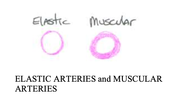
16. What is the largest type of blood vessel?
17. What do they contain a lot of?
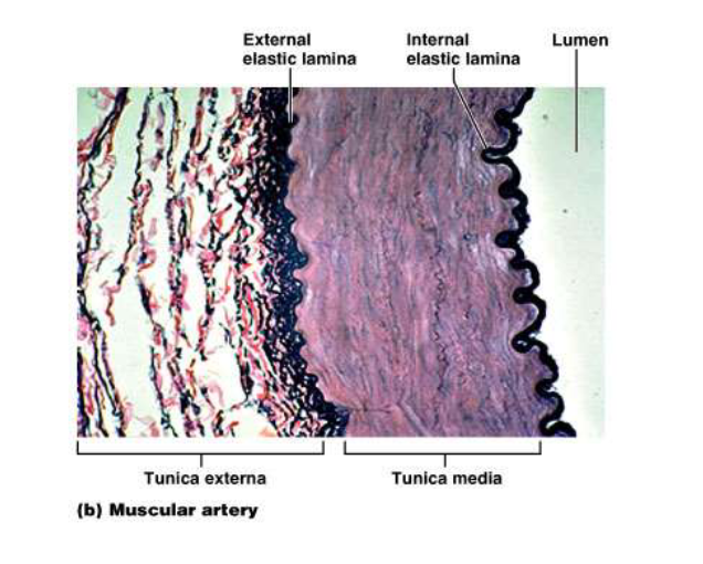
18. What type of artery is distal to elastic
arteries, and consists of most of the
19. named arteries?
20. What layer in these arteries is thick?
ELASTIC ARTERIES
There of lots of elastic fibers in the tunica media
Muscular Arteries
Tunica media is thick
21. What type of blood vessel can close the
lumen completely when it contracts?
What is a sac-like outpouching of an artery
called?
Aneurysm
What are three causes of an aneurysm?
– Defect in part of the artery wall
– High blood pressure
– Congenital (present at birth)
How to Recognize a Stroke (“STROKE”)
S * Ask the individual to SMILE.
T * Ask the person to TALK and SPEAK A
SIMPLE SENTENCE (Coherently; i.e. It is
sunny out today)
R * Ask him or her to RAISE BOTH ARMS.
O * Open the mouth and stick out the tongue
K * Keep them comfortable and still
E * Get EMERGENCY help (911)
If one side of the body responds differently
than the other side, or if they have trouble
with the task, call 911.
Which artery in the thigh is a good place to
take a pulse?
Femoral artery: good place to take a pulse since it is
superficial, but that also makes it susceptible to injury.
The circle of Willis forms a loop around which
structures?
Circle of Willis forms a loop around the pituitary gland
and the optic chiasma
27. Which are the smallest blood vessels, and are
found everywhere?
28. Which layer do they have?
CAPILLARIES
They only have an endothelium
Where is the only site of nutrient, gas exchange, and waste exchange in the cardiovascular system?
capillaries
The diameter of a typical capillary is similar to
that of what?
Capillary diameter is similar to an erythrocyte
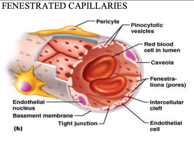
31. What are the three types of capillaries?
32. Which are the most common?
33. Which have pores?
34. Which have very large gaps?
Continuous – most common
Fenestrated (“window”) – have pores
Discontinuous (Sinusoids) – have very large gaps
Which capillaries are found where lots of
fluids need to be moved back and forth, such
as in the small intestine?
What type of capillaries are found in the red
marrow because their gaps are so large, a
RBC can fit through?
What type of capillaries are found in the red
marrow because their gaps are so large, a
RBC can fit through?
37. What is found at the start of each capillary?
38. What is the function of this structure?
PRE-CAPILLARY SPHINCTER controls the flow of
blood to individual capillaries. It directs the blood flow
to specific cells.
If one cell is starving, the capillary next to it will open.
The sphincter opens and closes depending on the needs
of individual cells.
Blood always flows to those cells and tissues that
need it. There is not enough blood to go around.
Name the 2 varieties of VEINS
VENULE: This is the smallest. It takes blood from the capillary to the vein.
VEIN: takes blood towards the heart.
How is blood able to get uphill and return to
the heart without backing up?
Skeletal muscle pushes on the vein to move the blood uphill, and the valves keep it from falling backwards and backing up.
What are the only BLOOD vessels that have
valves
Veins are the only BLOOD vessels that have valves (although LYMPH vessels also have valves).
Name 3 BLOOD PUMPS
the heart
elastic arteries
muscles constricting the veins
What vessel is often used to bypass a
damaged coronary artery in coronary bypass
surgery, and is the most likely vein to
become varicose?
Greater Saphenous vein
What vessel drains from the danger triangle
into the dural sinuses of the brain?
facial vein
What vessel is oxygen poor, but contains the lowest concentration of nitrogen waste?
renal vein
Name 3 Veins that are rich in oxygen and
nutrients:
umbilical vein
hepatic portal vein
pulmonary vein
47. What is the term for a vein that has
incompetent valves?
48. What is the name of the condition when a
person has inflamed veins?
49. What are three treatments for this
condition?
VARICOSE VEINS
PHLEBITIS
LASER, SCLEROSING, SURGERY
What is edema?
the accumulation of excess tissue fluid in loose
connective tissue
51. What are the two types of EDEMA?
52. Which is more severe?
pitting: worse in the evening
non-pitting: This is more severe because it does not go away easily. Just as swollen in the morning as in the evening.
53. What causes VENOUS STASIS ULCERS?
54. Who gets them most often?
55. What are three issues that must be treated
when one gets a venous stasis ulcer?
Varicose veins with excess acid products from
the blood plasma (sugar, carbon dioxide, etc),
which eventually erode all the way to the skin.
Common in diabetics.
Treatment must address sugar levels, vein
problem, and the open wound.
Why is PHLEBITIS dangerous?
Phlebitis can be associated with the
formation of blood clots (thrombosis),
usually in the deep veins of the legs, the
condition is called Deep Vein
Thrombophlebitis (DVT).
What are the signs and Symptoms of DVT?
Redness (erythema) and warmth with a
temperature elevation of a degree or more above
the baseline
Pain or burning along the length of the vein
Swelling (edema)
Vein being hard, and cordlike
Need to go to the emergency room if all
symptoms are present
58. What are SPIDER VEINS?
59. Are they dangerous?
60. What are two treatments?
61. What vessel in the fetus connects the
pulmonary trunk to the aortic arch so that
most of the blood bypasses the immature
lungs?
Small superficial veins become varicose and do
not function properly.
Unsightly appearance but are not dangerous.
Injections of alcohol or saline into the vein will
sclerose them (scar them shut), or laser
Ductus arteriorsis
What structure in the fetus connects the
right atrium to the left atrium?
Foramen ovale
what does the foramen ovale become shortly after birth?
Fossa Ovalis