Neuroanatomy Labs 3 & 4
1/81
There's no tags or description
Looks like no tags are added yet.
Name | Mastery | Learn | Test | Matching | Spaced |
|---|
No study sessions yet.
82 Terms
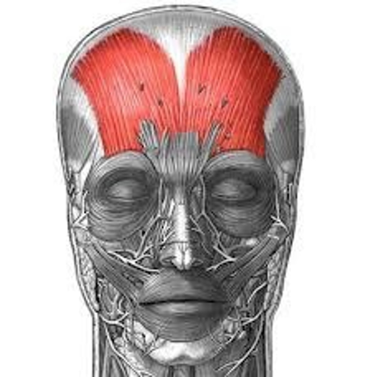
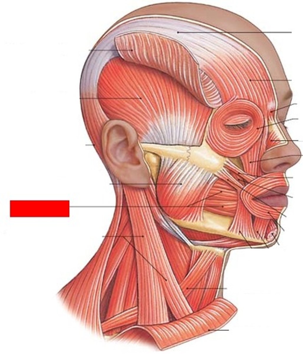
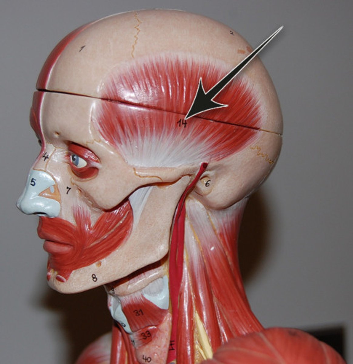
frontalis muscle
raises eyebrows
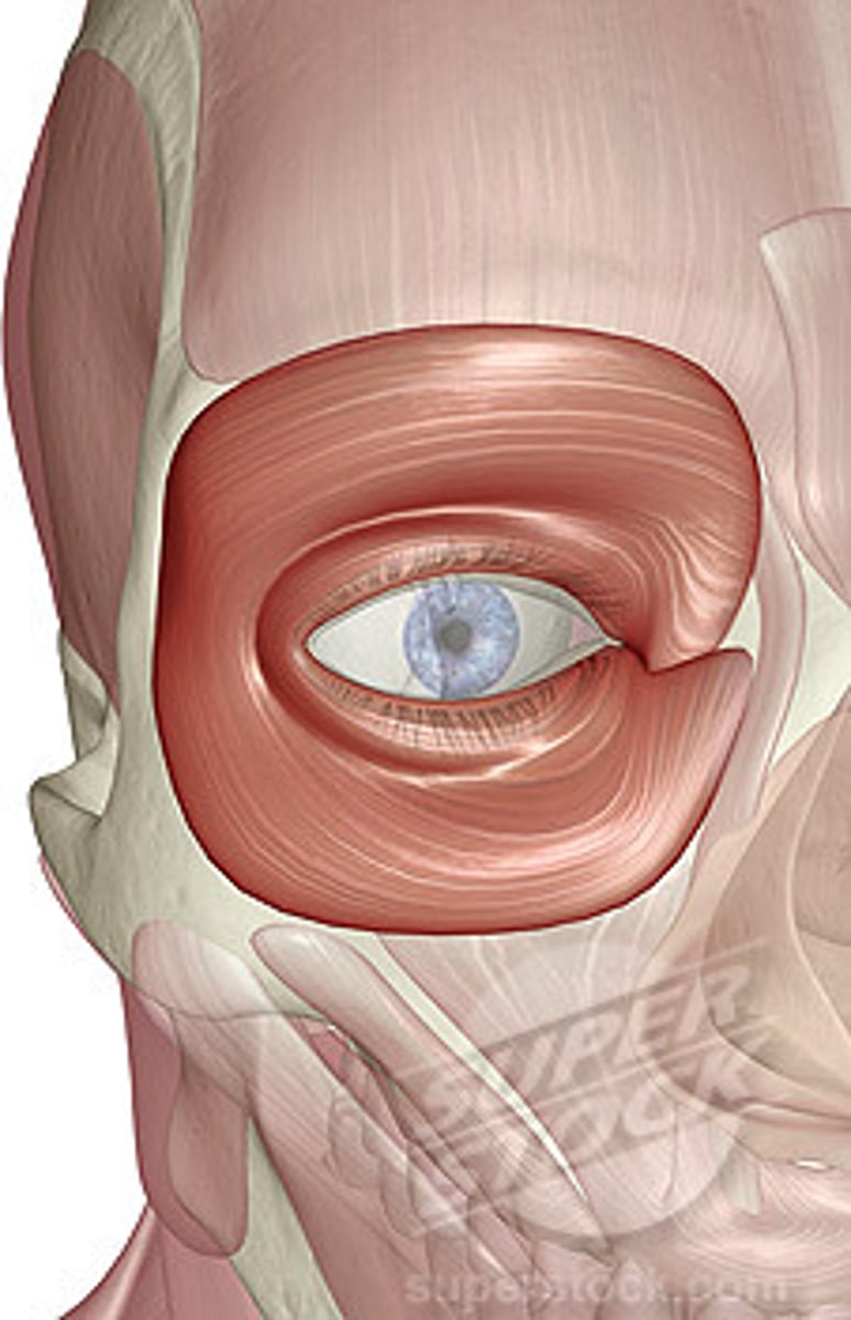
orbicularis oculi muscle
around the eye, closes the eyelids
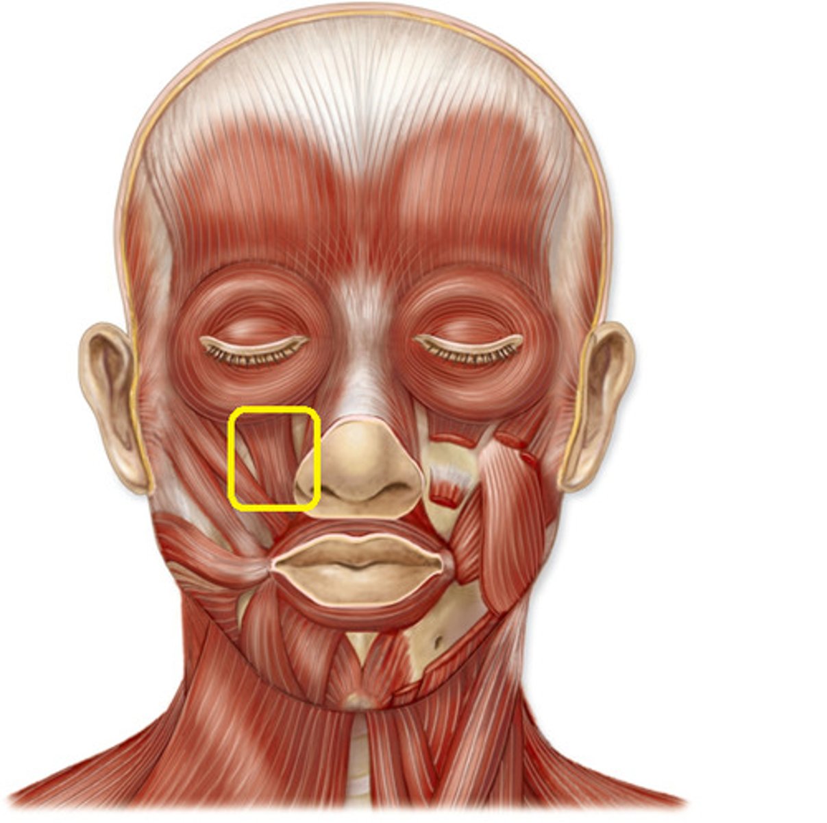
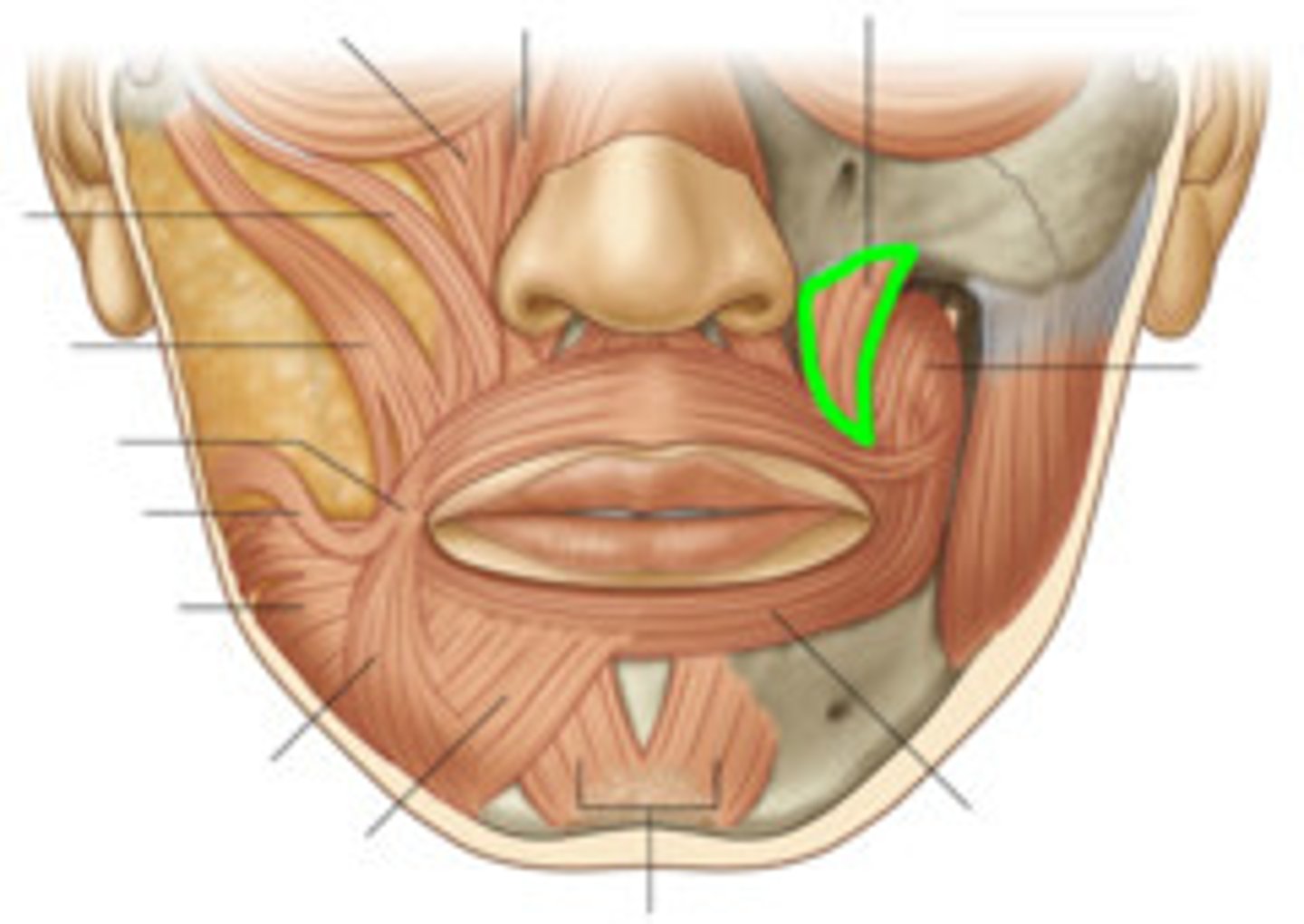
levator labii superioris muscle
elevates upper lip
levator anguli oris muscle
raises the angle of the mouth (look for muscle going to angle of mouth)
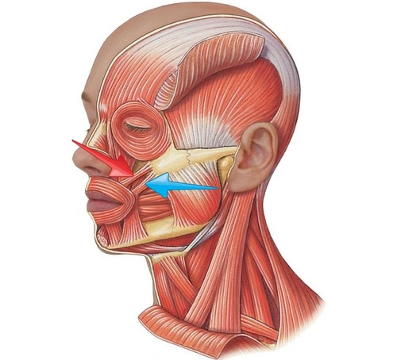
zygomaticus major muscle
contributes to laughing and smiling (pulls angles of mouth superolaterally)
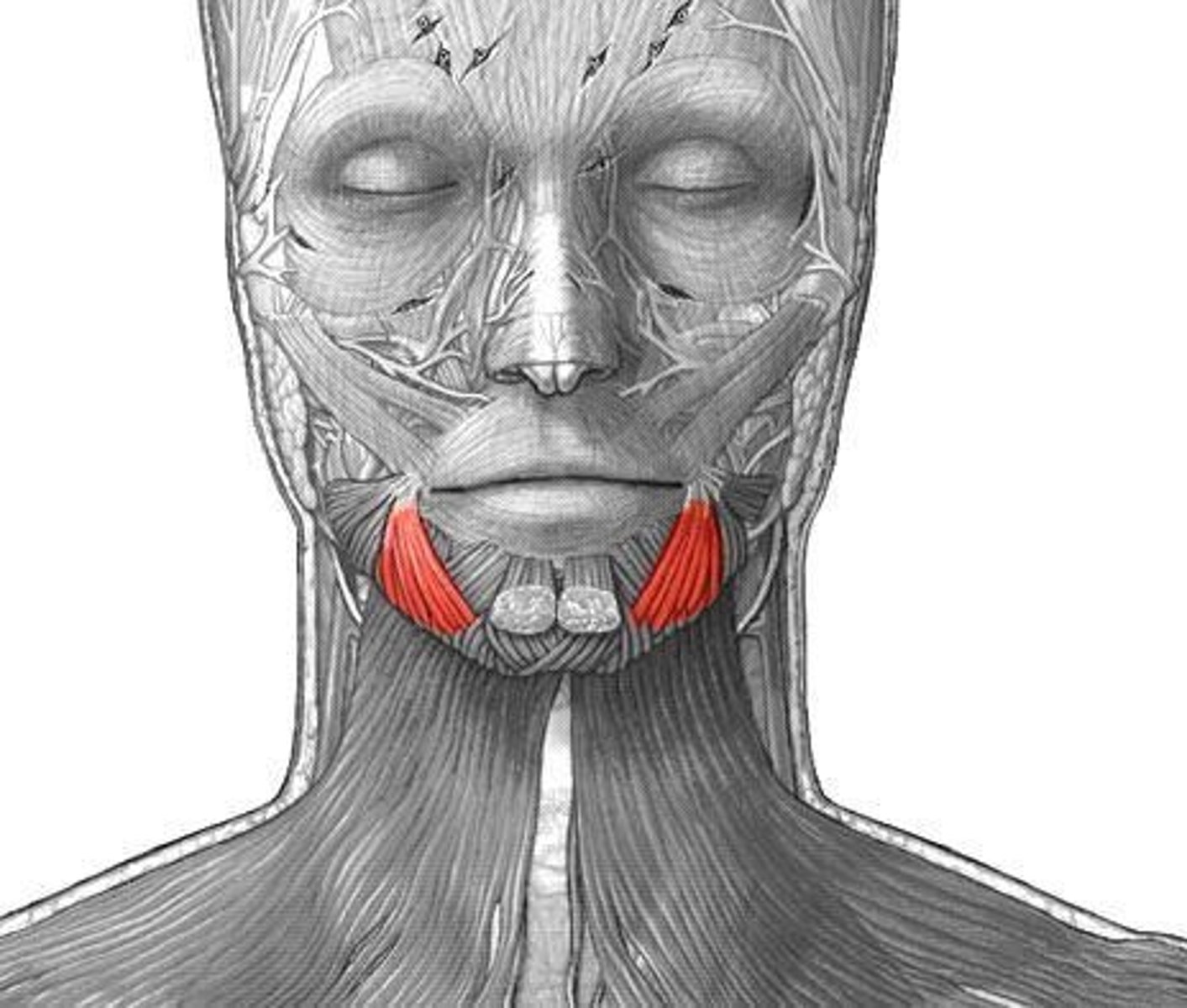
depressor labii inferioris muscle
depresses inferior lip
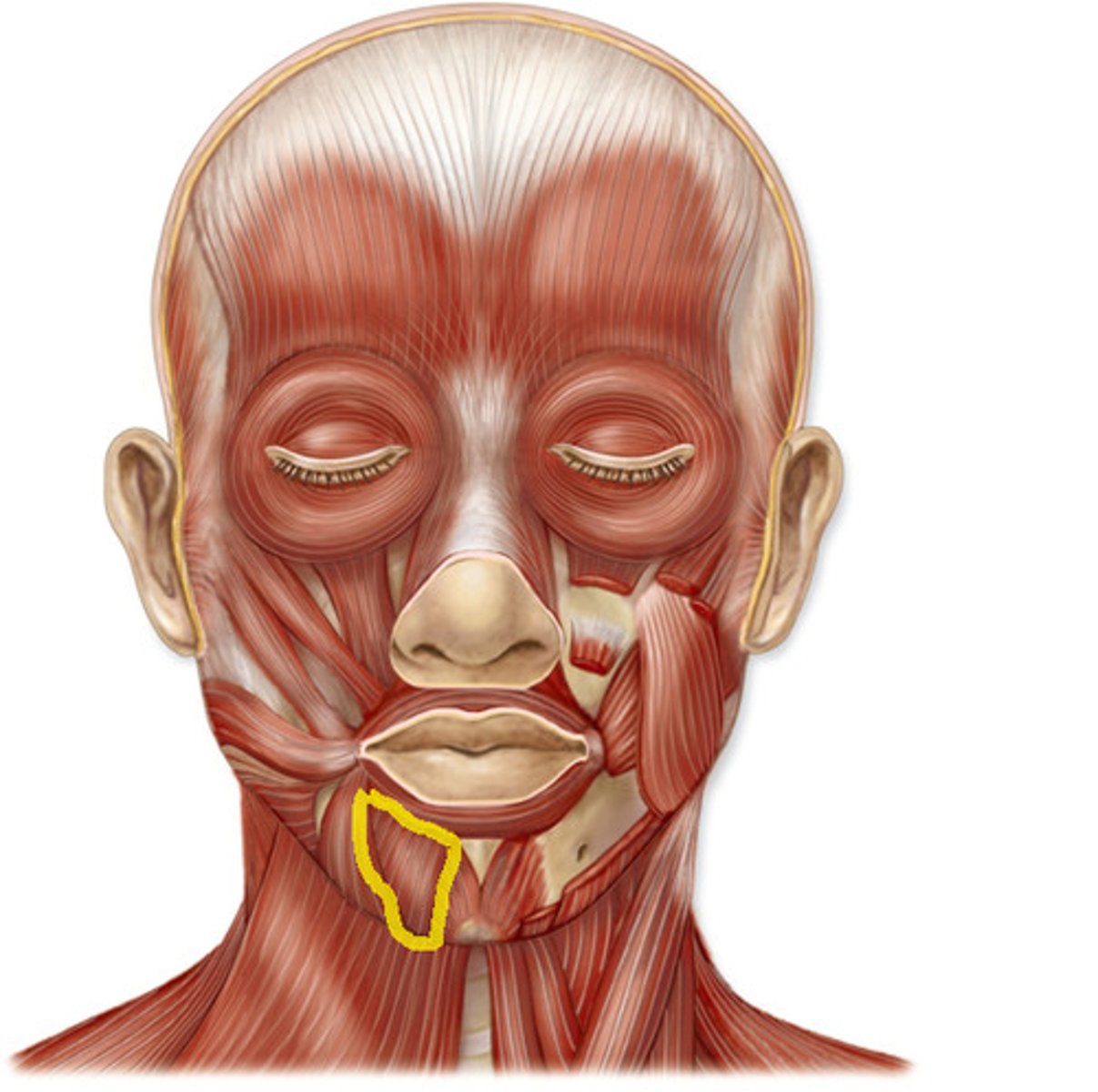
depressor anguli oris
depresses angle of mouth (look for muscle going to angle of mouth)
buccinator muscle
used for blowing air & positioning food when chewing
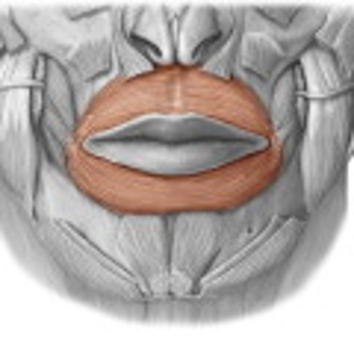
orbicularis oris muscle
closes lips, used for speech, eating, kissing, playing instruments, puckering
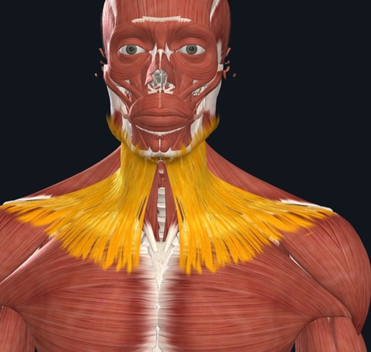
platsyma muscle
moves the mandible down
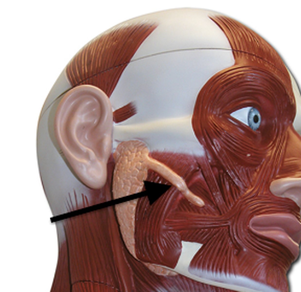
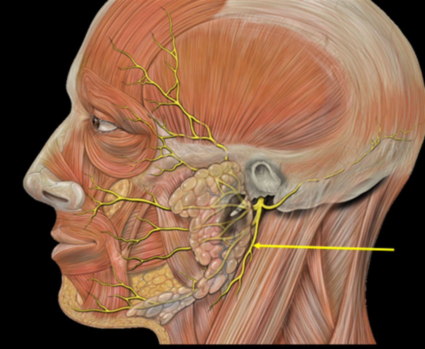
parotid gland
parotid duct
empties saliva
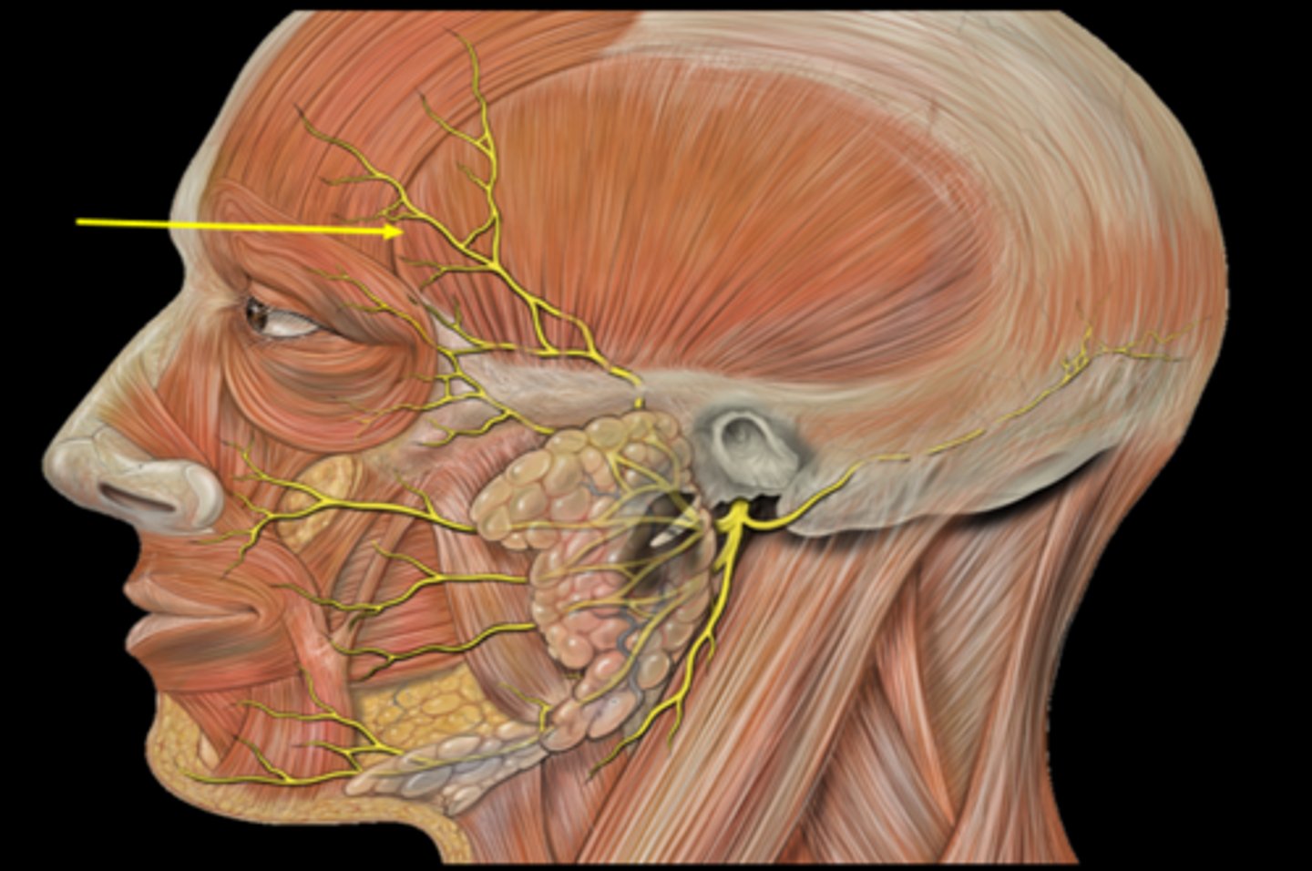
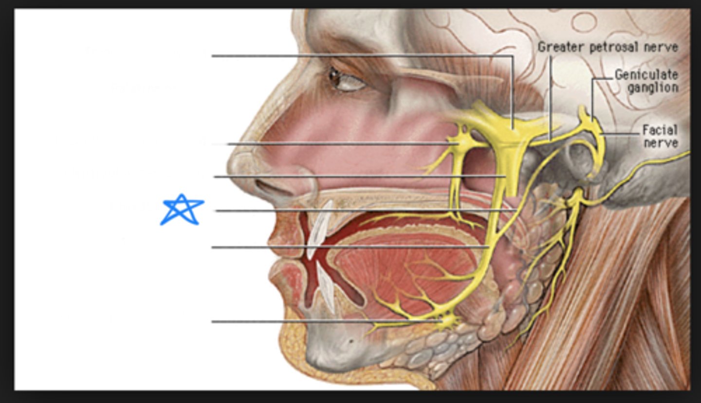
temporal branch of facial nerve
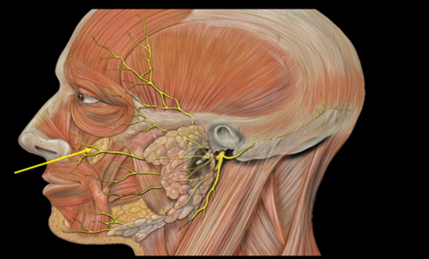
zygomatic branch
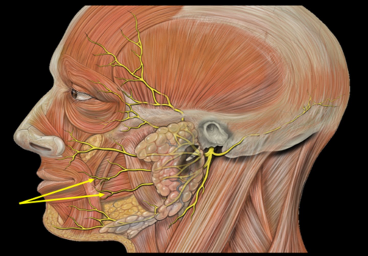
buccal branch of facial nerve
mandibular branch of facial nerve
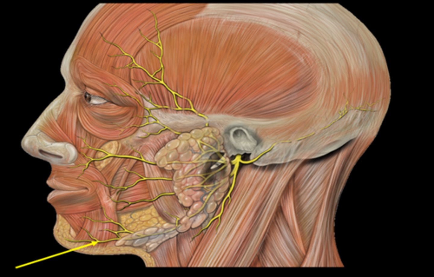
cervical branch of facial nerve
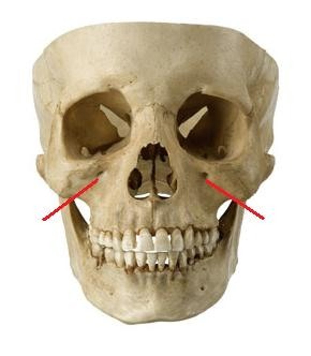
infraorbital neurovasculature (continuation of CN V2)
what bundle of nerves, arteries, and veins go through here?
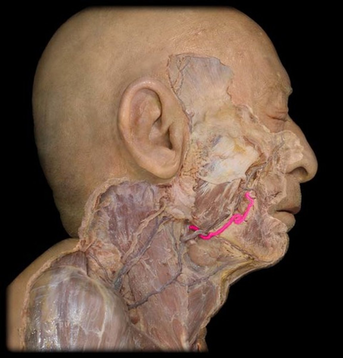
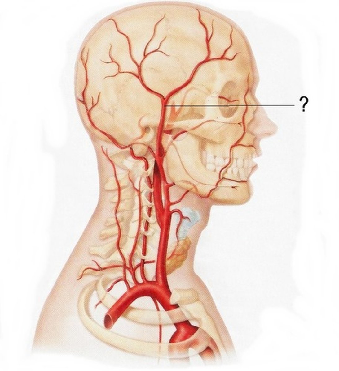
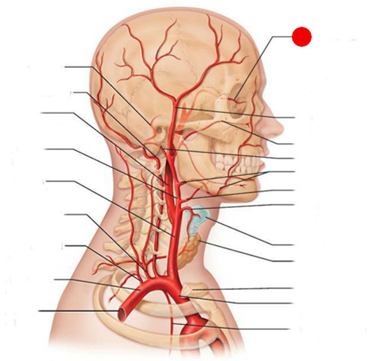
facial artery
mental neurovasculature (continuation of infraorbital neurovasculature)
what bundle of nerves, arteries, and veins go through here?
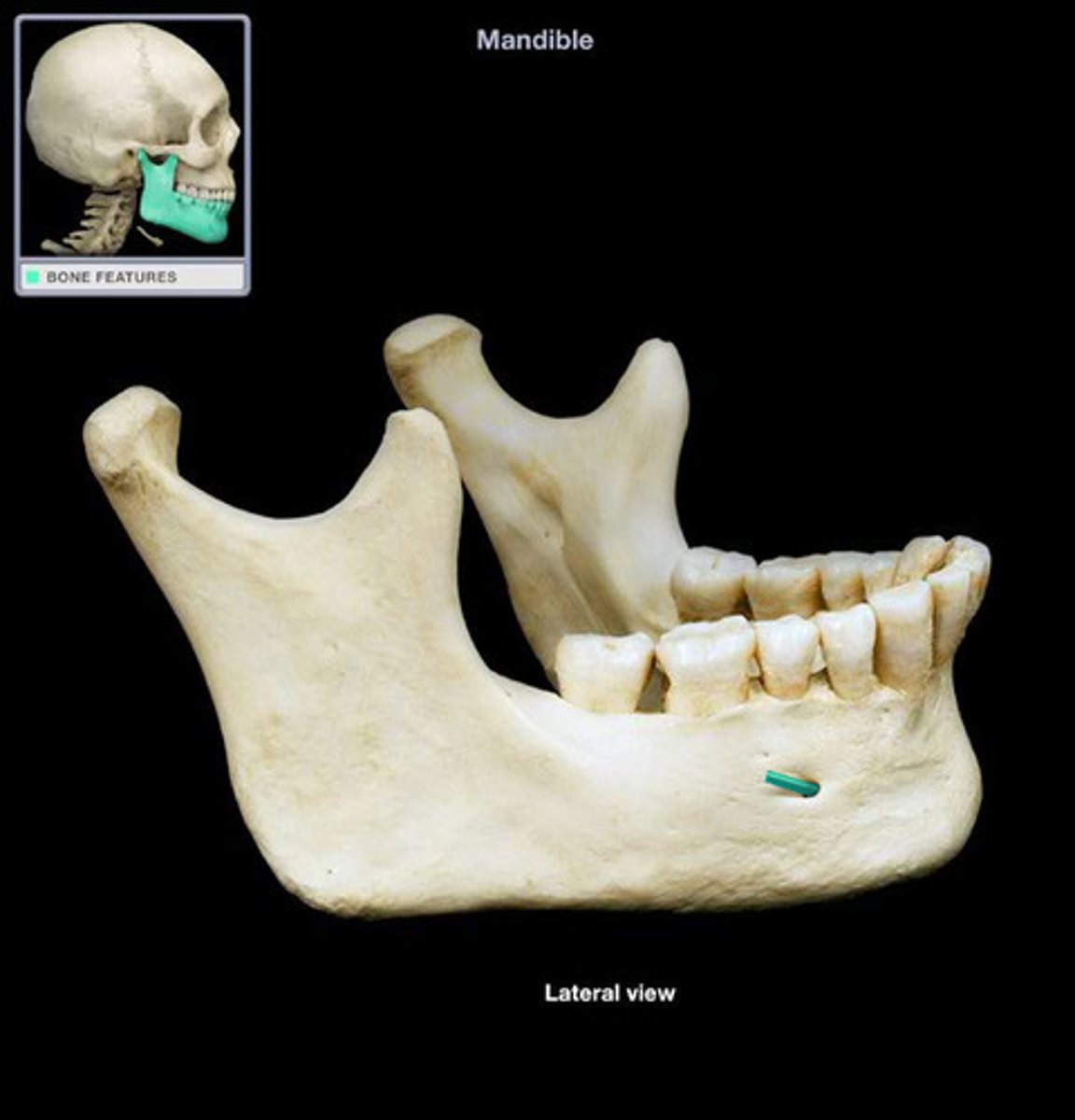
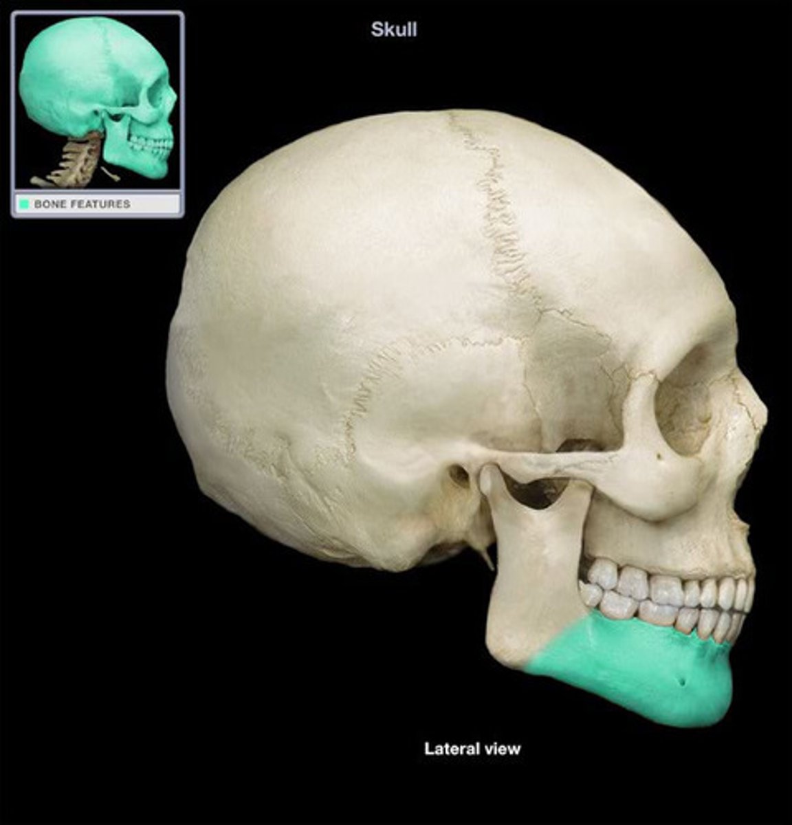
body of mandible
mental foramen
inferior alveolar neurovasculature become mental neurovasculature here

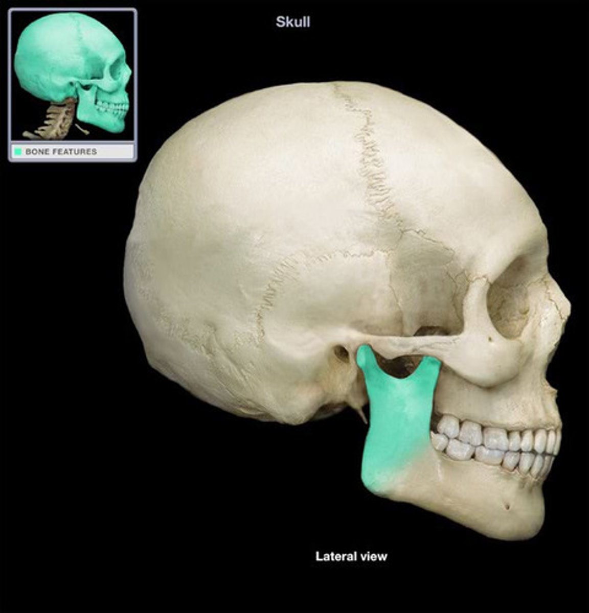
ramus of mandible
coronoid process
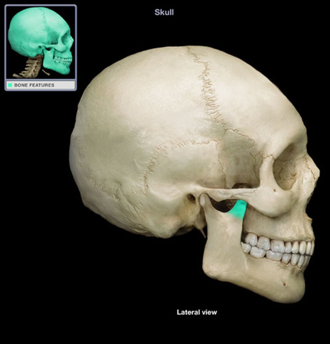
condylar process

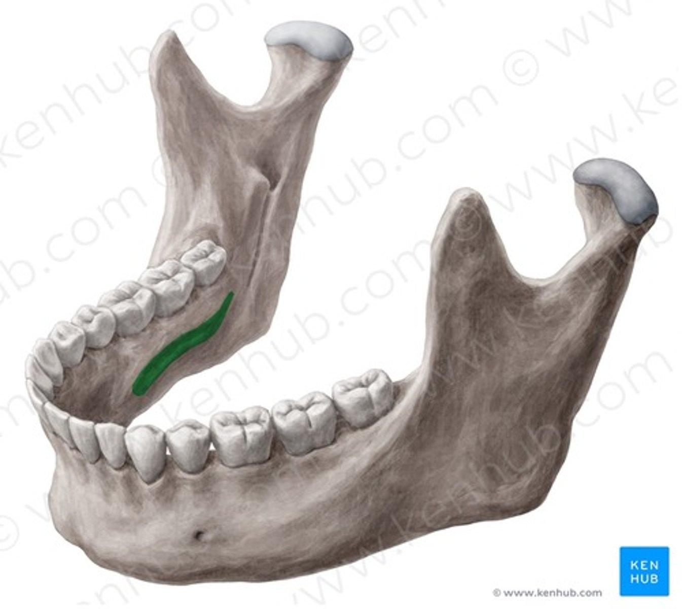
mylohyoid line (mylohyoid muscle attachment site)
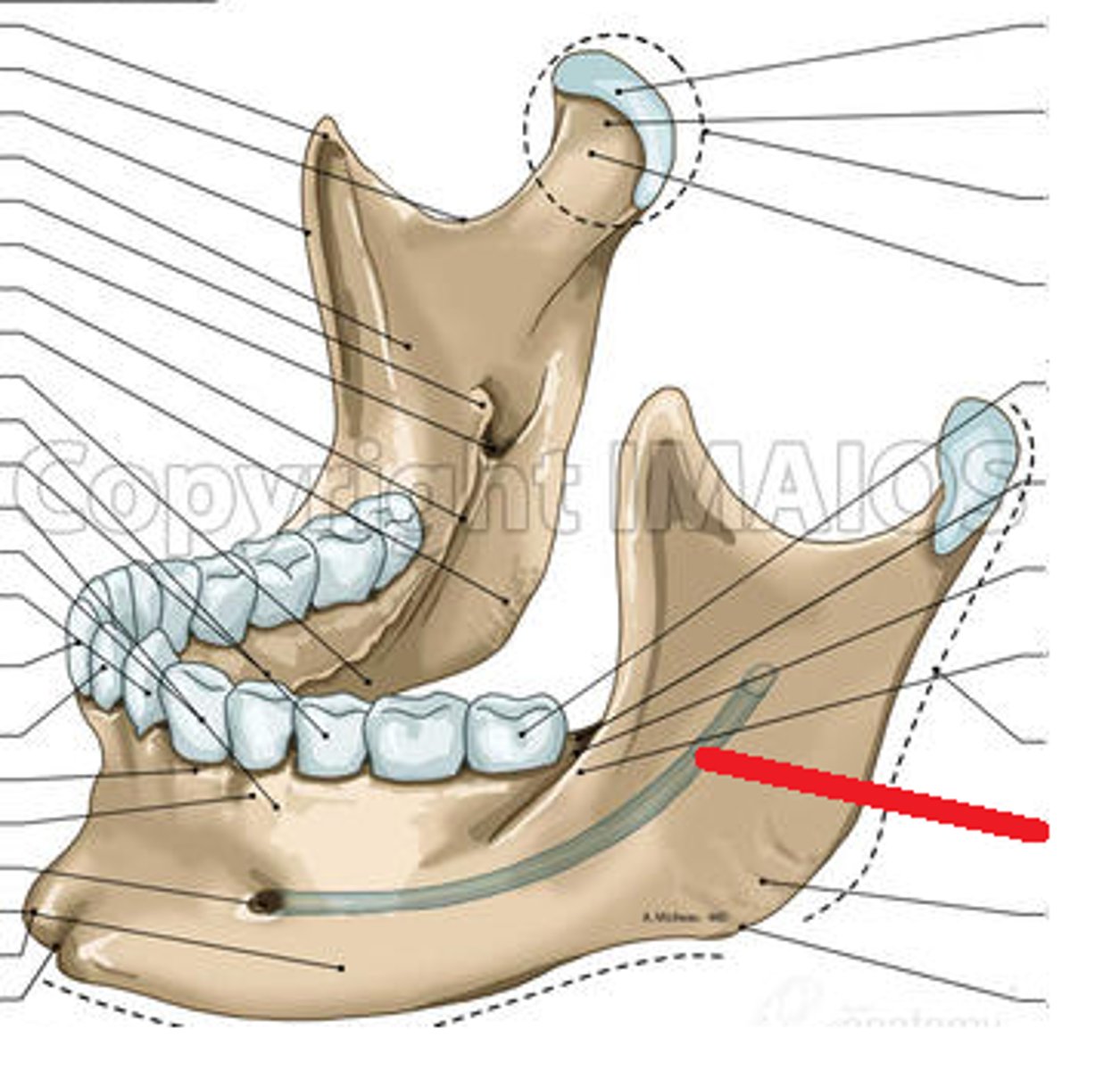
mandibular canal
temporalis muscle
closes mouth, innervated by V3
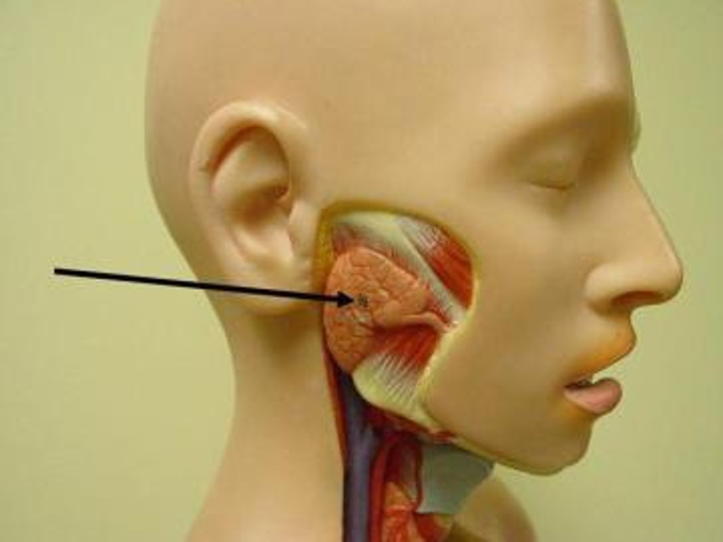
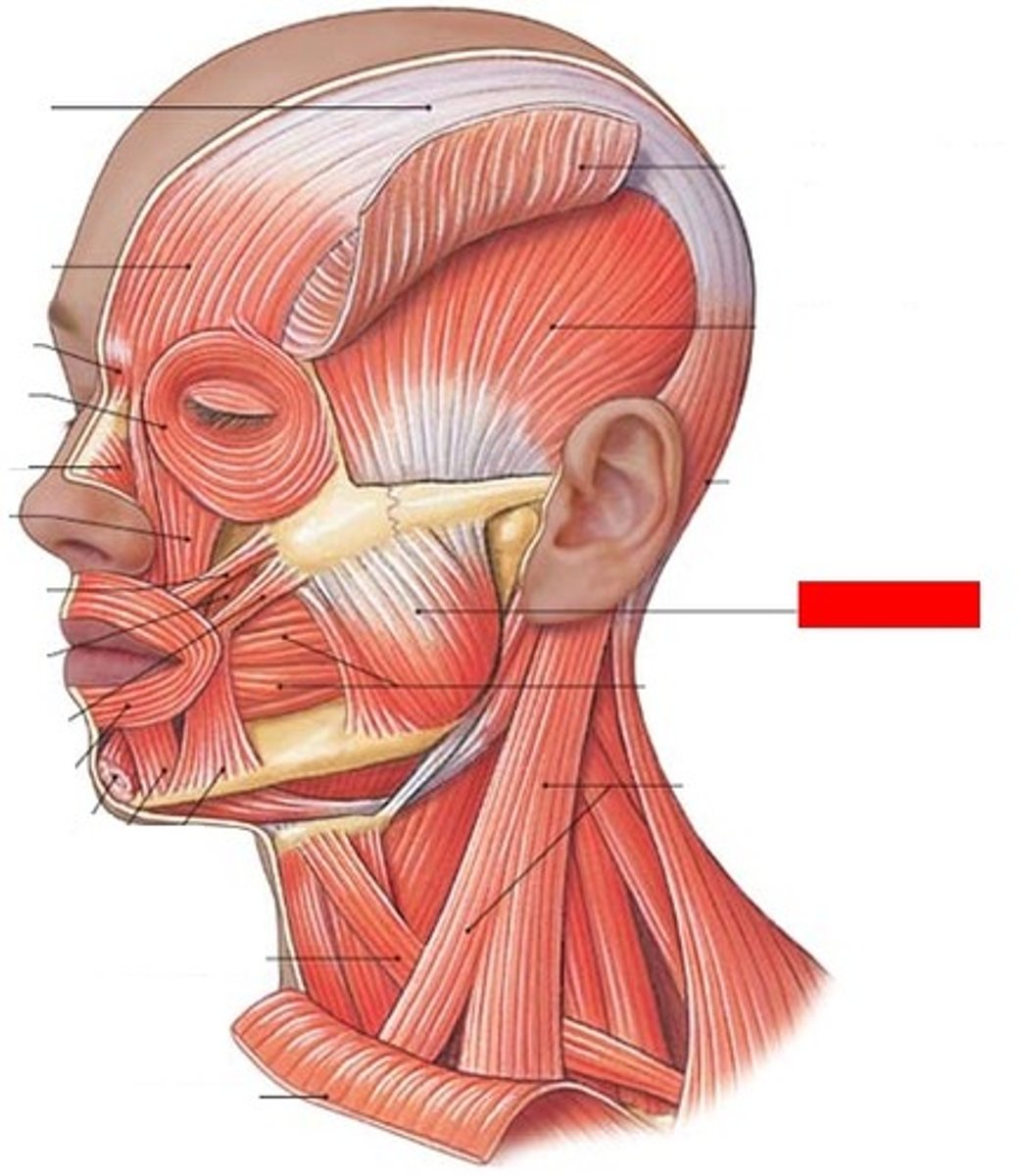
masseter muscle
innervated by V3
medial pterygoid muscle
elevates mandible, closes mouth, innervated by V3
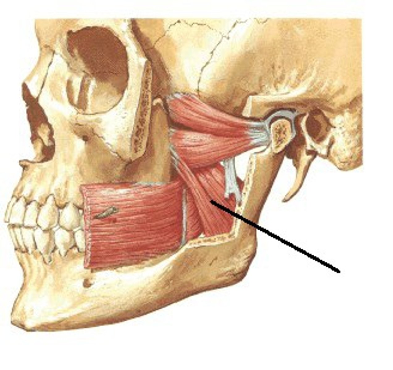
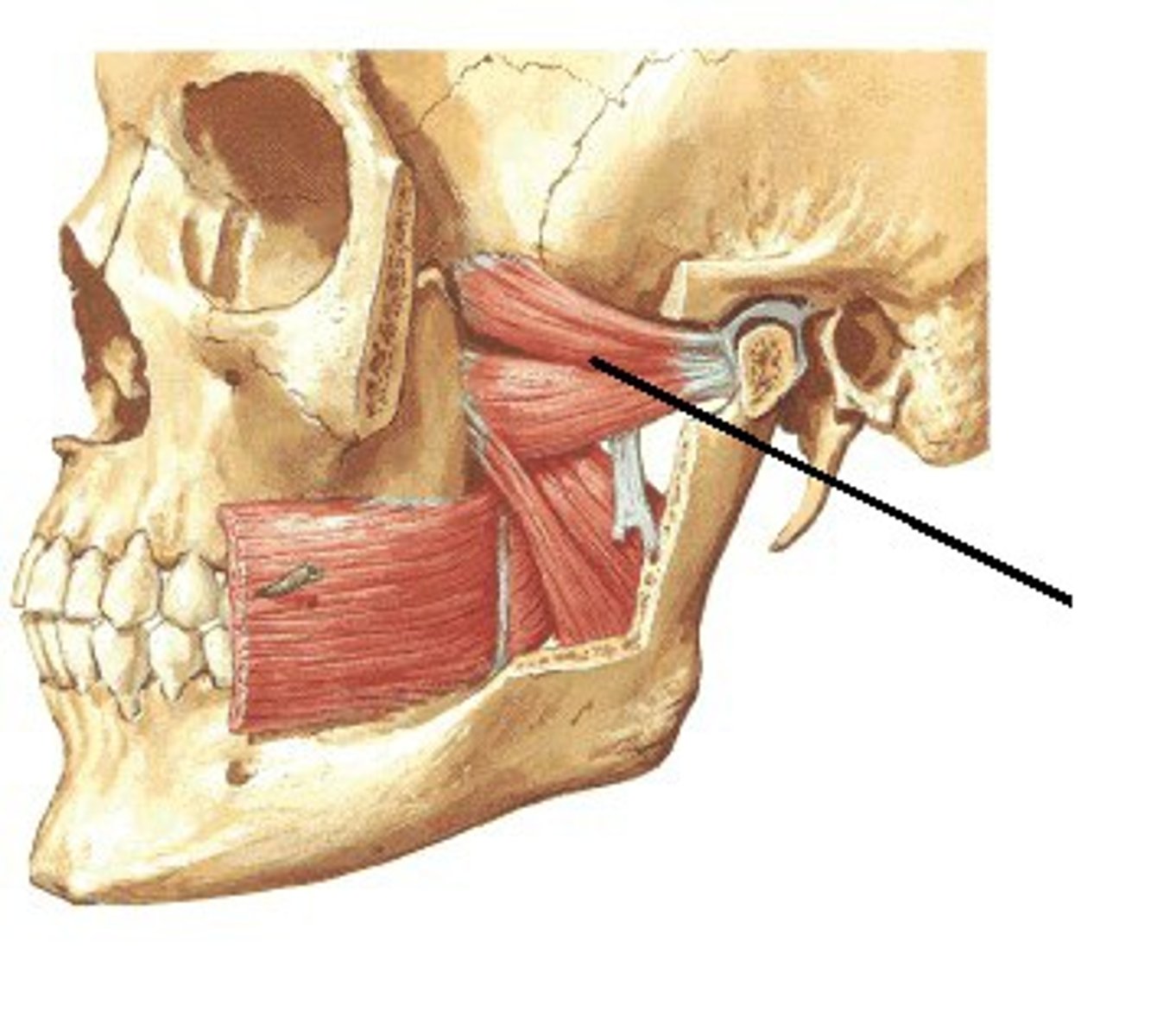
lateral pterygoid muscle
opens mouth, innervated by V3
maxillary artery

superficial temporalis artery
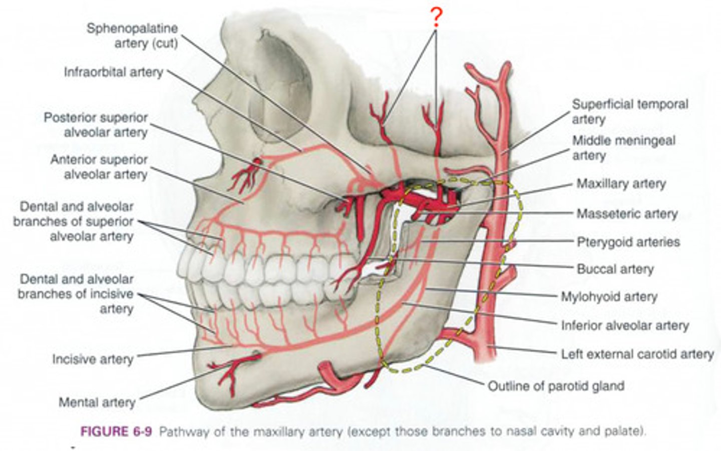
deep temporal arteries
middle meningeal artery
inferior alveolar artery

inferior alveolar nerve
branch of V3
nerve to mylohoid
branch of inferior alveolar nerve
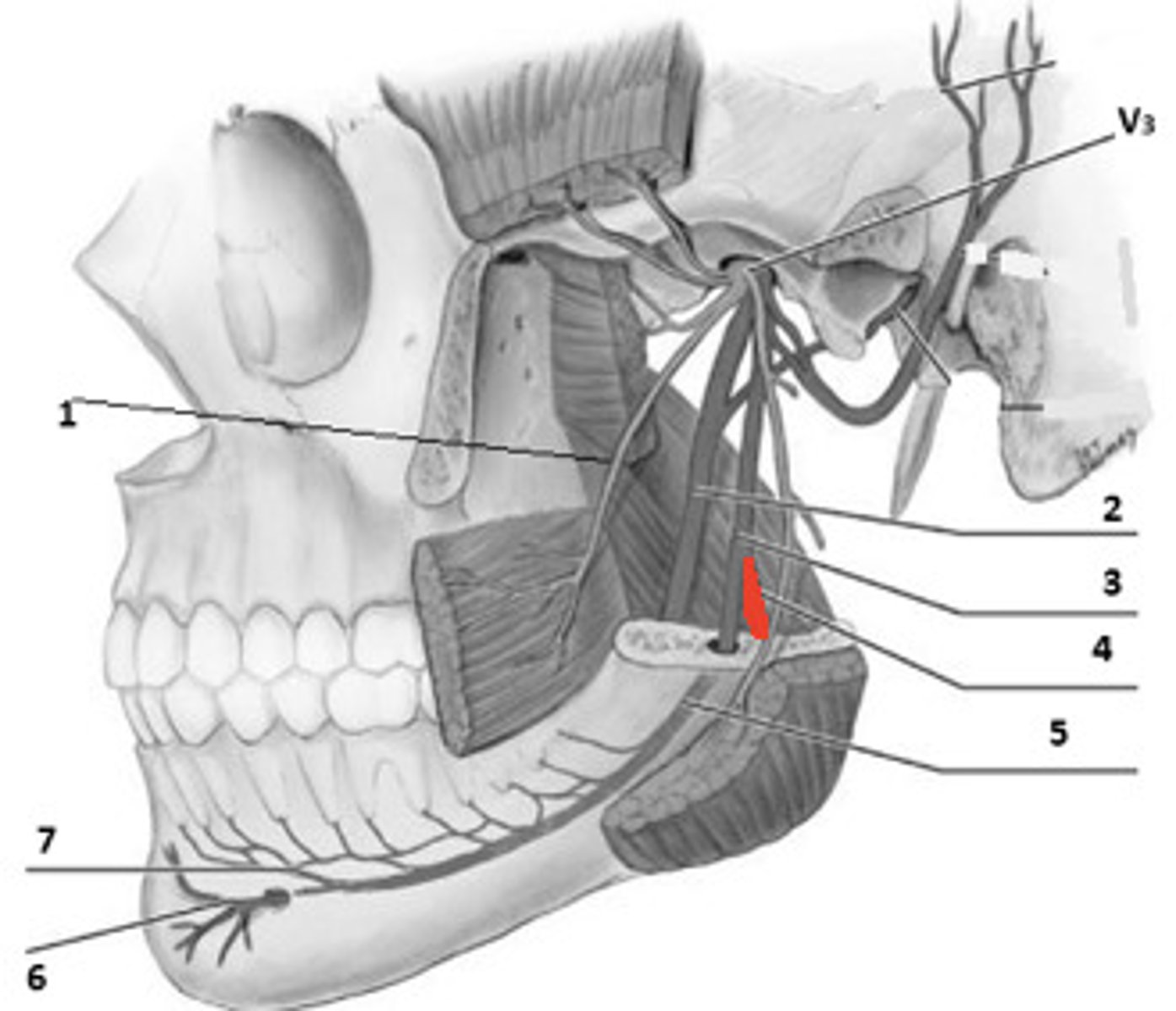
lingual nerve
Branch of V3, which supplies sensory innervation to the anterior 2/3 of the tongue
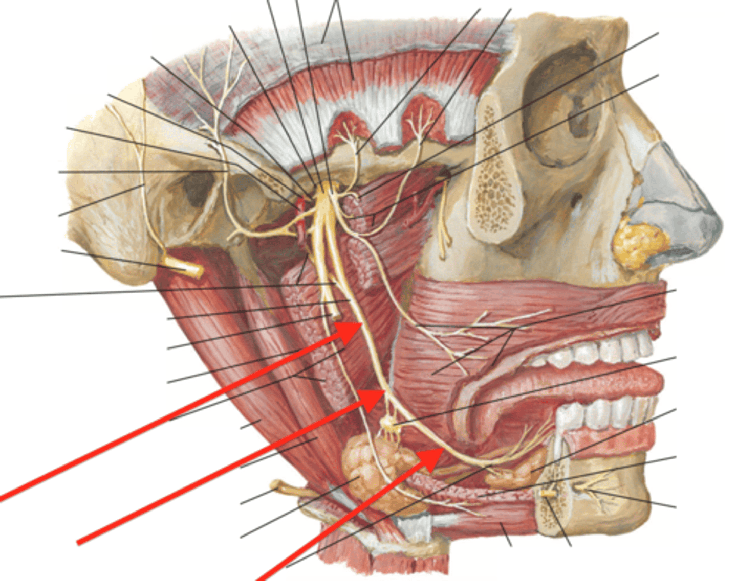
chorda tympani (joins lingual nerve)
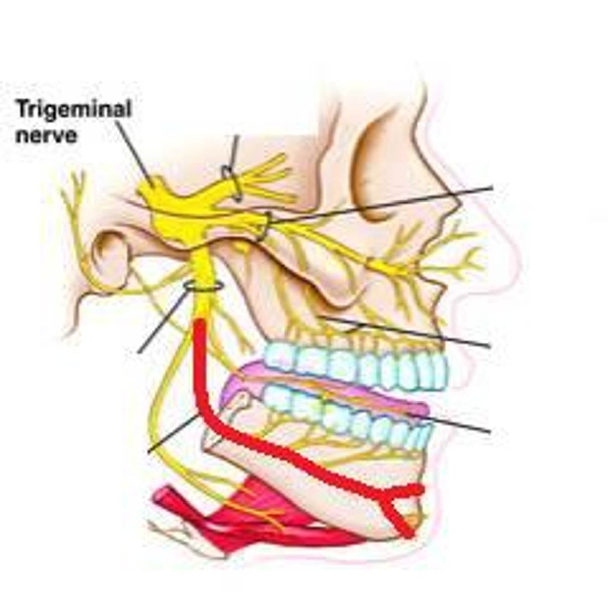
Mandibular nerve (V3)
retromandibular vein
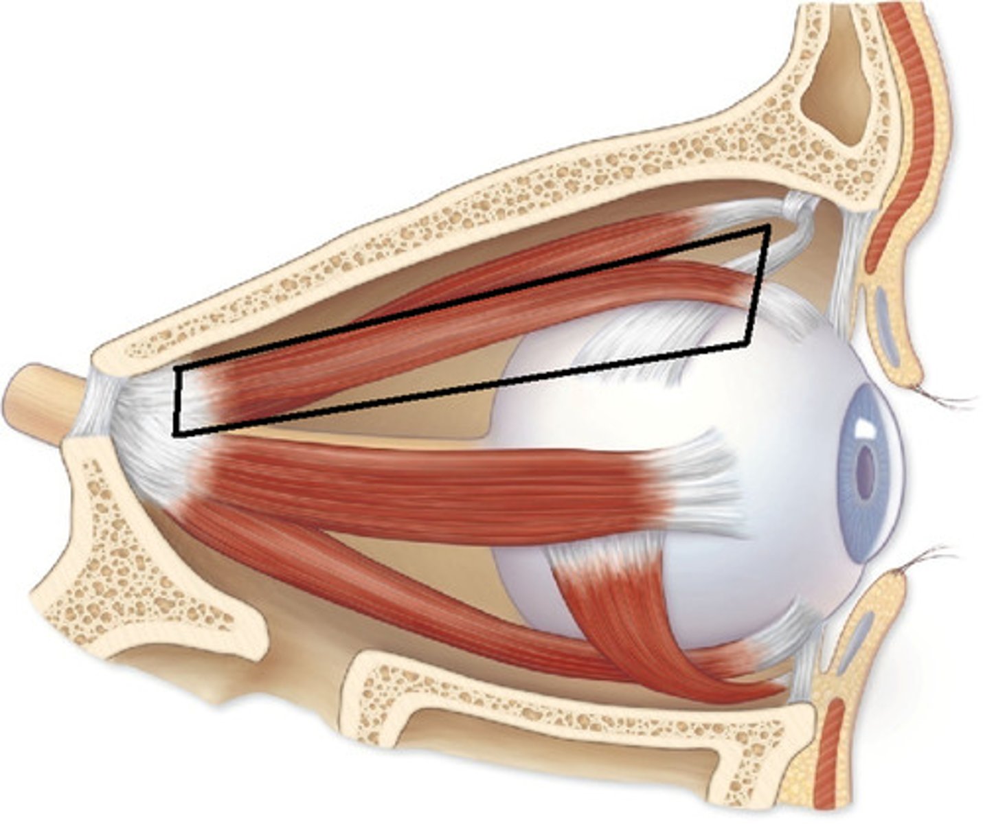
levator palpebrae superioris muscle
elevates upper eyelid, opens eye
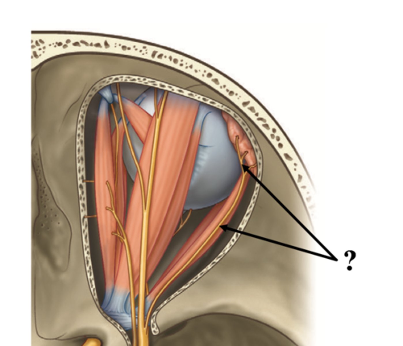
superior rectus muscle
elevates eye
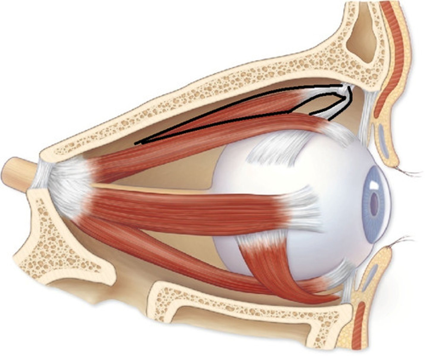
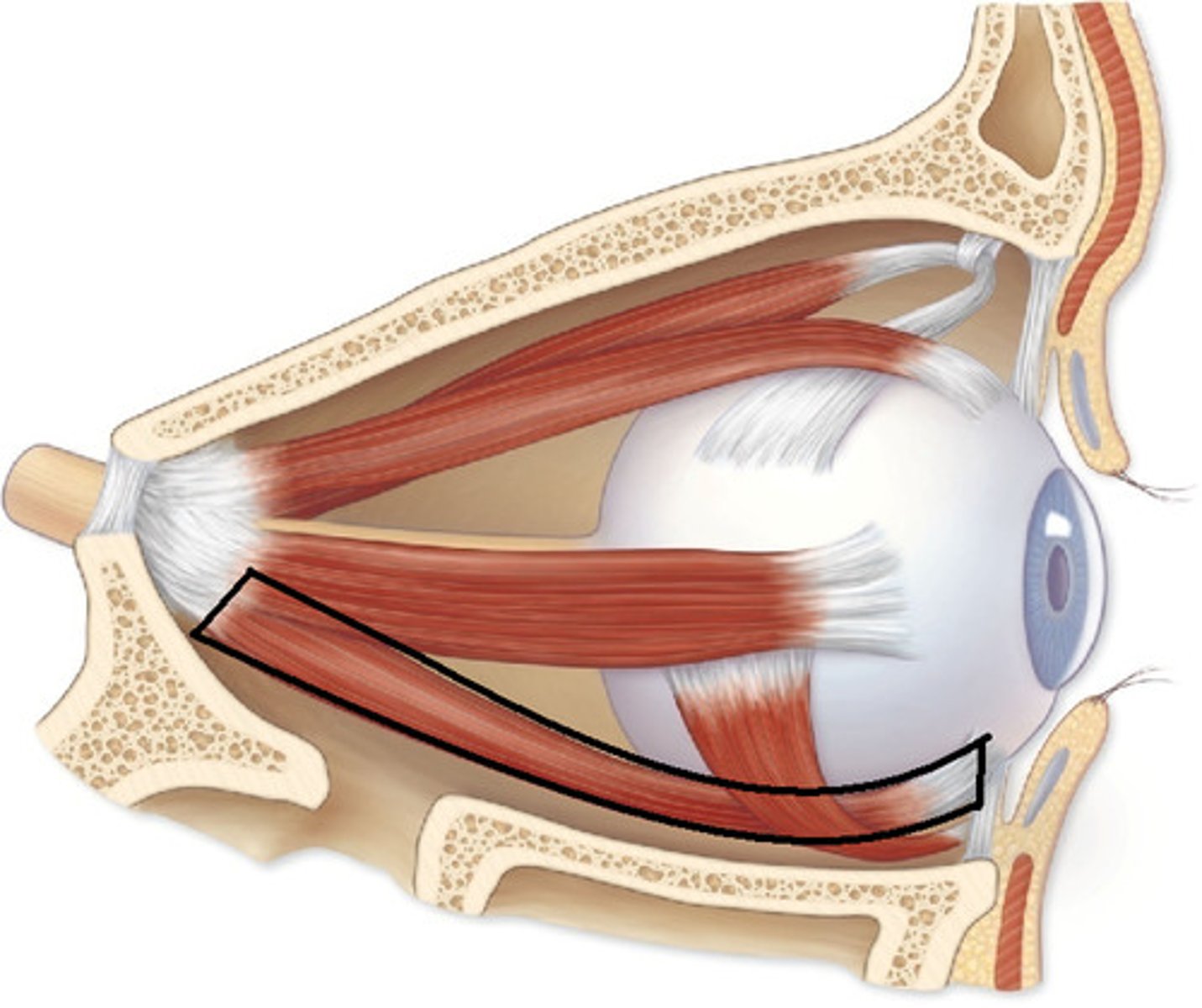
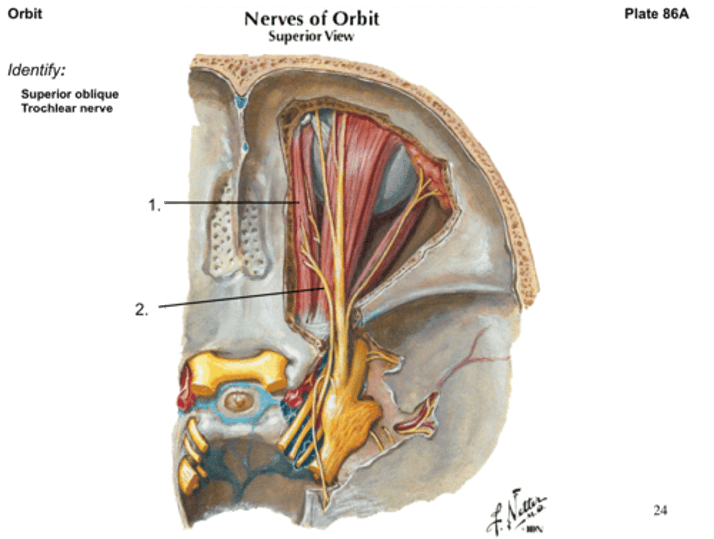
superior oblique muscle
depresses eye and turns it laterally
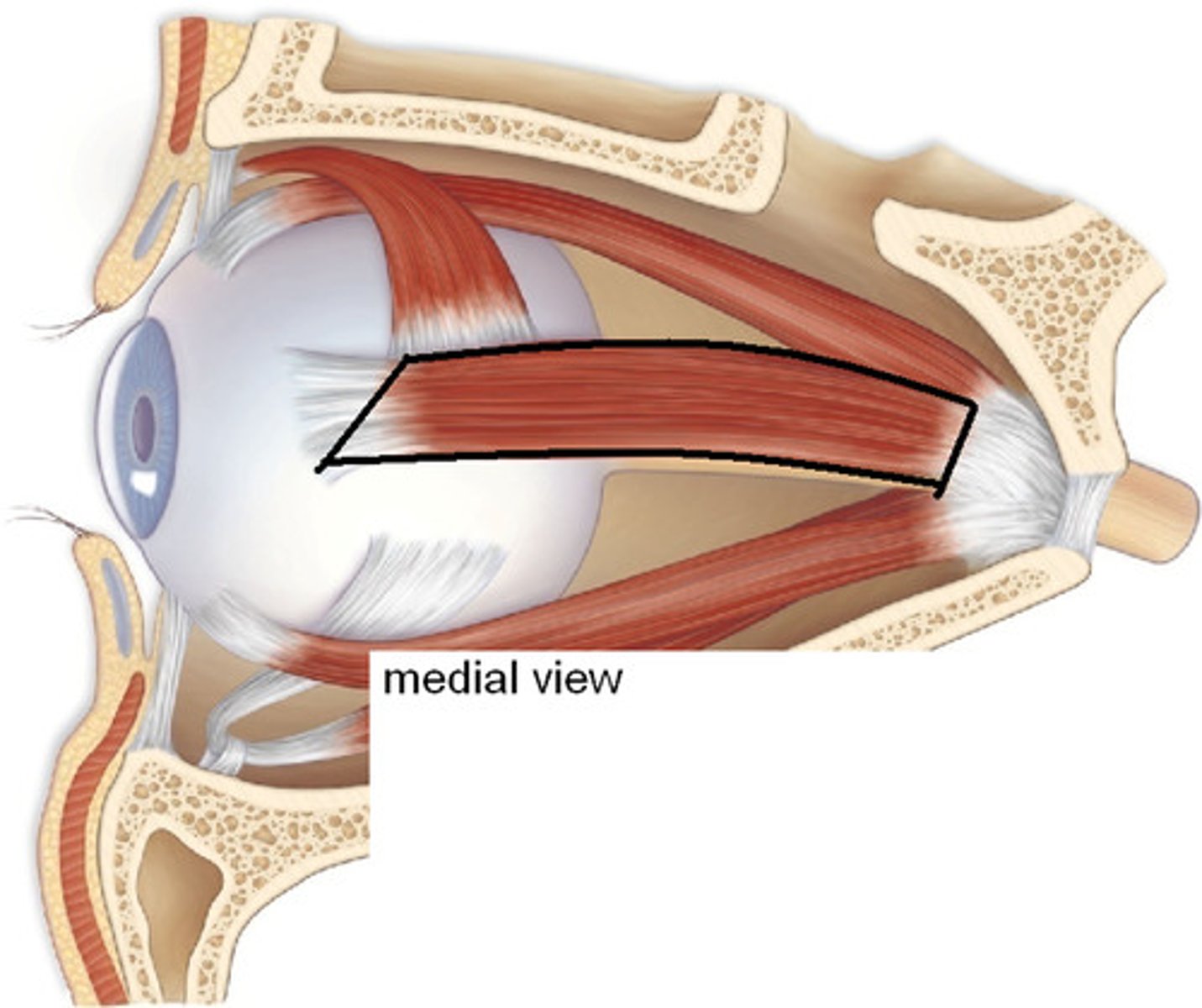
medial rectus muscle
moves eye medially
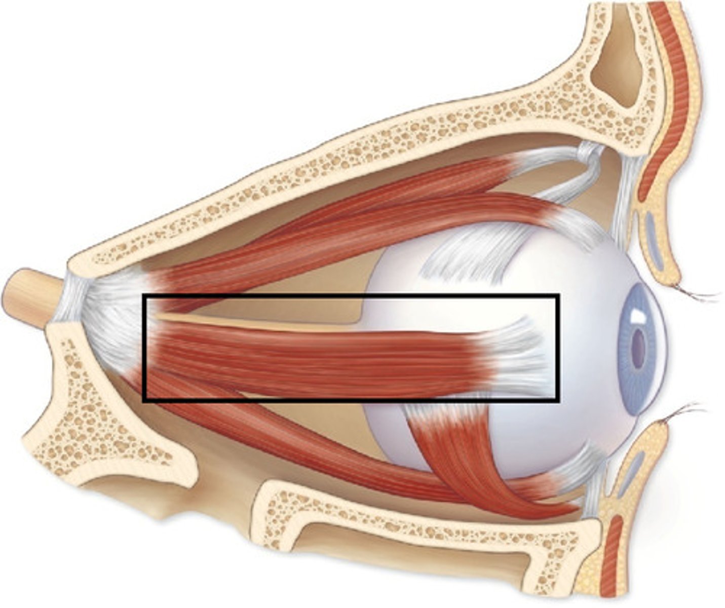
lateral rectus muscle
moves eye laterally
inferior rectus muscle
depresses eye
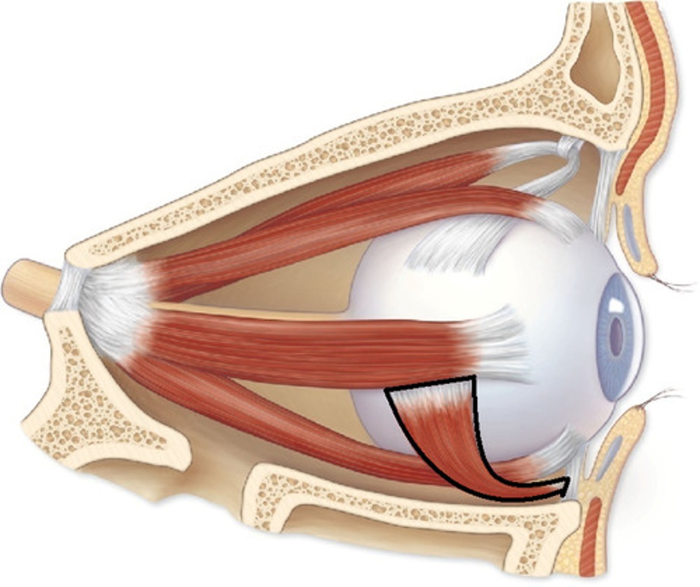
inferior oblique muscle
elevates eye and turns it laterally
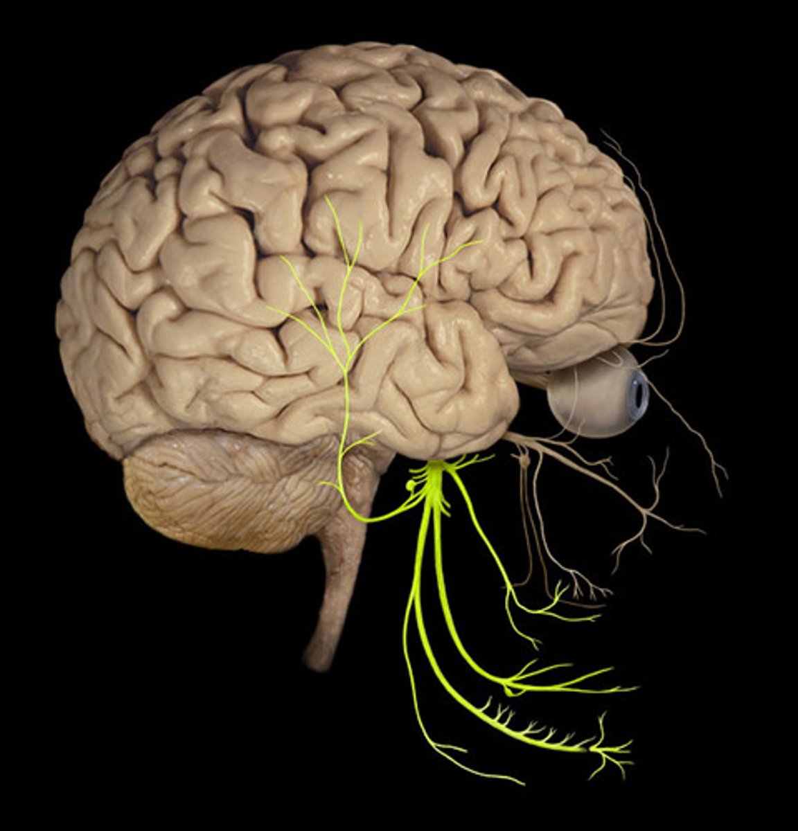
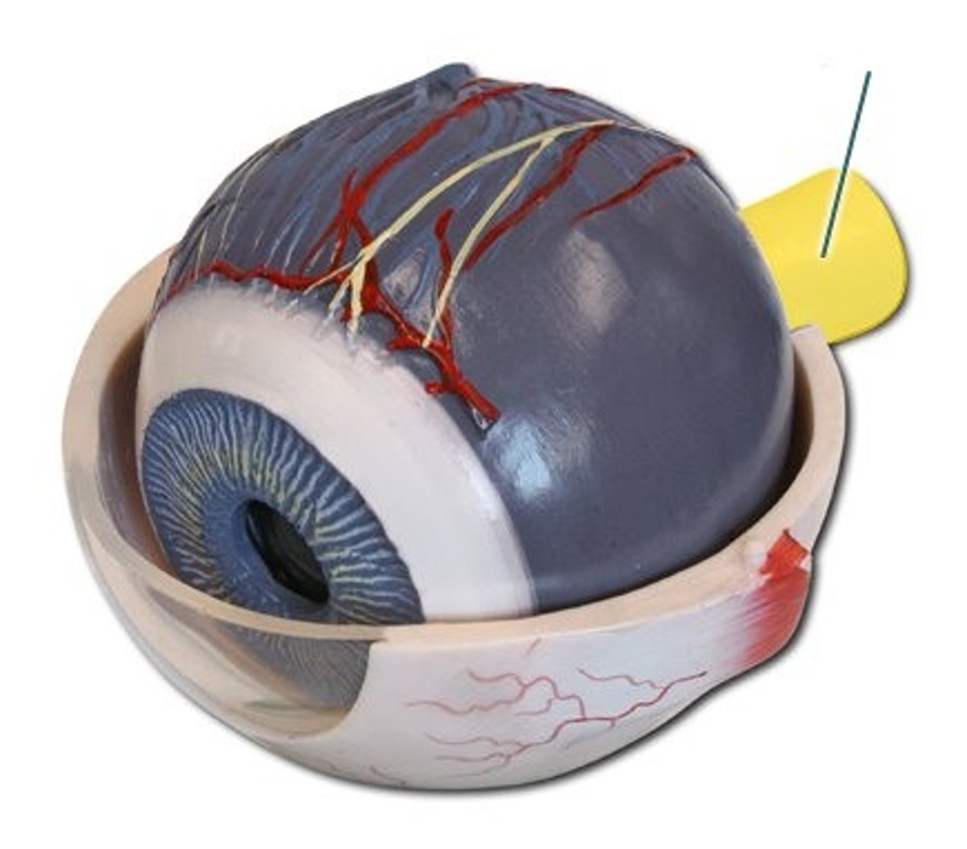
optic nerve
label the big yellow nerve projecting off the back of the eye
oculomotor nerve eye
which nerve innervates the superior rectus, medial rectus, inferior rectus, levador palpabrae superioris, and inferior oblique (sorry, couldn't find a good picture of this on the internet)
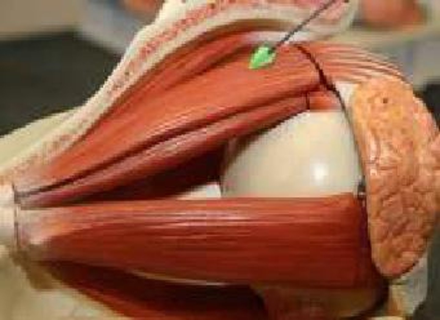
trochlear nerve orbit
Which nerve?
Frontal nerve (V1)
Supratrochlear nerve
Label Nerve 1

Supraorbital nerve
Label Nerve 2
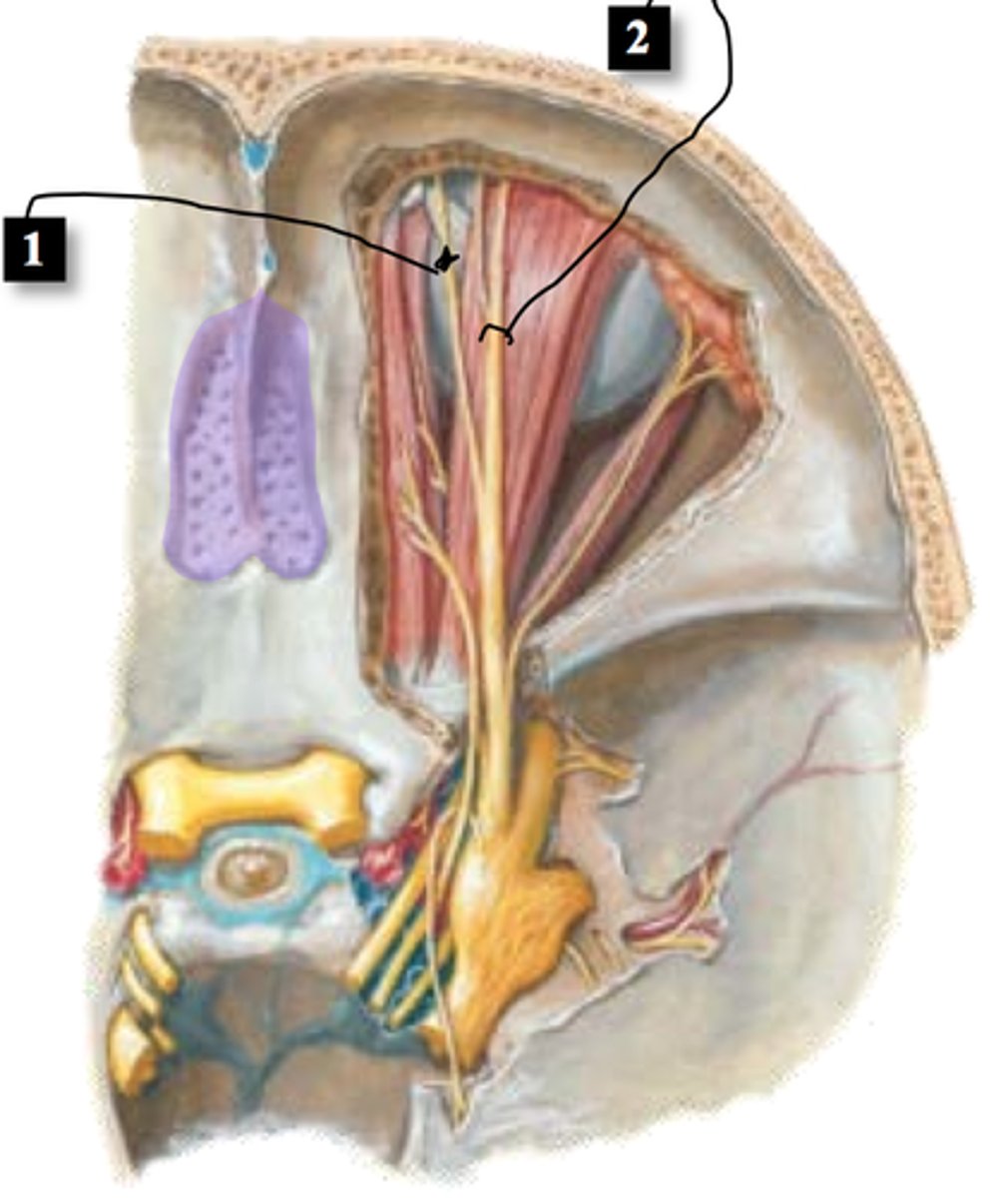
Supraorbital nerve
ophthalmic artery orbit
Name the artery that supplies the blood to the eye
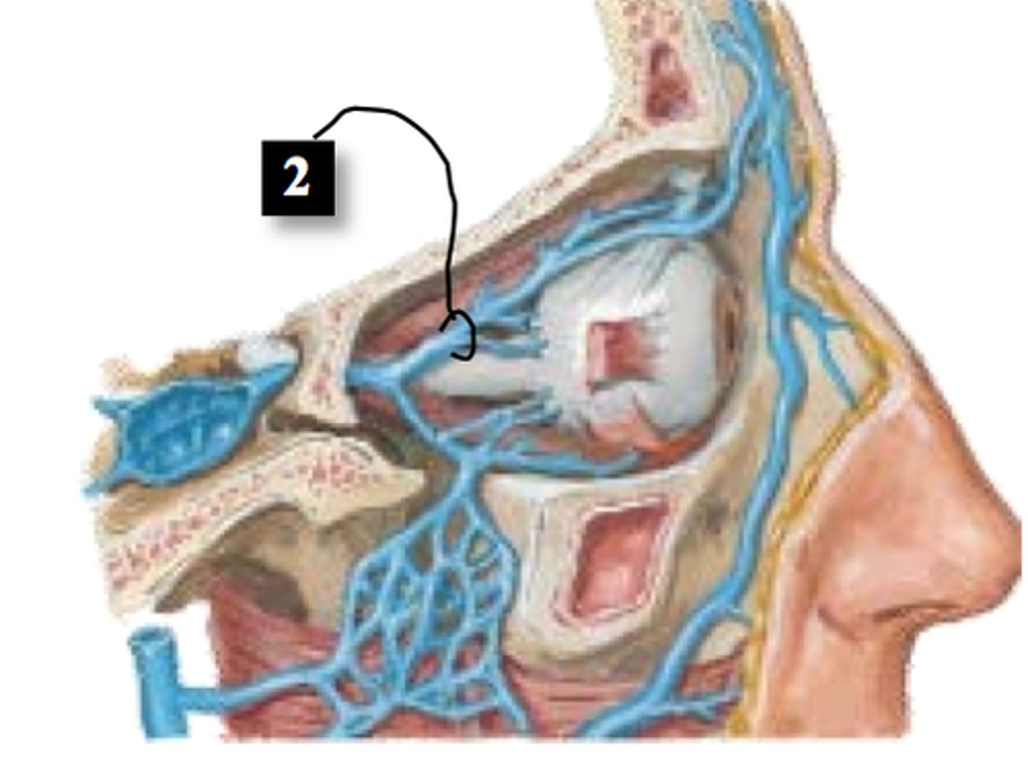
superior ophthalmic vein
Lacrimal nerve (V1) & gland
Label the gland and the nerve that innervates it
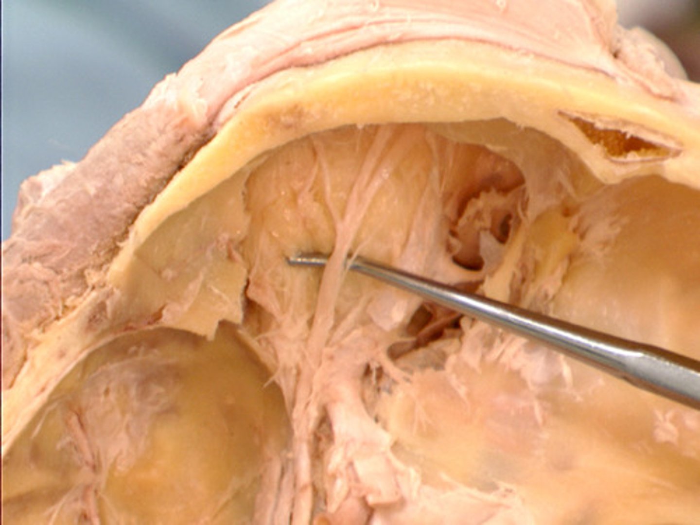
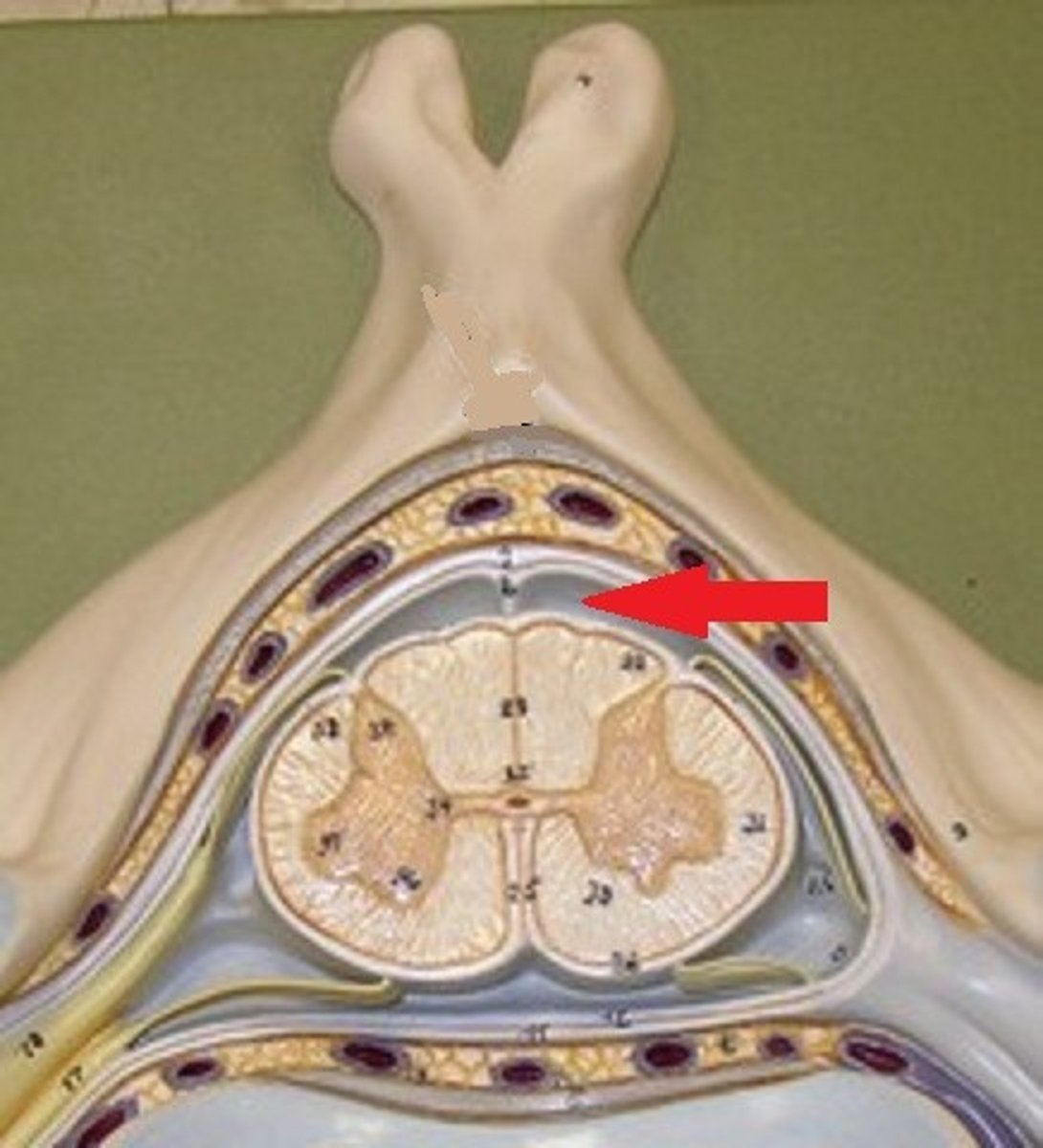
spinal epidural space
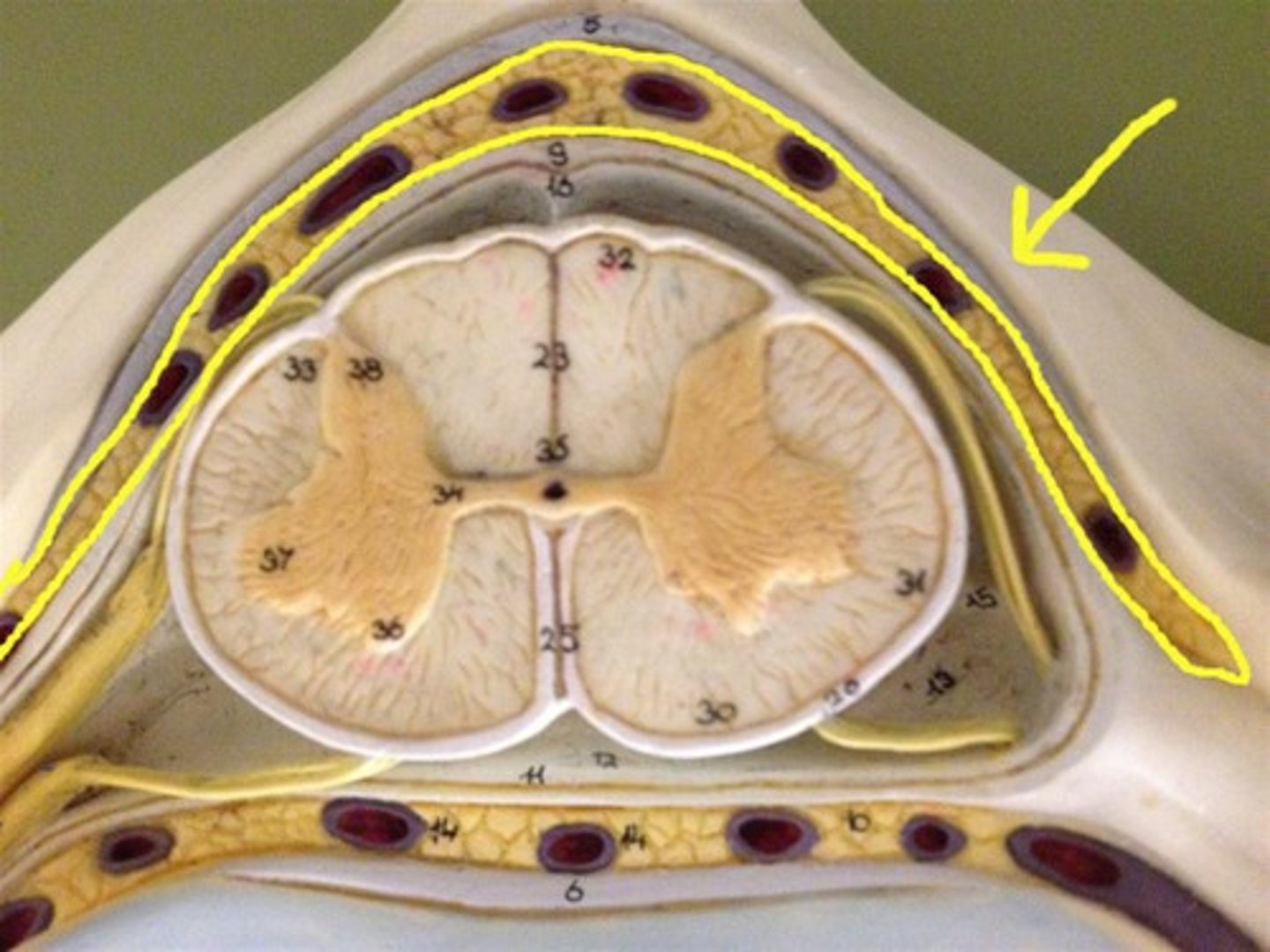
spinal dura mater
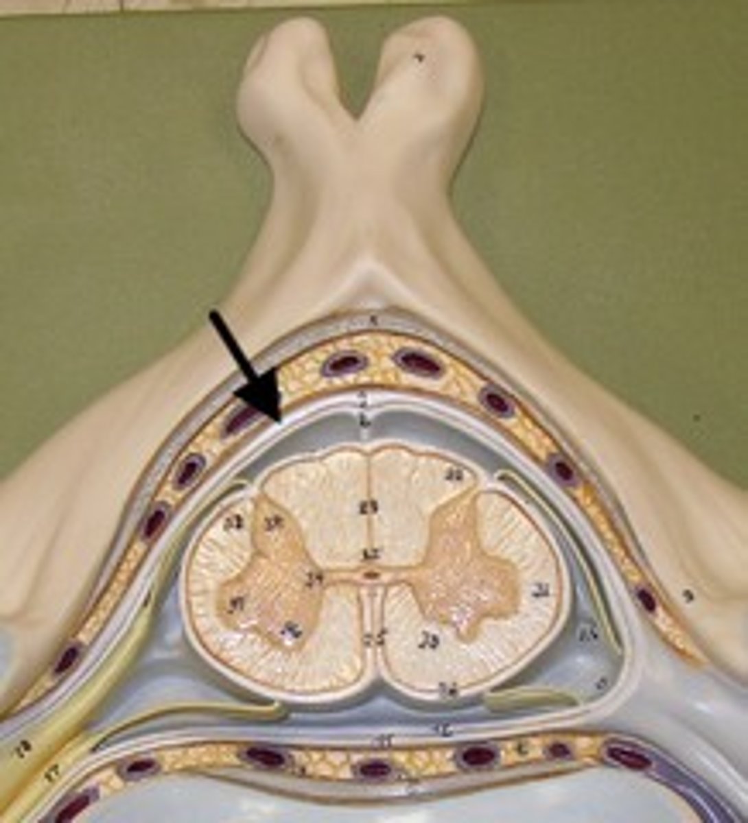
spinal subarachnoid space
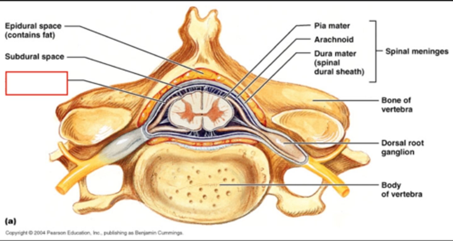
spinal subarachnoid space
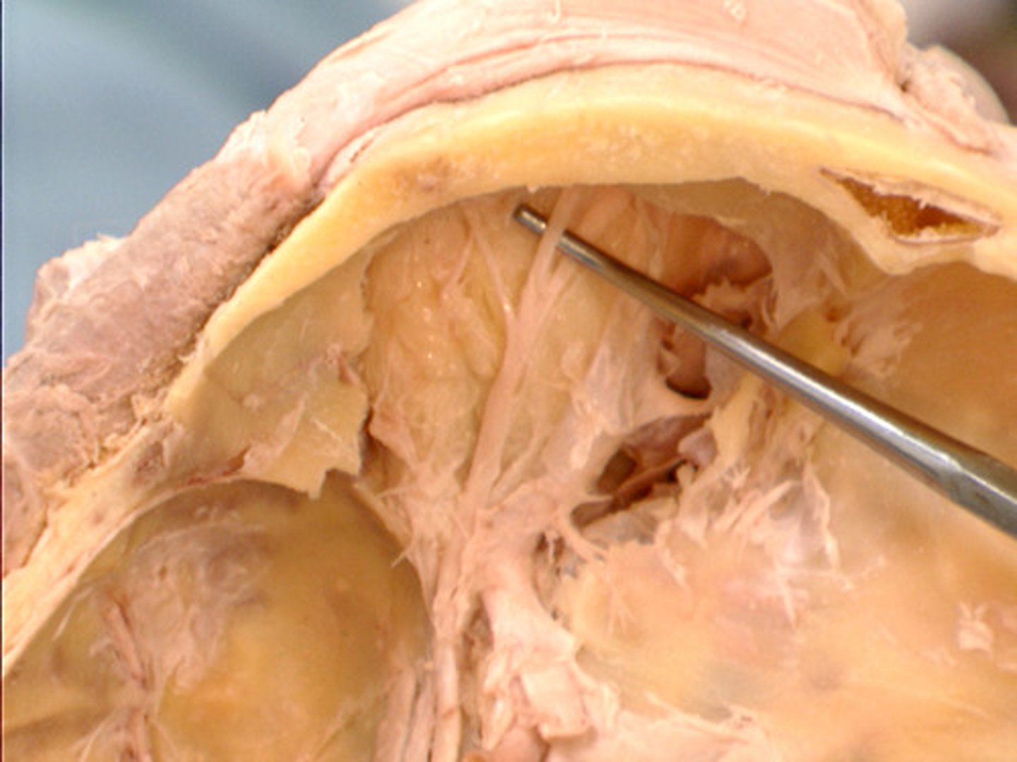
denticulate ligaments
dorsal rootlets
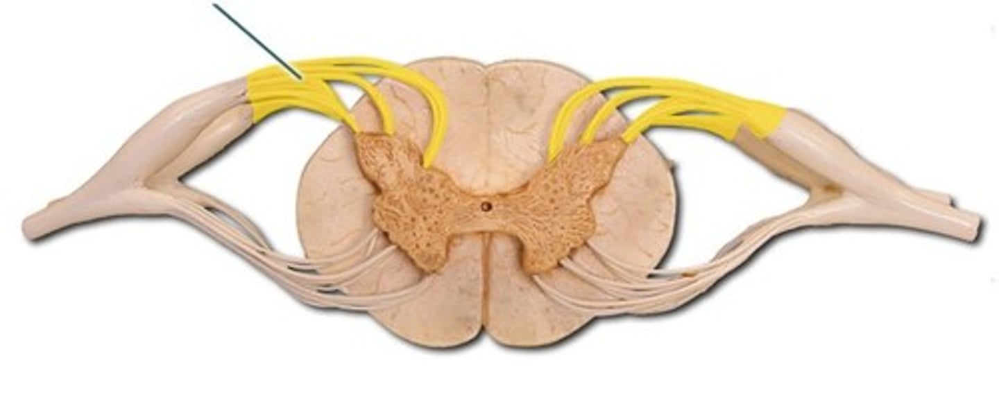
dorsal root
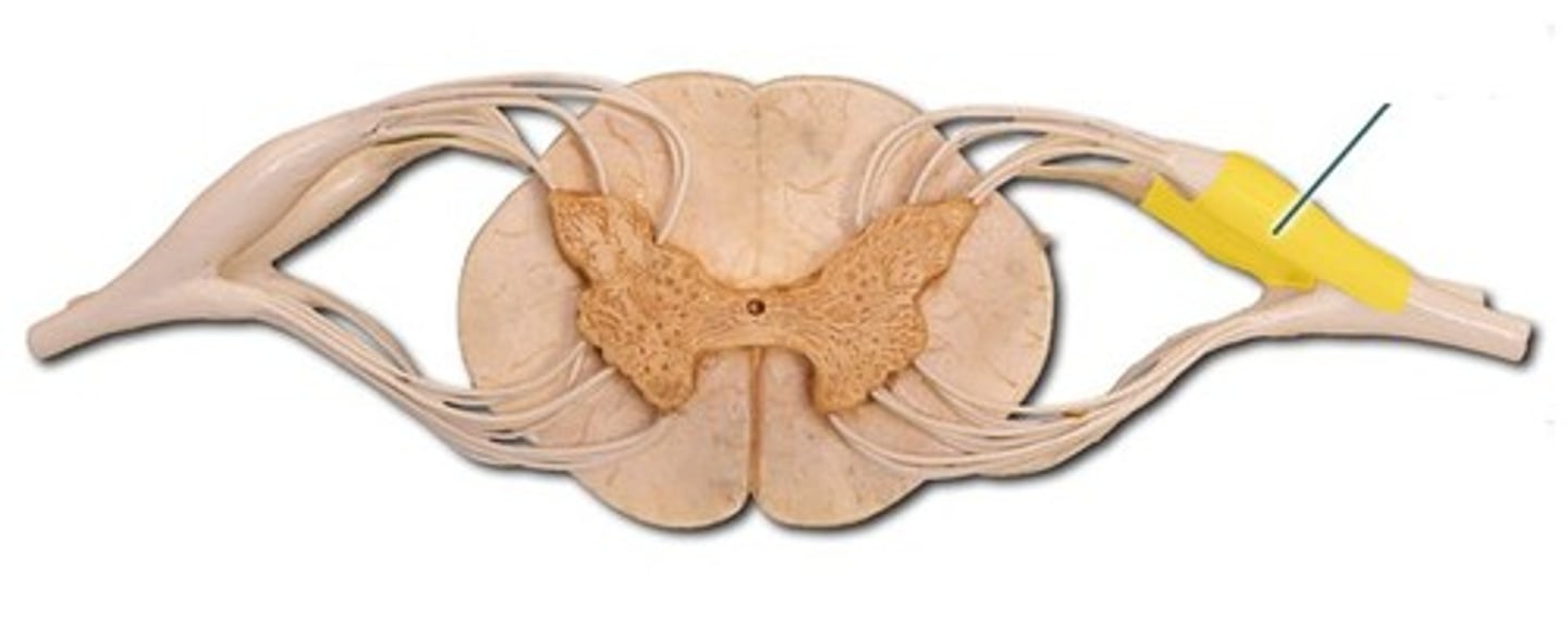
ventral rootlets
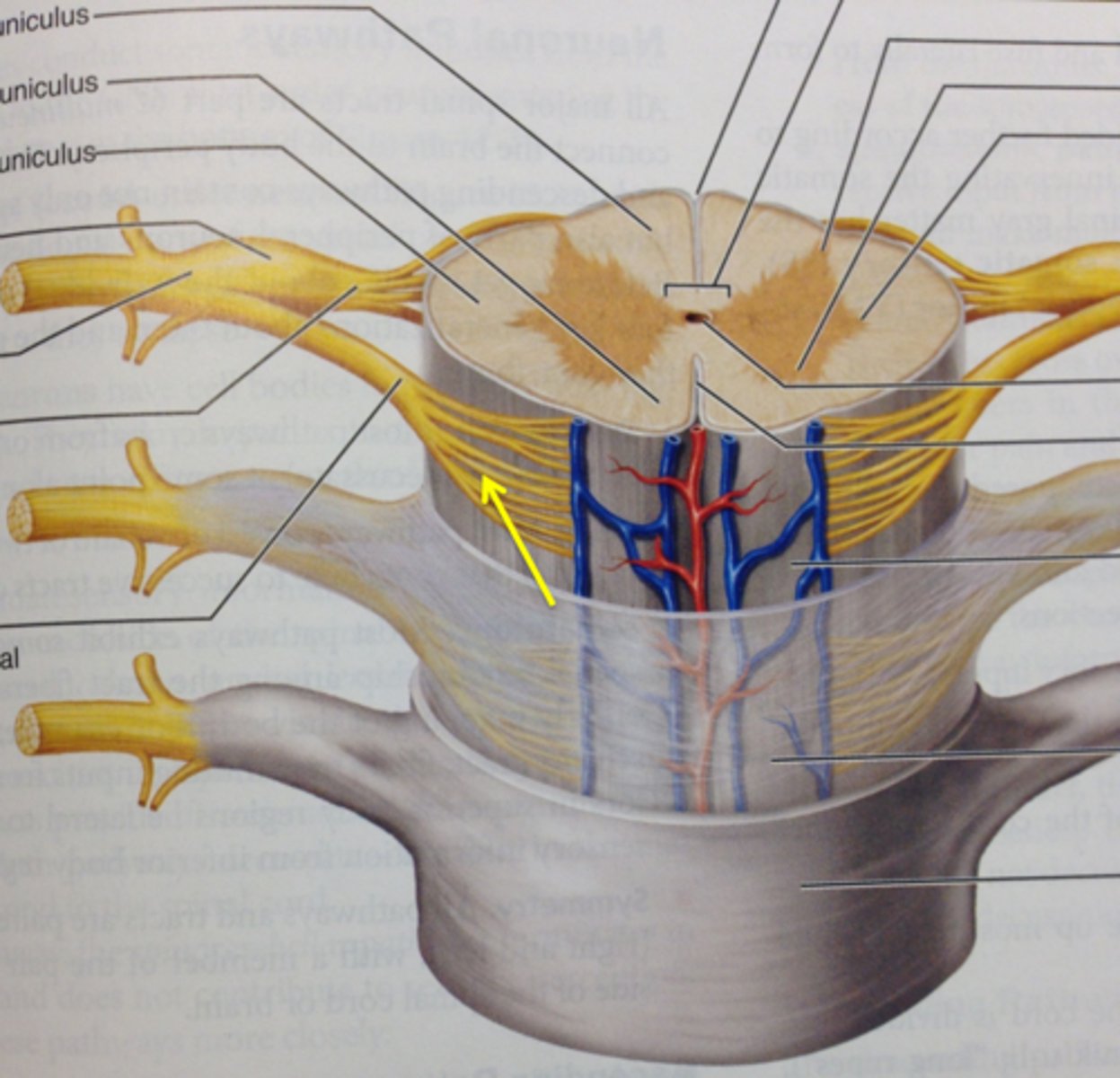
ventral root
dorsal root ganglion

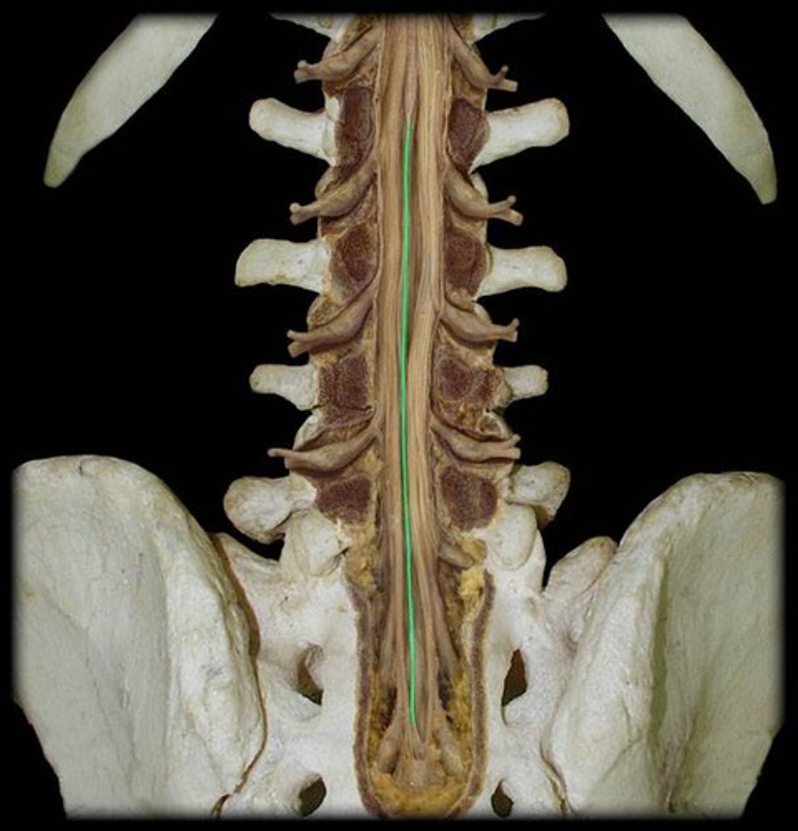
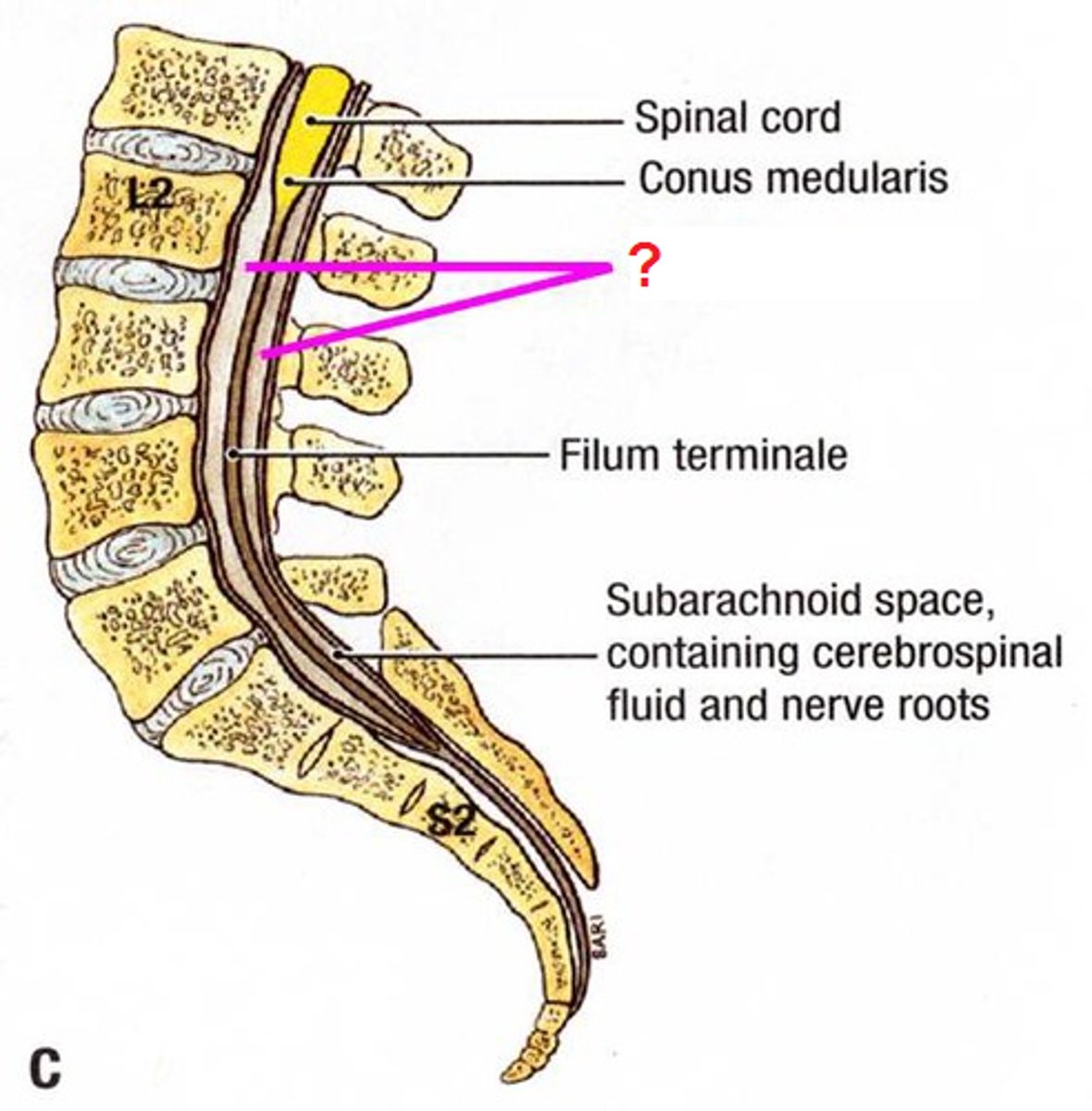
conus medullaris
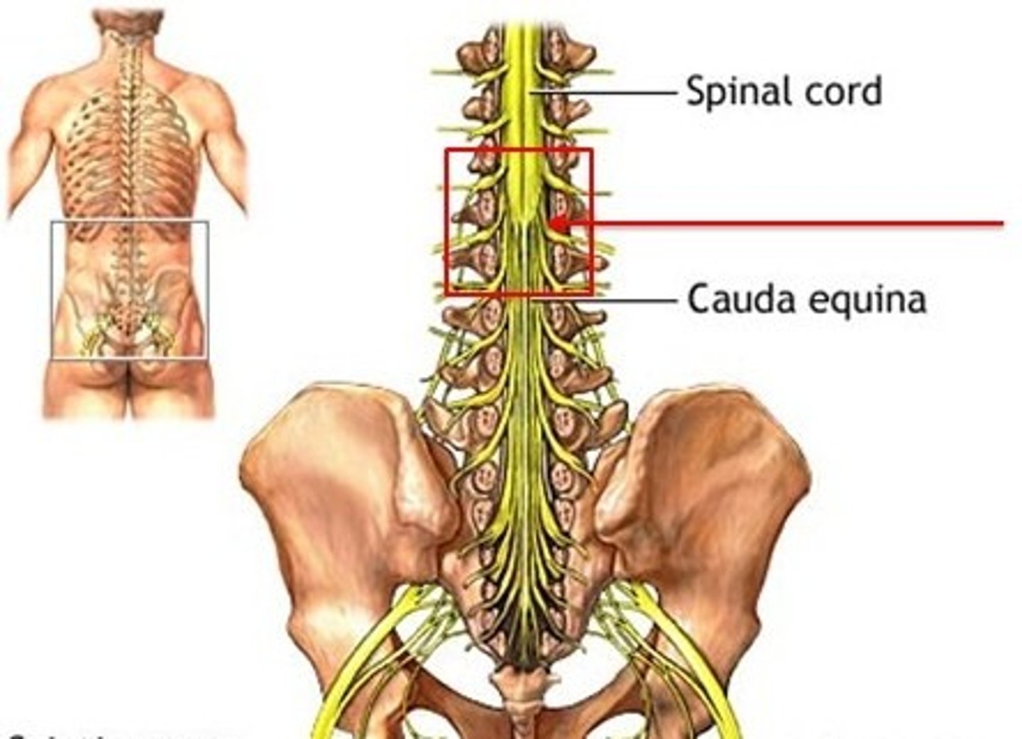
cauda equina
below cona medullaris
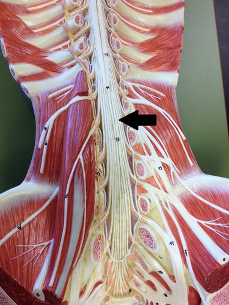
filum terminale
anchors spinal cord to coccyx
lumbar cistern
subarachnoid space inferior to conus medullaris
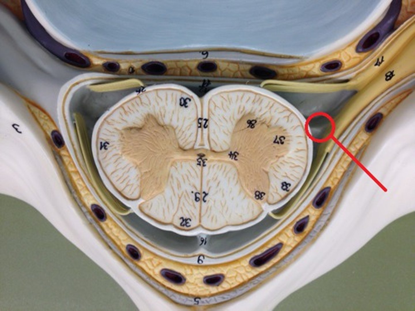
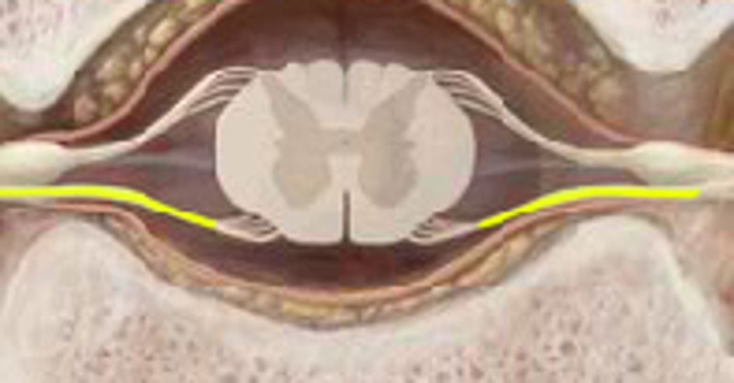
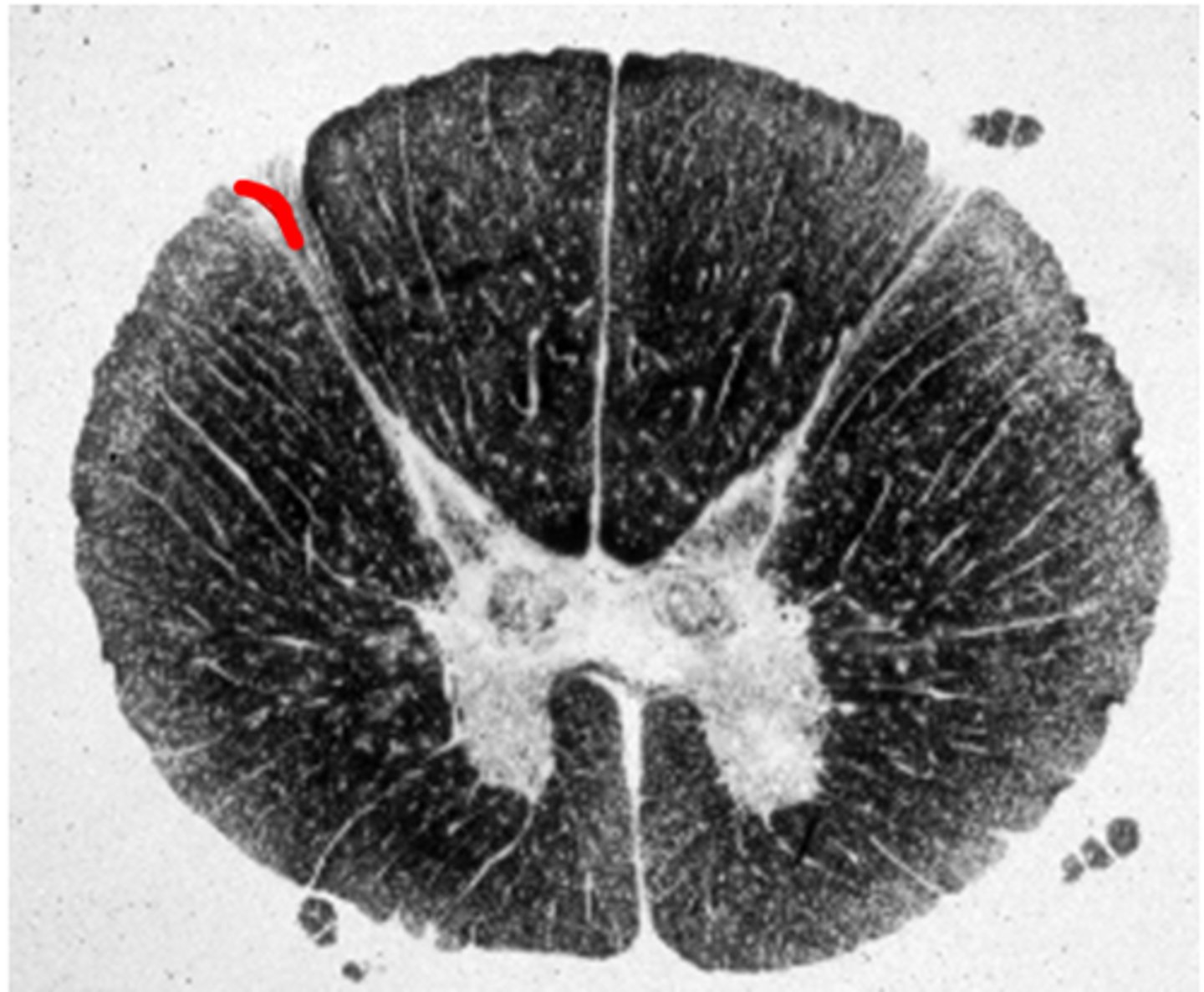
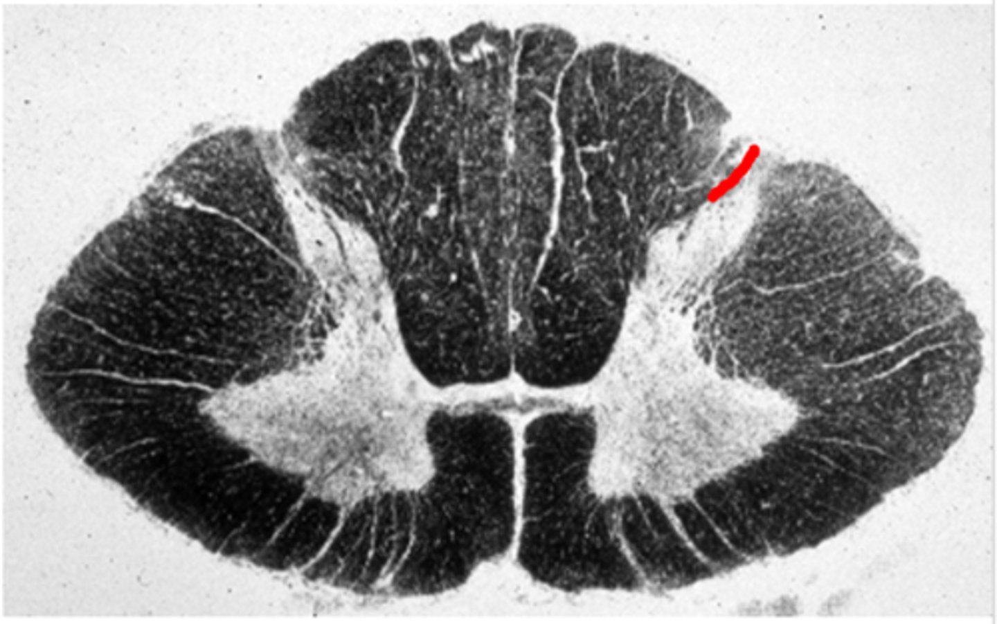
dorsolateral sulcus
Entry zone for the dorsal roots
dorsolateral sulcus
Entry zone for the dorsal roots
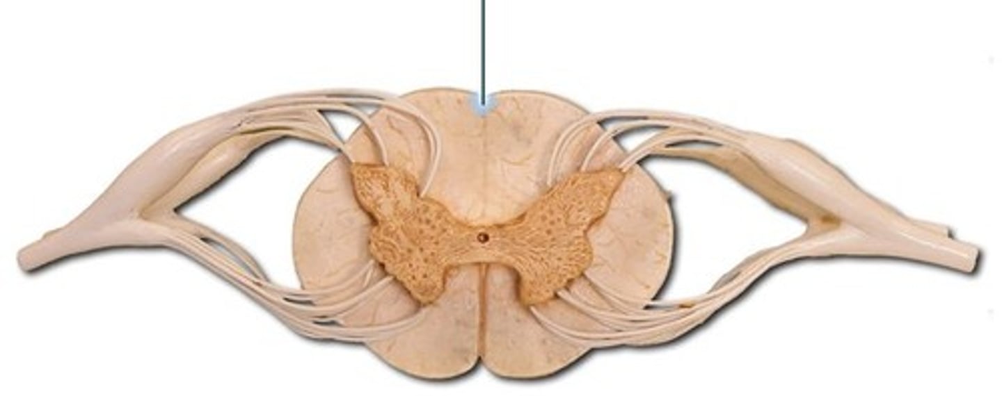
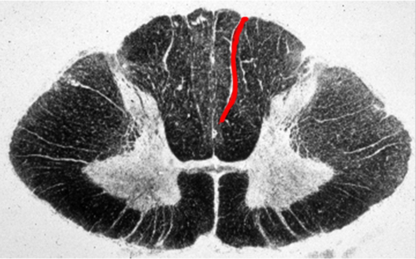
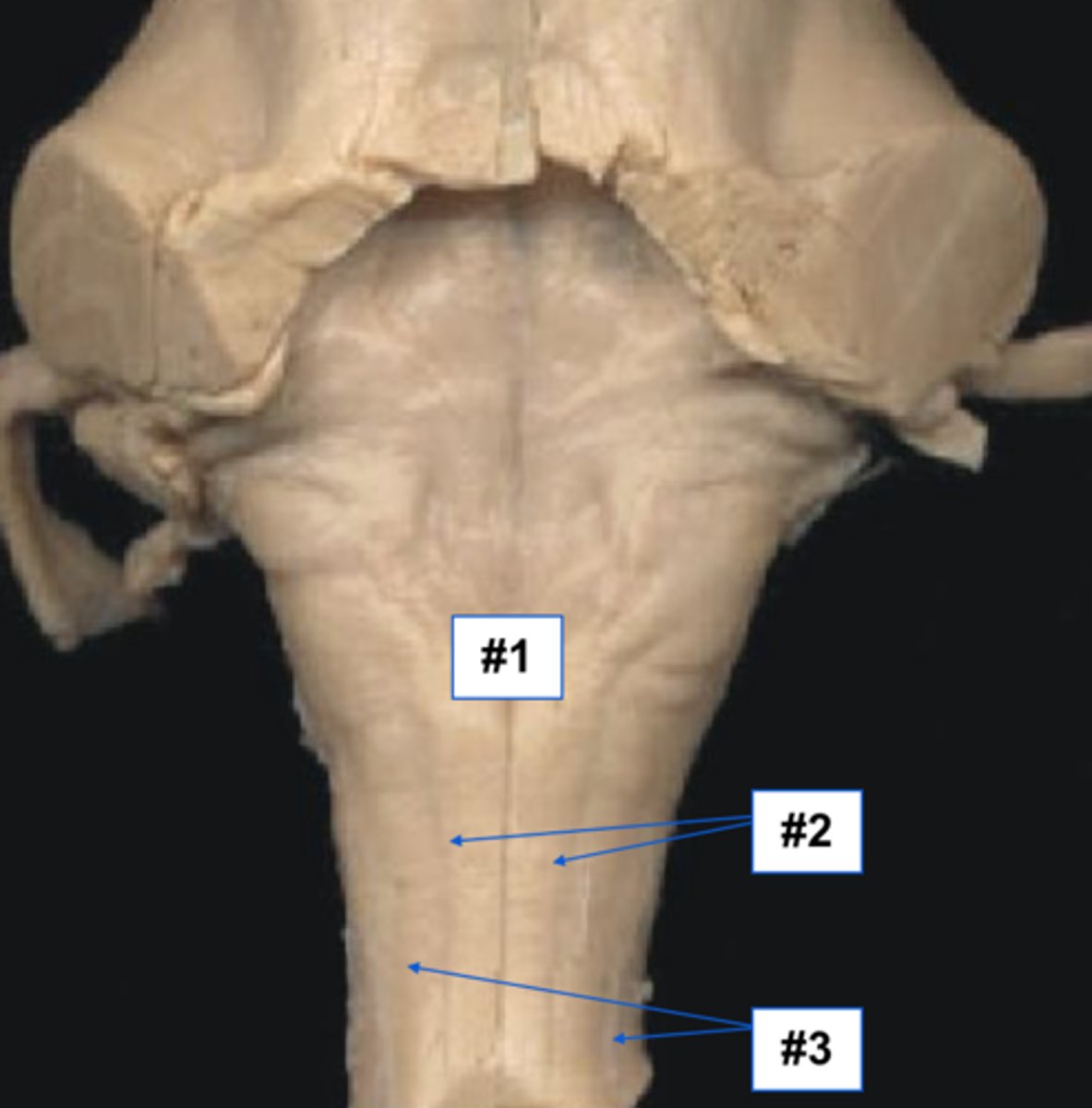
Dorsomedian Sulcus
between dorsal columns
Dorsomedian Sulcus
between dorsal columns
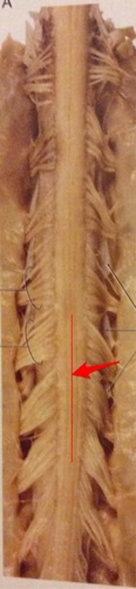
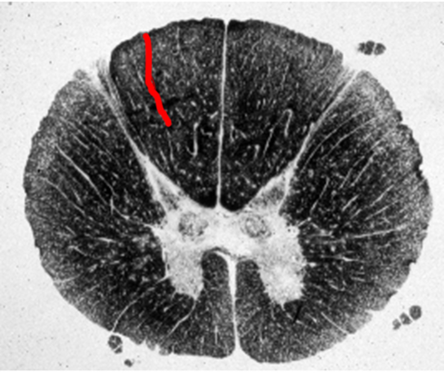
dorsal intermediate sulcus
Groove on the dorsal surface of the rostral part of the spinal cord, between the gracile and cuneate fasciculi, contains posterior spinal arteries
dorsal intermediate sulcus
Groove on the dorsal surface of the rostral part of the spinal cord, between the gracile and cuneate fasciculi, contains posterior spinal arteries
dorsomedian sulcus, dorsal intermediate sulcus, dorsolateral sulcus
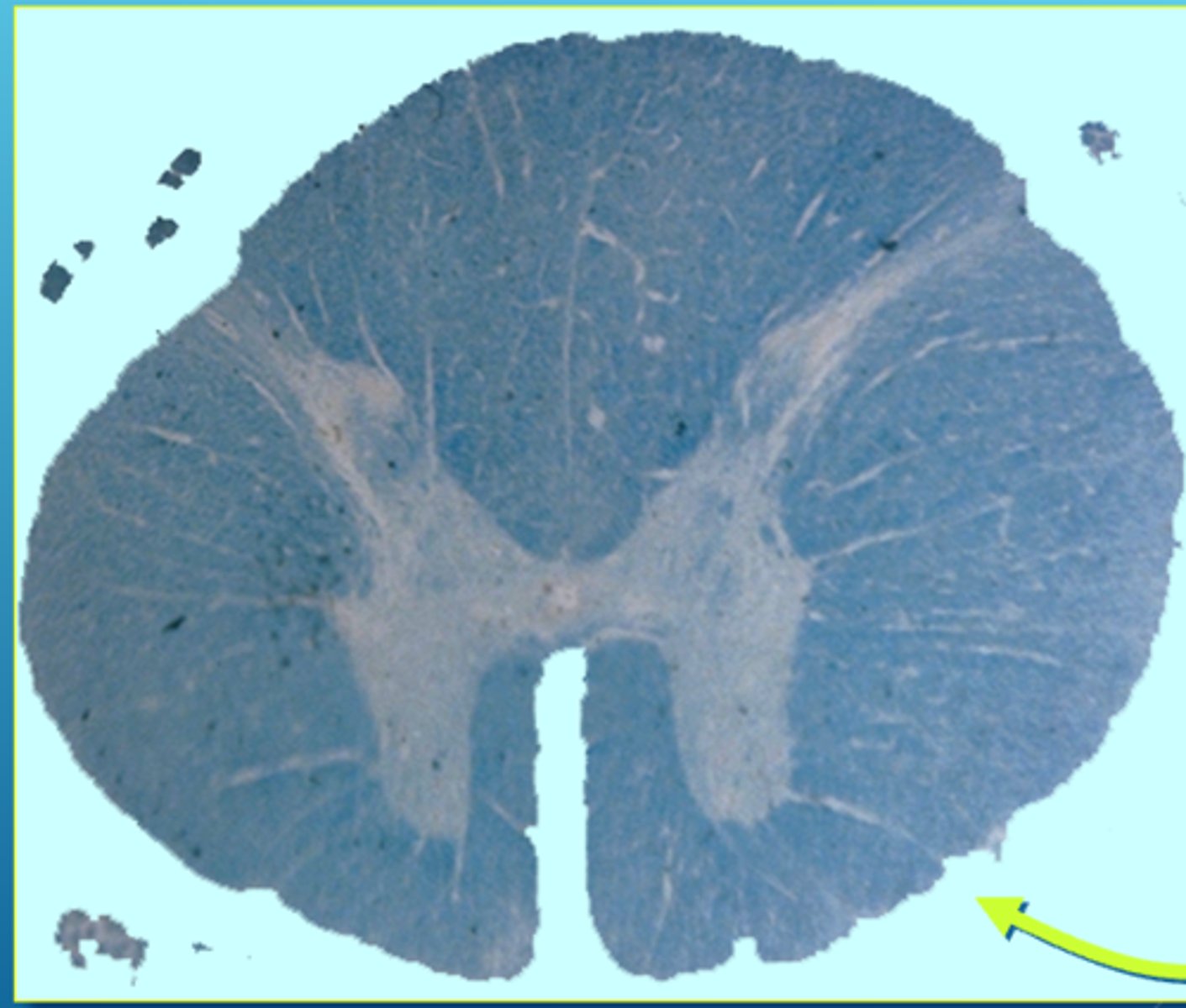
ventral median fissure
contains anterior spinal artery

ventral lateral fissure
Ventral root exit here