Unit 6 - Homeostasis
1/124
Earn XP
Description and Tags
Name | Mastery | Learn | Test | Matching | Spaced |
|---|
No study sessions yet.
125 Terms
[3.6.1.1] What is a stimulus?
A detectable internal/external change in the environment that brings about a response from an organism.
[3.6.1.1] What is a response?
A reaction to a stimulus that increases an organism’s chance of survival.
[3.6.1.1] What is a taxis?
A directional movement in response to a stimulus.
An animal performs a taxis when it orientates itself and moves towards/away from a stimulus which is coming from a particular direction.
[3.6.1.1] What is a kinesis?
A non-directional movement in response to a stimulus.
An animal does not respond to a stimulus by moving in a particular direction, instead the rate of movement and turning changes.
[3.6.1.1] What is a positive taxis?
Moving towards the stimulus.
[3.6.1.1] What is a negative taxis?
Moving away from the stimulus.
[3.6.1.1] What is a tropism?
A growth movement of part of a plant in response to a directional stimulus.
[3.6.1.1] What is phototropism?
Growth AWAY from light in the roots.
Growth TOWARDS light in the shoots.
[3.6.1.1] What is gravitropism?
Growth TOWARDS gravity in the roots.
Growth AWAY from gravity in the shoots.
[3.6.1.1] Why is negative phototropism and positive gravitropism an advantage in the roots?
The root grows further into the soil so there is a larger area available for nitrate ion, water and nutrient uptake. It also acts as an anchor point for the plant.
[3.6.1.1] Why is positive phototropism and negative gravitropism an advantage in the shoots?
The shoot grows towards sunlight so there is an increased rate of photosynthesis.
[3.6.1.1] What are auxins?
Plant growth factors.
[3.6.1.1] How does indoleacetic acid lead to uneven growth of the plant?
Stimulates cell elongation and the shoot grows, but inhibits elongation in root cells.
[3.6.1.1] Why does the shoot grow upwards in terms of IAA?
Negative Gravitropism in the Shoot - IAA diffuses to the lower side of the shoot, stimulating elongation. They elongate faster than on the light side, causing the shoot to bend towards the light.
Positive Gravitropism in the Shoot - IAA diffuses to the shaded side of the shoot, stimulating elongation. They elongate faster on the light side, causing the shoot to bend towards the light.
[3.6.1.1] Why does the root grow downwards in terms of IAA?
Positive Gravitropism in the Root- IAA diffuses to the lower side of the root, inhibiting elongation. The cells on the upper side elongate more, causing the root to bend downwards.
Negative Gravitropism in the Root - IAA diffuses to the shaded side of the root, inhibiting elongation. The cells on the lighter side elongate more, causing the shoot to be downwards (away from light).
[3.6.1.1] What is a reflex?
A rapid, automatic response to a stimulus, serving a protective function.
[3.6.1.1] What is an effector?
A muscle or gland that brings about a response to a stimulus.
[3.6.1.1] Why are hormonal responses localised and short-lived?
Neurotransmitters are secreted directly into the cells.
Neurotransmitters are quickly removed when they have done what they need to.
[3.6.1.1] What is the reflex-arc pathway?
Sensory Neurone —> Relay Neurone —> CNS —> Motor Neurone —> Effector
[3.6.1.2] How do receptors work?
Changes in the electrical charge across the plasma membrane.
[3.6.1.2] What are the characteristics of a resting potential?
Inside of the cell is more negative than the outside.
The cell is polarised.
[3.6.1.2] What are the characteristics of a generator potential?
Inside of the cell becomes more positive; the size of the potential difference is dependent on size of stimulus.
The cell begins to depolarise.
[3.6.1.2] What are the characteristics of an action potential?
If the generator potential is big enough, an action potential is generated, which causes the nerve impulse to be transmitted along a neurone.
[3.6.1.2] What is a Pacinian corpuscle?
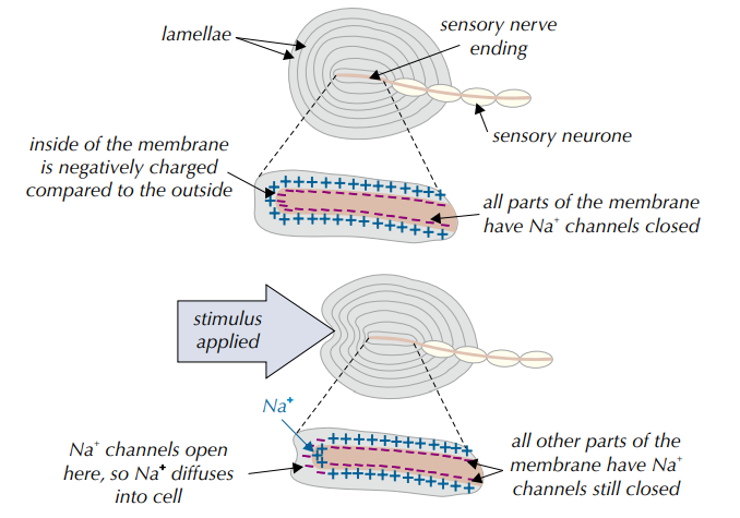
A very large mechanoreceptor that is almost 1mm long, and is sensitive to vibrations/large changes in pressure.
They consist of lots of lamellae, are made of thin flat connective tissue cells with some gel between each lamella and has a sensory nerve ending at its centre.
[3.6.1.2] How does a Pacinian corpuscle function?
When a Pacinian corpuscle is stimulated by pressure on the skin, its lamellae are deformed and the membrane of the sensory neurone inside it becomes stretched. This stretches the stretch-mediated sodium ion channels, which causes them to open, allowing sodium ions to diffuse into the neurone. This produces a generator potential. If this generator potential is strong enough to reach the threshold, it triggers an action potential.
[3.6.1.2] What are the main components of the eye?
Iris
Retina
Fovea
Optic Nerve
Blind Spot
Pupil
Lens
Cornea
[3.6.1.2] Where does light enter the eye?
The pupil.
[3.6.1.2] What controls the amount of light entering the eye?
The muscles of the iris.
[3.6.1.2] What are the two photoreceptors found in the eye?
Cones and Rods.
[3.6.1.2] What is the light sensitive pigment found in rod cells?
Rhodopsin.
[3.6.1.2] What is the light sensitive pigment found in cone cells?
Iodopsin.
[3.6.1.2] What is the distribution of rods vs cones in the retina?
Rods are more concentrated at the periphery, and absent at the fovea.
Cones are concentrated at the fovea.
[3.6.1.2] What is the difference in visual acuity of rods and cones?
Rods have a poor visual acuity.
Cones have a good visual acuity.
[3.6.1.2] How many connections do rods and cones have to one bipolar cell?
Bipolar cells have multiple connections to rods, whereas they only have one connection to cones.
[3.6.1.2] How do rod cells allow for vision?
Rhodopsin absorbs light, splitting into opsin and retinal. Opsin causes a chance in the permeability of the rod to sodium, which induces a generator potential.
In the absence of further light stimulation, rhodopsin will reform.
[3.6.1.2] How do cone cells allow for vision?
In bright light, iodopsin is broken down, generating an action potential in the ganglion cell.
[3.6.1.2] What are the types of cone cell?
Red Sensitive
Blue Sensitive
Green Sensitive
[3.6.1.2] How can cone cells become fatigued?
A yellow colour is perceived when there is equal stimulation of the red and green sensitive cones.
This is a constant stimuli to the red sensitive cones, which fatigue or adapt. As they cease to function, a yellow colour is perceived as green only.
[3.6.1.2] What is visual acuity?
The ability to tell apart two points that are close together.
[3.6.1.2] What is retinal convergence?
Several rods are connected to one bipolar cell, leading to summation.
[3.6.1.3] Why is the heart described as being myogenic?
It can stimulate its own contraction and relaxation.
[3.1.6.3] What are the main components of the electrical system of the heartt?
Sinoatrial Node (SAN)
A bundle of cells found in the right atrium which sets the rhythm.
Atrioventricular Node (AVN)
Bundle of His
Branches of the Bundle of His
Purkyne Fibres
Non-Conducting Tissue
Apex
[3.1.6.3] What are the main stages of the control of the cardiac cycle?
The Sinoatrial Node sends impulses across the atria, causing the muscle to contract.
Non-conducting tissue between the atria and ventricles prevents them from contracting at the same time.
The impulse is delayed at the Atrioventricular Node which allows the atria to fully empty of blood before the ventricles contract.
The impulse then passes down the Bundle of His to the apex of the heart.
The impulse travels along the Purkyne fibres from the apex, causing the ventricles to contract from the bottom upwards.
This pushes blood up and out of the heart.
[3.1.6.3] Where are baroreceptors and chemoreceptors located?
The walls of the aorta and the carotid artery.
[3.1.6.3] What are chemoreceptors sensitive to?
Hydrogen ion, O₂ and CO₂ concentration change.
[3.1.6.3] What are baroreceptors sensitive to?
Changes in blood pressure.
[3.1.6.3] Where do chemoreceptors and baroreceptors send nerve impulses?
To the medulla in the brain via sensory neurones (where the two centres are located which increase and decrease heart rate).
[3.1.6.3] What type of nervous system does the cardiovascular centre in the medulla fall into?
Autonomic.
[3.1.6.3] What is the cardio-acceleratory centre pathway?
Chemoreceptors detect a decreased pH caused by a increased CO₂ concentration in the blood OR baroreceptors detect a decreased blood pressure.
More impulses are sent to the medulla.
A nerve impulse is passed from the cardio-acceleratory centre along the accelerator nerve (sympathetic).
Noradrenaline is then released to the sinoatrial node.
This increases the rate of impulses, and heart rate increases.
This increased blood flow removes CO₂ from the blood more quickly, so blood pH increases OR blood pressure increases (so more oxygen is available for respiration).
[3.1.6.3] What is the cardio-inhibitory centre pathway?
Chemoreceptors detect an increased pH caused by a decreased CO₂ concentration in the blood OR baroreceptors detect an increased blood pressure.
Impulses are sent to the medulla.
A nerve impulse is passed from the cardio-inhibitory centre along the vagus nerve (parasympathetic).
Acetylcholine is then released to the sinoatrial node.
This decreases the rate of impulses, and heart rate decreases.
This decreased blood flow removes CO₂ from the blood more slowly, so blood pH decreases OR blood pressure decreases.
[3.6.2.1] What are the main components of a myelinated motor neurone?
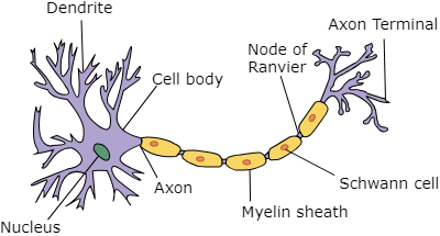
The components of a myelinated motor neurone:
Nucleus
Dendrites
Axon
Node of Ranvier
Myelin Sheath
Terminal Dendrites
Synaptic Knob
[3.6.2.1] What type of cell makes up the membrane of the myelin sheath?
Schwann Cells
[3.6.2.1] How are neurones adapted to their function?
Dendrites provide a large surface area for multiple connections to other cells.
Long axon allows impulses to travel longer distances continuously.
Myelin sheath provides insulation and increases impulse speed (ions can only be transported at the nodes of ranvier).
They contain many mitochondria to provide ATP for active transport of ions.
[3.6.2.1] What are the stages of a nerve impulse?
Resting Potential
Depolarisation
Repolarisation
Hyperpolarisation
[3.6.2.1] What is the resting potential of a nerve cell?
The difference in electrical energy across the cell surface membrane of a resting nerve cell, it is about -70mV and the membrane is said to be polarised.
[3.6.2.1] How is the resting potential established?
In the nerve cell membrane, there are intrinsic carrier proteins called sodium-potassium pumps which actively transport 3 sodium ions OUT of the axon, and 2 potassium ions INTO the axon, which requires ATP.
However, the membrane is very permeable to K⁺ ions which move back out via facilitated diffusion.
It is less permeable to the Na⁺ ions as there are fewer sodium ion channels open.
As a result, the inside of the axon becomes more negative than the outside, which establishes the resting potential.
[3.6.2.1] What is depolarisation?
The change in the electrical potential across the membrane of a neurone as a nerve impulse passes across it.
[3.6.2.1] What is the threshold for an action potential?
+40mV
[3.6.2.1] How is an action potential created?
Many voltage-gated sodium ion channel proteins in the nerve cell membrane open and sodium ions move into the axon by facilitated diffusion down their concentration gradient.
This causes the inside of the axon to become temporarily more positively charged compared to the outside of the axon, and this section is said to be depolarised.
Adjacent sections of the axon are similarly affected by the action potential resulting in a wave of depolarisation.
[3.6.2.1] What is repolarisation?
The return of a cell membrane to its resting potential.
[3.6.2.1] How is a membrane repolarised?
Voltage-gated sodium ion channel proteins in the membrane CLOSE and the voltage-gated potassium ion channel proteins OPEN — more K⁺ leaves the axon by facilitated diffusion.
Briefly the outside of the axon is even more positive than during the resting potential, and the membrane is hyperpolarised.
Voltage-gated potassium ion channels then CLOSE to allow the membrane potential to return back to the resting potential.
The net positive charge outside the membrane is restored.
[3.6.2.1] What other type of ion moving into the neurone could also cause hyperpolarisation?
Chloride.
[3.6.2.1] What is the refractory period and when does it occur?
The period of time when the membrane cannot transmit another impulse.
Takes place when the membrane is repolarising and the voltage-gated sodium ion channels are closed.
[3.6.2.1] What are the purposes of the refractory period?
Ensures the action potential is propagated in one direction only.
Produces discrete (separate) nerve impulses.
Limits the number of action potentials.
[3.6.2.1] What are the principles of waves of depolarisation?
The wave of depolarisation has to pass through every section of the membrane.
The movement of sodium ions INTO the neurone causes more voltage-gated Na⁺ channels to OPEN in the next part of the membrane, causing depolarisation.
Behind the depolarised region, the voltage-gated Na⁺ channels CLOSE and the voltage-gated K⁺ channels OPEN, allowing this part of the membrane to repolarise to reach its resting potential.
The impulse passes along the whole membrane like this until it reaches the end of the neurone.
[3.6.2.1] In which type of neurone does every section of the membrane need to be depolarised?
Unmyelinated Neurones
[3.6.2.1] In which type of neurone does saltatory conduction occur?
Myelinated Neurones
[3.6.2.1] What is saltatory conduction?
The impulse ‘jumps’ between nodes of Ranvier because ions cannot pass through the phospholipid layer of the Schwann cells.
Depolarisation only occurs at the nodes of Ranvier (where Na⁺ can pass through) — the neurone’s cytoplasm conducts enough electrical charge to depolarise the next node, so the impulse ‘jumps’.
[3.6.2.1] What are the benefits of saltatory conduction?
Increased speed of transmission.
It requires less ATP as there are less Na⁺/K⁺ pumps required to return to the resting potential.
[3.6.2.1] What is the all-or-nothing principle?
Action potentials are always the same size. However, we can discriminate between different sized stimuli because a stronger stimulus produces more frequent action potentials.
Each neurone has a threshold - the level of stimulus which triggers an action potential.
If depolarisation doesn’t reach the threshold, there will be no action potential because the voltage-gated Na⁺ channels stay closed.
If the threshold is reached, the voltage-gated Na⁺ channels OPEN by positive feedback - the more depolarisation occurring, the more channels that open.
[3.6.2.1] How does axon diameter affect the speed of conduction?
Wider neurones transmit impulses faster because axons with smaller diameters have a smaller surface area to volume ratio which means more ions can lead out of the axon, making it more difficult for the action potential to propagate.
[3.6.2.1] How does temperature affect the speed of conduction?
Temperature increases the speed of conduction.
Ions have more kinetic energy so they diffuse faster.
Increased rate of respiration, so there is more ATP available for Na⁺/K⁺ pumps.
[3.6.2.2] What is the process of synaptic transmission in a cholinergic synapse?
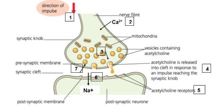
1) A nerve impulse travels down the presynaptic neurone and arrives at the synaptic knob.
2) Calcium ion channel proteins in the presynaptic membrane OPEN and Ca²⁺ ions diffuse INTO the neurone.
3) Vesicles containing acetylcholine move towards the membrane and Ca²⁺ is moved out of the nerve cell by active transport.
4) The vesicles fuse with the membrane and release acetylcholine which diffuses across the synaptic cleft.
5) Acetylcholine attaches to complementary receptors on the postsynaptic membrane.
6) This causes sodium ion channel proteins to OPEN and Na⁺ ions diffuse INTO the postsynaptic neurone, depolarising the membrane and causing an action potential if the threshold is reached.
7) Acetylcholine is then hydrolysed by acetylcholinesterase in the synaptic cleft. The components are then taken back into the presynaptic neurone and used to resynthesise acetylcholine.
[3.6.2.2] Why is acetylcholinesterase important within a cholinergic synapse?
Prevents acetylcholine from continually stimulating the postsynaptic neurone, keeping impulses as discrete events.
Hydrolysis acetylcholine into its components so it can be be taken back up by the presynaptic neurone for resynthesise.
[3.6.2.2] Why do synapses exhibit unidirectionality?
Vesicles containing neurotransmitters are only released from the presynaptic neurone.
Receptors for the neurotransmitter only exist on the postsynaptic neurone.
[3.6.2.2] What is spatial summation?
Involves impulses from several neurones converging on the same postsynaptic neurone.
The neurotransmitter builds up in the synaptic cleft, causing the threshold to be reached in the postsynaptic neurone if enough sodium ion channels are open to cause depolarisation.
[3.6.2.2] What is temporal summation?
Involves several impulses arriving in rapid succession from the same presynaptic neurone.
The neurotransmitter builds up in the synaptic cleft, causing the threshold to be reached in the postsynaptic neurone if enough sodium ion channels are open to cause depolarisation.

[3.6.2.2] What is the purpose of summation?
Allows for differentiation between strong and weak stimuli.
Allows processing of stimuli from different receptors.
[3.6.2.2] What is the impact at excitatory synapses?
Depolarises the membrane by opening sodium ion channels.
[3.6.2.2] What is the impact at inhibitory synapses?
Hyperpolarises the membrane by K⁺ diffusing OUT or GABA/Cl⁻ IN.
[3.6.2.2] What is the neuromuscular junction?
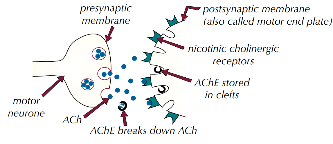
Where a motor neurone meets a skeletal muscle fibre.
Neuromuscular junctions are between motor neurones and muscle fibres — they use acetylcholine which binds to nicotinic cholinergic receptors on the muscle cell membrane.
[3.6.2.2] What is the structure of a neuromuscular junction?
[3.6.2.2] What are the similarities between cholinergic transmission and neuromuscular junction transmission?
Both release acetylcholine.
Acetylcholine binds to cholinergic receptors on the postsynaptic cell.
Acetylcholinesterase breaks down acetylcholine at the synaptic cleft/neuromuscular junction.
[3.6.2.2] What are the differences between cholinergic transmission and neuromuscular junction transmission?
The neuromuscular junction has folds forming clefts to store acetylcholinesterase, whereas synapses don’t.
The neuromuscular junction has more receptors.
Acetylcholine is always excitatory at a neuromuscular junction, but can be excitatory or inhibitory at a synapse.
[3.6.2.2] What effect on the synapse can a drug which has a similar shape to the neurotransmitter have?
Blocks receptors on the postsynaptic membrane so the neurotransmitter can’t bind, e.g. atropine.
Mimics the neurotransmitter by opening sodium ion channels on the postsynaptic membrane, e.g. nicotine.
[3.6.2.2] What effect on the synapse can a drug which is an inhibitor of acetylcholinesterase have?
Acetylcholine cannot be broken down and continues to act on the postsynaptic neurone, causing paralysis, e.g. organophosphate insecticides and nerve gases.
[3.6.2.2] What effect on the synapse can a drug which stimulates the release of a neurotransmitter have?
Most postsynaptic receptors are activated, e.g. amphetamines.
[3.6.2.2] What effect on the synapse can a drug which is an inhibitor of the release of neurotransmitters have?
Fewer postsynaptic receptors are activated, e.g. alcohol.
[3.6.2.2] What effect on the synapse can a drug which blocks postsynaptic receptors have?
Less neurotransmitter is taken back up, and more remains in the synaptic cleft, e.g. prozac.
[3.6.3] What is meant by antagonistic muscles?
Muscles that work in pairs to move a bone:
As one contracts, the other relaxes.
[3.6.3] What is the structure of skeletal muscle?
Skeletal muscles (striated muscles) are made up of sarcomeres.
Each muscle fibre is surrounded by a sarcolemma, and contain more than one nucleus and many mitochondria.
The cytoplasm of muscle fibres is the sarcoplasm.
Within each fibre are myofibrils, which are slender threads containing the proteins required for muscle contraction.
[3.6.3] Why are mitochondria NOT found in the sarcomere?
They would be in the way during muscle contraction.
[3.6.3] Why can more detail of muscles be seen with an electron microscope than an optical microscope?
Electron microscopes have a higher resolving power as the electron beam wavelength is shorter than the wavelength of light, and has a higher magnification.
[3.6.3] What is the structure of a myofibril?
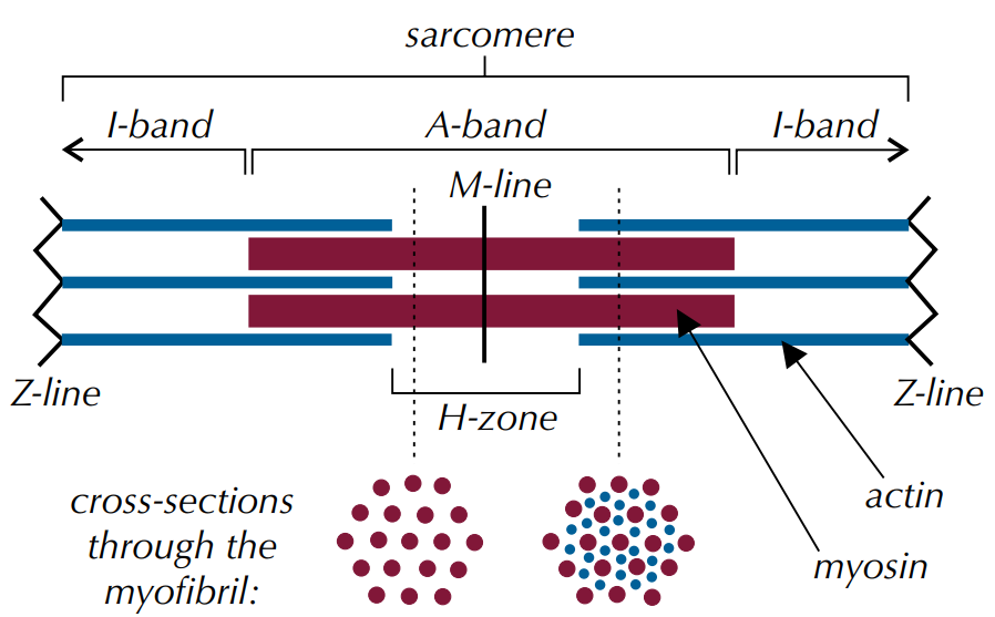
[3.6.3] What is the A band?
Anisotropic Band (the dark band).
[3.6.3] What is the I band?
Isotropic Band (the light band).
[3.6.3] Which protein makes up the thick muscle filaments?
Myosin.
[3.6.3] Which protein makes up the thin muscle filaments?
Actin.
[3.6.3] What are the light bands composed of?
Actin ONLY.
[3.6.3] What are the dark bands composed of?
Myosin and Actin.