Advanced MedSurg Exam 1
 Call Kai
 Learn
Learn Practice Test
 Spaced Repetition
 Match
Match Flashcards
 Knowt Play
1/53

Earn XP
Description and Tags
Dysrhythmias
Name | Mastery | Learn | Test | Matching | Spaced | Call with Kai | Chat |
|---|
No analytics yet
Send a link to your students to track their progress
54 Terms
What is a dysrhythmias?
Disorder of formation of electrical impulses of heart of conduction (hemodynamic instability)
What is the difference between the two types of cardiac cells?
Pacemaker cells: automatic, generate impulse
Muscle cells: contract/relax, no impulses generally, pump blood
How does the cell conduction system work?
Impulse from SA node—> left atrium—> left ventricle—> AV node—> purkinje fibers—> all cells
What is the role of an EKG?
Information ONLY about electrical events NOT contractile events (always check pt)
Why should you always check the patient even if EKG is normal?
pulseless electrical activity can occur
Why can tachycardia be detrimental?
Increase myocardial oxygen demand, decreases ventricular filling and coronary artery perfusion time
Why can bradycardia be detrimental?
Decrease in cardiac output can cause syncope and chest pain
How is cardiac output calculated?
HR x Stroke volume (amount of blood pumped out with every beat)
What is the process of impulse formation?
Polarized state (resting negative charge of myocardial cell)—> depolarized state (becomes positive)—> repolarization (cell is restored to resting state with ATP)
How is depolarization shown on an EKG?
atrial depolarization: P wave
Ventricular depolarization: ST segment and T wave
What is the PR interval on an EKG?
Amount of time for SAN stimulation, and atrial depolarization through AVN before ventricular depolarization) 0.12-0.20 seconds
What does the QRS complex represent (and normal range)?
0.06-0.10 seconds (ventricular depolarization)
What is the ST segment on an EKG indicate?
normal isoelectric, if above or below could be ischemia or infarction
What does the T wave represent?
Ventricular repolarization following QRS complex
What does the QT interval represent?
ventricular depolarization and repolarization
What can a prolonged QT interval indicate?
ventricular dysrhythmia
How is HR calculated on an EKG?
count number of QRS complexes in 6 second strip and multiply by 10
What is Normal Sinus Rhythm (NSR)?
Eletrical impulse starts in SA node and travels through normal conduction pathway
What is normal criteria for sinus rhythm on EKG?
1 P wave per QRS
What are the 6 kinds of SA node dysrhythmias?
(Sinus) tachy, brady, arrhythmia, pause, arrest, sick sinus syndrome
What is sinus tachy?
SAN creates an impulse at a faster than normal rate >100 bpm
What are causes/ tx for sinus tachy?
cause: shock, anemia, stress, hypovolemia, blood loss, infection, hypoxia etc.
tx: treat underlying cause, monitor

What dysrhythmia is this EKG showing?
Sinus tachy
What is sinus brady?
SAN creates impulse slower than normal rate <60 bpm

What dysrhythmia is this EKG showing?
sinus brady
What are causes and tx of sinus brady?
cause: decreased metabolic needs, intracranial pressure, MI, vagal stimulation, meds, hypothyroidism
tx: only if symptomatic, atropine for vagal cause, pacemaker
What are the types of atrial dysrhythmias?
Premature atrial contractions, afib, atrial flutter
What is premature atrial contraction (PAC)
An early electrical impulse discharges in atrium before next normal impulse of SAN
What is an ectopic beat?
pacemaker site outside of the SAN (like atrium)
What is the cause and tx for PAC?
Causes: HF, hypokalemia (<3.5) , atrial ischemia, meds (albuterol)
Tx: no tx needed generally, assess, remove causing agent (ex. caffeine)
What is atrial flutter?
Atria is depolarized at rates of 250-400 times per minute from re-entry circuit

What is this EKG showing?
atrial flutter
What are causes for atrial flutter?
underlying cardiac disease, HTN, CAD, hypoxia, HF, SAN damage
What can be caused by the lack of atrial contraction from atrial flutters?
blood stasis and thromboembolisms
What are the treatments for atrial flutters?
control HR (diltiazem, beta blockers), create NSR (amiodarone, cardioversion), Vagal maneuvers (carotid sinus massage, cough) anticoagulation
What is afib?
Uncoordinated atrial electrical activation from many areas in atria
What is the ventricular rate dependent on?
the ability of AV node to conduct atrial impulses
What are characteristics of Afib?
NO P WAVE, very irregular EKG
What is the clinical significance of Afib?
Loss of systolic volume, reduced cardiac output, risk for thromboembolism
What are the tx goals for afib?
decrease ventricular rate, convert to NSR, prevent thromboembolism
What meds can be used to treat afib? (meds for rate control / NSR conversion)
Rate control: CCBs, Beta blocker, dioxin
NSR conversion: amiodarone, dofetilide, flecanide
Where is the common origin of thromboembolisms caused by afib?
left atrial appendage closure
What is tx for afib caused thromboembolism?
Antiplatelets (aspirin), anticoags (warfarin), Factor Xa inhibitors (-ban)
If your pt is dizzy, unstable BP what is the tx?
cardioversion (synchronized defibrillation)
What is a premature ventricular contraction (PVC)?
Premature impulse that originates in the ventricle before normal sinus impulse
What are characteristics of PVC?
QRS is wide, ST and T wave changes, irregular rhythm
What are the causes of PVC?
hypoxia, acidosis, electrolyte imbalances (hypokalemia), OSA
What is the tx for PVC?
remove causing agent, lidocaine
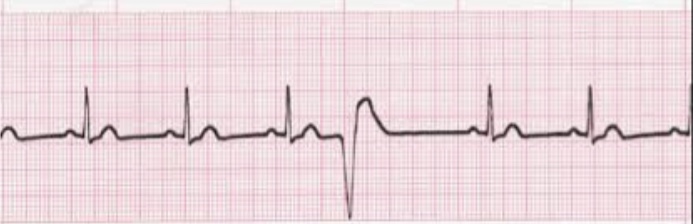
What is this EKG showing?
premature ventricular contraction (PVC)
What is ventricular tachycardia (VT)?
3 or more PVCs in a row, rate >100, EMERGENCY, can be life threatening
What are characteristics of VT?
high rate, regular rhythm, QRS wide and no P waves
What are causes of VT?
MI, ischemia, hypoxia, electrolyte imbalance, CAD, acidosis, HF, valve disease
What are the treatments for VT? (stable, unstable, pulseless
Stable: antiarrhythmic drugs (amiodarone)
unstable: sedate, cardioversion, drugs
Pulseless: defibrillation/ CPR
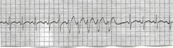
What does this EKG show?
ventricular tachycardia (VT)