Med Surg II week 3 (ch 32)
 Call Kai
 Learn
Learn Practice Test
 Spaced Repetition
 Match
Match Flashcards
 Knowt Play
1/102

There's no tags or description
Looks like no tags are added yet.
Name | Mastery | Learn | Test | Matching | Spaced | Call with Kai | Chat |
|---|
No analytics yet
Send a link to your students to track their progress
103 Terms
COPD consists of ____, it is a ______ that ______ and is chracterized by ___
chronic bronchitis and pulmonary emphysema
it is a group of lower airway disorders that disrupt airflow and gas exchange
characterized by tissue damage, that is irreversible, increases in severity and eventually leads to respiratory failure
chronic bronchitis problem
airway problem
emphysema problem
alveolar problem
COPD is characterized by
bronchospasms and dyspnea
emphysema two major changes
loss of lung elasticity and hyperinflation of the lung
therefore decreased gas exchange causing an increase in RR
emphysema patho
air trapping caused by loss of elastic recoil in alveolar walls, overstretching and enlargement of the alveoli into bullae, and collapse of small airways (bronchioles)
breathing in COPD
breathing is more work, the lungs are hyperinflated and become weak and don’t work properly
turn to the use of accessory muscles (neck, chest wall, abdomen) instead, but they are not as efficient
become air hungry, inhalation begins before completing exhalation
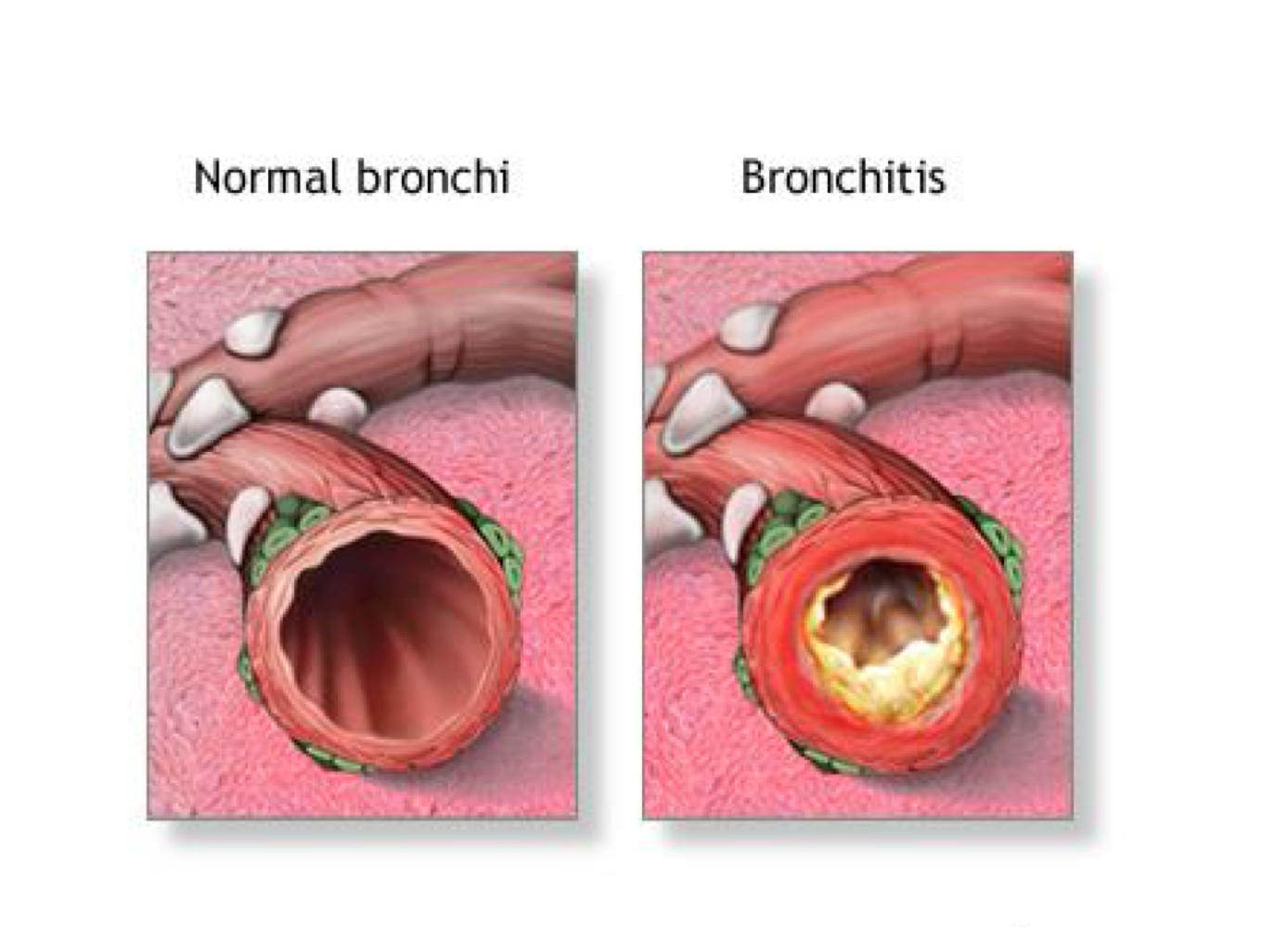
Chronic bronchitis patho
inflammation of the bronchi and bronchioles caused by chronic exposure to irritants, especially tobacco
inflammation results in vasodilation, congestion and large amts of thick mucous, mucosal edema, and bronchospasm
impacting only the airways
complication of bronchitis
the large amounts of mucous from inflammation is a breeding ground for bacteria resulting in chronic infections
complications of COPD
hyoxemia, tissue anoxia
acidosis due to poor gas exchange
respiratory infections
cardiac failure, COR PULMONALE
cardiac dysrhythmias
Cor pulmonale
right sided HF caused by chrnoiic pulmonary diseases (COPD)
patho- air trapping increases pressure in lung tissue, narrows lung BV, making BF difficult
increase pressure and narrowed vessels in pul vasculature= heavy work for right side to pump, resulting in R sided HF
COPD history
breathing problems, SOB, can they speak a full sentence without needing a break
wheezing, coughing, history of smoking, dyspnea
COPD assessment
general appearance- wt loss, enlarged neck muscles, tripod position, grooming
respiratory changes- chest size large, respiratory muscle fatigue (short/rapid/shallow RR, abdominal breaths), increased RR, barell chest, ascultate for wheezing
cardiac changes- signs of R sided HF
Barell chest
AP ratio (lateral diameter of chest) is 1:1 rather than 1:5
Dyspnea assessment tool- visual, analog, dyspnea scale
indicate the amount of SOB you have at this time by marking the line
only quantifies one period in time

bronchitis s/s
BLUE BLOATER (r/t cor pulmonale)
cyanotic, clubbing, chronic cough
sputum production
increased rr, increased hgb
cardiac enlargement, right sided heart failure

emphysema charcteristics
pink puffer
increased co2 retention (acid)
pursed lip breathing
orthopnia
Barrel chest, thin appearance
prolonged expiratory time, jerky sentences
no cough
hyperresonance on percussion
Labs for COPD (8)
ABG values- obtain baseline first, assess for oxygenation, ventilation, acid-base status
sputum samples
CBC (wbc confirms infection)
hemoglobin/hct= polycythemia, bone marrow tries to inc RBC to improve hypoxemia
serum electrolytes= acidosis can mean hyperkalemia
serum AAT levels= when unsure of cause of COPD, if its low it can mean COPD
chest xray= see hyperinflation of alveoli, see flat diaphragm
pulmonary function test= assess lung volume and diffusion capacity before/after inhalating a bronchodilatior (re-evaluate as needed)
complication of increased paCo2
pt can go into metbaolic alkalosis via kidney compensation for low o2
COPD longterm impacts
weight loss
anxiety
decreased endurance
potential for pneumonia
airway management of COPD
breathing techniques- use diaphragm, abdominal, pursed lip, to help manage acute dyspneic episodes
positioning= HOB upright, get OOB to chair
suctioning, hydration
*pt should participate
cough enhancement for COPD
effective cough clears mucous membranes
use incentive spirometer first thing in AM and before bed
Pulmonary rehabilitation with COPD
exercise improves indurance and function
teach patient to pace activity with rest, use oxygen
COPD- problems
imbalanced nutrition- tired, fatigued, so need help to get fresh meals and cook
anxiety
activity intolerance- pace activity w rest
potential for pneumonia or other res infections- avoid large crowds, pneumonia vaccination, yearly influenza vaccine
medications for COPD- corticosteroids (exemplars, form, use)
fluticasone= inhaler, must rinse mouth out after to prevent thrush
prednisone= pilll
not longterm, for exacterbations
Advair= combination of fluticaosne and salmeterol, use IV in hospital
medications for COPD- Mucolytics
acetylcysteine (mucomyst)= nebulizer, has rotten odor
guaifenesin
medications for COPD- Short Acting Beta Agonists
albuterol= ventolin
bronchodilator, s/e is tachycardia, tremors, palpitations
medications for COPD- Long acting beta agonists
Salmeterol= severent
given alone or with fluticaone/adavair
medications for COPD- Cholinergic Antagonists
ipratropium= atrovent
tiotropium= spiriva (s/e= dry mouth, urinary retention)
come as pills that are crushed with an inhaler, inhaled powder
COPD evaluation
achieve effective breathing patterns that decrease work of breathing (diaphragmatic, abdominal, pured lip)
maintain body weight within 10% of idea weight
decreased anxiety
increase activity to tolerable level
avoid serious respiratory infections
Pulmonary embolism- definition
a collection of solids/liquid/or air enters venous circulation and lodges in pulmonary vessels
abrupt or slow onset
pulmonary embolism types
fat embolism from fracture of femur
bolus of air from central line
risk factors of pulmonary embolism
prolonged immobilization
Central venous catheters
surgery
pregnancy, oral BCP
obestiy
age
genetic conditions that inc blood clotting
hx of thromboembolism
PT NEED TO BE ANTICOAGULATED TO PREVENT
PE diagnosis
angiogram with dye
MRI with dye
PE highway
from VTE up leg to right atrium, right ventricle, thru pul artery and to the lungs, where it loged
PE respiratory s/s
dyspnea, tachypnea, tachycardia
pleuritic chest pain (sharp/stabbing pain on inspiration)
dry cough, hemopotysis
PE cardiac s/s
distended neck veins
syncope
cyanosis
systemic hypotension
abnormal heart sounds, abnormal ecg
general s/s of PE
low grade fever due to inflammatory response
petichiae
flu like symtoms
PE labs
ABG= hypoxia, resp alkalosis to resp acidosis
paO2-fiO2 (ratio falls= start hyperventilating causing resp alk, lactic acidosis, met acid)
pulse oximetry- hypoxemia
signs to monitor of PE
hyoxemia, hypotension, potenial for excessive bleeding, anxiety
PE interventions
call rapid response
nonsurgical= monitor ABG, o2 sat, give heparin IV, warfarin (coumadin) PO
surgical= embolectomy, vena cava filter
heparin apTT time
1.5-2.5 times control
Vena cava filter
place in pt who tend to develop clots, blood passes thru filter in VC and captures clots
warfarin antedote
heparin antedote
vitamin K- phytonadione
protamine sulfate
What excludes a pt from receiving thrombolytics
recent surgery
PE prevention
stop smoking, decrease wt
increase physical activity
if traveling/sitting for long periods, get up frequently
drink water
refrain from massage/compressing leg muscles
s/e from anticoagulation
bleeding
ph levels, co2 levels, o2 levels
ph levels= 7.35-7.45
co2 levels= 35-45
o2 levels= 80-100
Acute respiratory failure- types
ventilatory/hypercapnic failure (Co2 is >50, pH <7.35)
oxygenation/hypoxemic failure (O2 is <60)
combined ventilatory/oxygenation failure (o2 <60, co2 >50 with pH <7.35)
Acute respiratory failure- Ventilatory failure (patho, what is the problem, specific examples?)
problem is with the actual movement of the air, the chest pressure does not change enough to allow air/o2 to flow in/out of lungs, co2 is retained
problems- physical issue with lungs/chest wall, defect in respiratory control center in brain, poor function of respiratory muscles (ESP DIAPHRAGM)
Acute respiratory failure- Ventilatory failure (extrapulmonary causes and intrapulmonary causes)
extrapulmonary causes= polio, SCI, opioids
intrapulmonary causes= COPD, PE, pneumothorac
Acute respiratory failure- Oxygenation failure (patho, what is the problem, specific examples?)
thoracic pressure changes are normal, air can be moved in/out) but does not oxygenate the pulmonary blood sufficiently
ventilation is normal, problem is decreased lung perfusion
impaired diffusion of oxygen at the alveolar level
ex= high altitudes, breahting air with low o2 levels, abnormal Hgb
Acute respiratory failure- Combined failure (patho, specific examples?)
Decreased bronchioles and alveoli cause oxygenation failure; work of breathing increases; respiratory muscles are unable to function effectively
occurs in patients with abnormal lungs…chronic bronchitis, emphysema, asthma attack
Acute resp failure interventions
oxygen
position of comfort- semi to high fowlers
relacation, diversion, guided imagery
energy conserving measures= relieve anxiety, relax, pursed lip breathing
Acute resp failure meds
analgesics
steriods
bronchodilators
Acute Respiratory Distress syndrome definition
hypoxia that persists even when oxygen is delivered at 100%
ARDS clinical manifestations
decreased pulmonary compliance
dyspnea
noncardiac associated bilateral pulmonary edema
dense pulmonary infiltrates seen on xray= see inflammation, cytokine storm
ARDS diagnostic assessment
lower paO2 value on ABG
refractory hypoxemia- not treatable with o2
whited out (ground glass) appearance to cxr (cytokine storm)
no cardiac involvement on ECG
low-to-normal PCWP
ARDS o2 interventions
endotracheal tube and mechanical ventilation with posititve end expiratory pressure (PEEP) or continous positive airway pressure (CPAP)
ARDS pharm interventions
inf= abx
bronchodilators
fluid therapy (BUT BECAREFUL! easy to overhydrate, monistor s/s of rtn)
ARDS nutrition therapy
risk for malnutrition
tube feeds
no bowel sounds use TPN
stabilizing ETT nursing interventions
assess tube placement- end tidal co2, breath sounds, portal cxr, see chest rise/fall, assess for stomach dstn (in stomach)
oral hygiene= chlorhexidine oral care, movement HOB
restraints
pain assessment
communication= look at pt first, not ventilator
mechanical sedation for ETT
diprivan (propofol)
use in assist control settings so pt does not wake up and yank it out
side effects of sedation= hypotensoin, dec rr,
types of positive pressure ventilators
pressure cycled ventilators
time cycled ventilators
volume cycled ventilators
(set by amount of pressure, time, and volume)
what are the ways patients recieves breath from ventilators
assist control ventilation= set RR, machine delivers whether the pt has control of breaths or not, full control
synchronized intermittent mandatory ventilation= machine set to a lower RR, if pt does not initiate enough of own breaths, machine kicks in, allows pt to do some breathing
pressure support=can initiate own breath
CPAP= continuous positive pressure
BiPAP= higher w inspiration and lower w exhalation, more comfortable
ventilator controls/setting- tidal volume
500 mL
volume of air with each breath, based on weight
ventilator controls/setting- rate
10-14 bpm
ventilator controls/setting- fraction of inspired oxygen
FiO2, notmal 21%
20-100%
100= really sick, want lower numbers
ventilator controls/setting- Peak airway (inspiratory) pressure (PIP)
highest pressure exerted in inspiration
ventilator controls/setting- Continuous positive airway pressure (CPAP)
mode of support,
patient breathes spontaneously, but pressure stays positive to keep alveoli open
ventilator setting- Positive end expiratory pressure (PEEP)
for pt who are really sick
increase oxygen and gas ecchange by maintaining a baseline pressure at the end of exhalation
consideration about delivering oxygen
it can dry mucous membranes causing breakdwon
want to humibidy and warm to body temp
complications of mechanical ventilation
cardiac problems
GI problems (always place NGT)
nutritional problems- want TPN/Tube feeds
musculoskeletal trauma
VAP
ventilator dependence
DOPE achrosyn to troublehsoot ventilator alarms
displaced
obstruction
pneumothroax
equipment failure
respiratory complications from mechanical ventilation
barotruama= damage to lungs from too much + pressure
volutrauma= damage to lungs from too much volume given only to one lung
extubation
hyperoxygenate pt
thoroughly suction ET and oral cavity to prevent sputum from dislodging
rapidly deflate cuff of ET
tell pt to inspire deeply, and remove during expiration
tell pt to cough
MONITOR AFTER, monitor o2, breath pattern, position i high fowersl and give face mask
Tracheotomy
surgical incision into trachea to create an airway to maintain gas exchange
Tracheostomy
stoma (opening) that results from tracheotomy
can be temporary or permanent
trach care
suctioning (do this before removing inner cannula)
cleaning and replacing- neck ties changed PRN, dressing and inner cannula replaced once a shift (Q12 hrs), clean around stoma w chlorhexidine
NEVER cut gauze pad and place around stoma, only use precut
ensure warmth and humidification
prevent accidental decannulation- have obturator at bedside
trach complications
tube obstruction
tube dislodgement and accidental decannulation
pneumothroac
subcutaneous emphysema
bleeding and infection
chest trauma- examples
50% of deaths of patients who experience unintentional traumatic injuries
pulmonary contusion= bruised lung
rib fracture
flail chest
pneumothorax, hemothroax, tension pneumothroax
flail chest- definition, cause, treat
paradoxical chest movement, sucking inward of the loose chest area during inspiration and puffing out during expiration
cause= 3 or more ribs broken in 2 or more places
prob will need mech ventilation w PEEP
what is a pneumothorax? what causes it?
air in the pleural space causes a loss of negative pressure, a rise in chest pressure, and a reduction in vital capacity, which can lead to lung collapse
Caused by blunt chest trauma
common s/s of a pneumothorax
assymetric lung expansion
tracheal deviation (tension pneumo)
sob, unequal rise and fall, absent breath sounds
hypoxia, hyperresonance on percussion on side
hemothorax
bleeding in chest cavity
closed pneumothorax
air enters the space between the lung and chest wall from inside the lung
ex= COPD
open pneumothorac
hole in the chest wall allows air to pass freely between the outside environment and the space around the lung
ex=stab wound
tension pneumothorax- ER!
air from ruptured lung enters pleural cavity without means of escape, air pressure builds up and effected lung is compressed
all mediastinal tissues are displaced to the opposite side of the chest
HALLMARK SIGN= tracheal deviatoin
chest tube
placed in the intrapleural space, apply suction
restore negative pressure to re-expand lungs
insertion is via a puncture wound, guided by finer

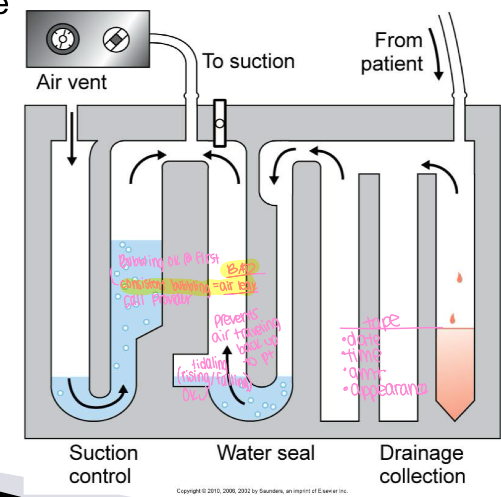
chest tube drainage system
chamber 1- collects from fluid draining from pt
chamber 2= water seal prevents air from entering pt pleural space
chamber 3= suction control of system
has portable options, some have dry suction, some have sterile water
nursing management of chest tube
assess cardiopulmonary and VS every 1-4 hours PRN
assess, treat, and reassess for pain
montior output every 1-4 hours and record amount/color
evalaute chest tube drainage system for rise/fall (tidaling) and check connections
maintain semifowlers 30-45 degree angle to assist in drainage/lung expansion/comfort
encourage deep breathing/cough/incentive spirometry to help lungs reexpand
chest tube reportable conditions
blood drainage of 70ml/hr or greater
sudden stopping of drainage
change in character of drainage
absence of tidaling in water-seal chamber or persistent bubbling
bubbling in chest tube
water levels will normally rise/fall w rsepiration until the lung is expanded
bubbling immediatelay after insertion signifies that air is being removed from the pleural space
bubbling with exhalation and coughing is ok
HOWEVER persistent bubbling indicates air leak in pt lung or ddrainage system
how should CDU be position to a pt
one foot below chest tube insertion site and free of dependent loops/kinks
supplies for chest tube
betadine swabs, clamps, 4×4 ABD, gauze, tape, sertile gloves and water
How do you handle tubes
do not strip tube, can lead to tension pneumothorax
milk with hand over hand method
disconnection of chest tube from CDU- management after
insert chest tbue end into sterile water (creates water seal, negative pressure to prevent air from entering)
call provider STAT
once connected, a full chest assessment is needed, CXR
dislodgment of chest tube
if tube is pulled out, promptly pressure dressing using several 4×4
stay with patient to observe for respiratory distress
call provider stat
prepare for new tube insertion
s/s of tension pneumothorax
acute respiratory distress
tachypnea, shallow respirations, decreased breath sounds
cyanosis, asymmetrical breathing
decreased BP, tachycardia
marked deviation of trachea
ER management of tension pneumothorax w chest tube
start o2 at 4 liters, call provider stat, monitor vs, prepare for new insertion
pt may need cxray and abg