APHY 102 M3-4 Study Guide Questions Flashcards
1/110
There's no tags or description
Looks like no tags are added yet.
Name | Mastery | Learn | Test | Matching | Spaced | Call with Kai | Chat |
|---|
No analytics yet
Send a link to your students to track their progress
111 Terms
What is MALT, and what are examples of it?
Mucosa-Associated Lymphoid Tissue (MALT) is unencapsulated lymphatic tissue found in the digestive, respiratory, urinary, and reproductive tracts. Examples include the tonsils, appendix, and Peyer's patches in the ileum.
what are the functions of lymph nodes?
Filter harmful particles and pathogens from lymph
Provide immune surveillance
House lymphocytes that attack pathogens
House macrophages that engulf foreign material and debris
Which lymph nodes drain different regions of the body?
Cervical: Head and neck
Axillary: Upper limbs, chest, breast
Supratrochlear: Forearm and hand
Inguinal: Lower limbs and external genitalia
Pelvic: Pelvic organs
Abdominal: Abdominal organs
Thoracic: Thoracic organs

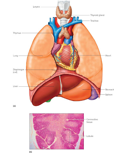
Where is the thymus located?
The thymus is located in the mediastinum, posterior to the upper sternum.
Which cells mature in the thymus?
T lymphocytes (T cells) mature in the thymus.
What hormones are produced by the thymus?
Thymosins, which stimulate T-cell maturation
What happens to the thymus as people age?
It is large during infancy and childhood, shrinks after puberty, and is gradually replaced by adipose and connective tissue in older adults.
Where is the spleen located?
In the upper left abdominal cavity, beneath the diaphragm and behind the stomach.

What are the two tissue types in the spleen?
White pulp – lymphocytes
Red pulp – red blood cells, lymphocytes, and macrophages
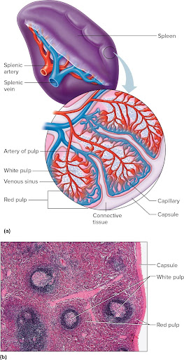
What are the functions of the spleen?
Filters blood
Removes worn-out red blood cells
Macrophages destroy foreign particles
Lymphocytes defend against infection
What is immunity?
The ability of the body to prevent pathogen entry or destroy pathogens that enter the body.
What is a pathogen?
A disease-causing agent such as bacteria, viruses, fungi, protozoa, or parasites.
Compare innate defenses and adaptive defenses.
Innate (Nonspecific) | Adaptive (Specific) |
|---|---|
Present at birth | Develops after exposure |
Nonspecific | Antigen specific |
Rapid response | Slower first response |
No memory | Has memory cells |
Includes skin, inflammation, fever, NK cells, phagocytes, complement | Includes B cells, T cells, antibodies |
What are examples of physical and mechanical barriers?
Skin
Mucous membranes
Cilia
Tears
Saliva
Urine flow
Coughing
Sneezing
What are the four signs of inflammation, and what causes them?
Redness – increased blood flow
Heat – increased blood flow
Swelling – fluid leakage into tissues
Pain – chemicals stimulate nerve endings
What are examples of chemical barriers?
Lysozyme (tears/saliva)
Gastric acid
Sebum
Interferons
Complement proteins
These destroy microbes or prevent their growth.
What is the function of Natural Killer (NK) cells?
NK cells destroy virus-infected cells and cancer cells without prior exposure.
What is phagocytosis, and which cells perform it most actively?
Phagocytosis is the engulfment and digestion of pathogens. The most active phagocytes are neutrophils and macrophages.
What is chemotaxis?
The movement of immune cells toward chemicals released from damaged tissues or pathogens.
Which cells become macrophages?
Monocytes leave the bloodstream and develop into macrophages in tissues.
Why is fever important?
Fever slows pathogen growth, increases metabolic rate, and enhances immune cell activity.
What is the difference between an antigen and an antibody?
Antigen: A foreign substance that triggers an immune response.
Antibody (Immunoglobulin): A protein produced by plasma cells that binds to a specific antigen.
What is the cellular (cell-mediated) immune response?
An immune response carried out by T lymphocytes, which directly attack infected or abnormal cells.
What is the humoral immune response?
An immune response carried out by B lymphocytes, which produce antibodies against antigens.
What are the two main types of lymphocytes, where do they mature, and what are their functions?
T cells: Mature in the thymus; provide cellular immunity.
B cells: Mature in red bone marrow; provide humoral immunity by producing antibodies.
How are T cells activated?
T cells are activated when an antigen-presenting cell (APC) displays an antigen on its Major Histocompatibility Complex (MHC) and the T-cell receptor recognizes it. Helper T cells then release cytokines that stimulate other immune cells.
What is the Major Histocompatibility Complex (MHC)?
MHC is a group of proteins on cell surfaces that display antigens to T cells, allowing the immune system to distinguish self from non-self.
What are the three main types of T cells and their functions?
Helper T cells (CD4): Activate B cells, cytotoxic T cells, and other immune cells by releasing cytokines.
Cytotoxic T cells (CD8): Destroy virus-infected cells, cancer cells, and transplanted cells.
Regulatory (Suppressor) T cells: Slow or stop the immune response after an infection to prevent excessive damage.
How are B cells activated?
B cells are activated when they bind to a specific antigen and receive signals (cytokines) from activated helper T cells. They then divide and differentiate into plasma cells and memory B cells.
What are the functions of plasma cells and memory B cells?
Plasma cells: Produce and secrete large amounts of antibodies.
Memory B cells: Remain in the body long-term and respond rapidly if the same antigen is encountered again.
What are the five classes of immunoglobulins (antibodies), and what are their major functions?
Immunoglobulin | Major Function |
|---|---|
IgG | Most abundant antibody; provides long-term immunity; crosses the placenta to protect the fetus. |
IgA | Found in breast milk, saliva, tears, mucus, and other body secretions; protects mucosal surfaces. |
IgM | First antibody produced during the primary immune response; excellent at activating complement. |
IgE | Involved in allergic reactions and defense against parasites; triggers histamine release. |
IgD | Found mainly on B-cell surfaces; helps activate B cells. |
Memory Trick:
IgG = Gestation (crosses placenta)
IgA = A for "All secretions" (tears, saliva, breast milk)
IgM = Made first
IgE = Environmental allergies
IgD = Develops B cells
What are the major actions of antibodies?
Neutralization: Blocks toxins and viruses from entering cells.
Agglutination: Clumps pathogens together to make them easier to remove.
Precipitation: Causes dissolved antigens to clump together.
Opsonization: Coats pathogens, making them easier for phagocytes to engulf.
Complement activation: Triggers the complement system to destroy pathogens.
What is the difference between the primary and secondary immune response?
Primary Response | Secondary Response |
|---|---|
First exposure to an antigen | Later exposure to the same antigen |
Slower response | Much faster response |
Fewer antibodies produced | Many more antibodies produced |
Memory cells are formed | Memory cells respond immediately |
What are the different types of acquired immunity?
Type | How It Is Acquired | Example |
|---|---|---|
Naturally acquired active | Infection stimulates your own immune system | Recovering from chickenpox |
Artificially acquired active | Vaccination stimulates your own immune system | Flu shot |
Naturally acquired passive | Antibodies received naturally from the mother | IgG through the placenta; IgA in breast milk |
Artificially acquired passive | Injection of preformed antibodies | Rabies immune globulin or antivenom |
Memory Tip:
Active = Your body makes antibodies.
Passive = You receive antibodies from another source.
What is an autoimmune disease, what are autoantibodies, and what are some examples?
Autoimmune disease: A disorder in which the immune system attacks the body's own tissues.
Autoantibodies: Antibodies that mistakenly target the body's own cells.
Examples:
Rheumatoid arthritis
Systemic lupus erythematosus (SLE)
Type 1 diabetes mellitus
Multiple sclerosis
Graves' disease
Hashimoto's thyroiditis
What are the main actions of the digestive system?
Ingests food
Mechanically and chemically digests food
Absorbs nutrients and water
Eliminates indigestible waste (defecation)
What is the difference between mechanical digestion and chemical digestion?
Mechanical digestion: Physically breaks food into smaller pieces (chewing, churning, segmentation).
Chemical digestion: Uses enzymes and acids to break food into molecules that can be absorbed.
Which organs make up the alimentary canal, and which are accessory organs?
Alimentary Canal (in order):
Mouth
Pharynx
Esophagus
Stomach
Small intestine
Large intestine
Rectum
Anal canal/Anus
Accessory Organs:
Teeth
Tongue
Salivary glands
Liver
Gallbladder
Pancreas
What are the organs of the digestive system and their main functions?
Mouth: Ingestion and chewing
Pharynx: Swallowing
Esophagus: Moves food to the stomach
Stomach: Stores food and begins protein digestion
Small intestine: Most digestion and nutrient absorption
Large intestine: Absorbs water and forms feces
Rectum: Stores feces
Anus: Eliminates feces
Liver: Produces bile
Gallbladder: Stores and concentrates bile
Pancreas: Produces digestive enzymes and bicarbonate
What are the four layers of the alimentary canal (from deepest to most superficial), and which layer absorbs nutrients?
Mucosa (contains the absorptive epithelium; nutrient absorption occurs here)
Submucosa
Muscularis externa
Serosa (or adventitia in some regions)
Memory Trick:
"My Stomach Makes Soup"
Mucosa
Submucosa
Muscularis externa
Serosa
What are the basic movements of the alimentary canal, and what controls them?
The digestive tract moves food by:
Peristalsis
Segmentation
These movements are controlled by the enteric nervous system, which is regulated by the autonomic nervous system.
What are peristalsis and segmentation?
Peristalsis: Wave-like contractions that move food forward.
Segmentation: Mixing contractions that break food apart and mix it with digestive juices
What are the functions of the mouth, and what is mastication?
Receives food
Begins mechanical digestion
Begins carbohydrate digestion with saliva
Forms a bolus for swallowing
Mastication = chewing.
What are the lingual frenulum, papillae, and lingual tonsils?
Lingual frenulum: Fold of tissue attaching the tongue to the floor of the mouth.
Papillae: Projections on the tongue that provide grip; many contain taste buds.
Lingual tonsils: Lymphatic tissue at the base of the tongue that helps fight infection.
How many primary and secondary teeth do humans have?
Primary (baby) teeth: 20
Permanent (adult) teeth: 32
What are the three pairs of salivary glands, and what do they secrete?
Parotid glands: Mostly watery (serous) saliva rich in amylase
Submandibular glands: Mixed serous and mucus secretions
Sublingual glands: Mostly mucus secretions
What is the difference between serous cells and mucous cells?
Serous cells: Produce thin, watery secretions containing digestive enzymes (especially salivary amylase).
Mucous cells: Produce thick mucus that lubricates and protects the mouth.
Describe the three regions of the pharynx.
Nasopharynx: Behind the nasal cavity; passageway for air only.
Oropharynx: Behind the oral cavity; passageway for food and air.
Laryngopharynx: Connects to the esophagus and larynx; passageway for food and air.
What is the function of the epiglottis?
The epiglottis closes over the larynx during swallowing to prevent food and liquids from entering the airway.
What are the functions of the esophagus?
The esophagus transports food from the pharynx to the stomach by peristalsis.
What are the functions of the stomach, and what are rugae?
Functions of the stomach:
Stores food
Mixes food with gastric juice
Begins protein digestion
Produces chyme
Kills many microorganisms with acid
Rugae: Folds of the stomach lining that allow it to expand after eating.
What are the layers of smooth muscle in the stomach?
Inner oblique layer
Middle circular layer
Outer longitudinal layer
Memory Trick: "OCL"
What is the purpose of the pyloric sphincter?
It regulates the movement of chyme from the stomach into the duodenum and prevents backflow into the stomach.
Describe the four regions of the stomach.
Cardia: Receives food from the esophagus.
Fundus: Dome-shaped upper portion; stores food and gas.
Body: Largest region; mixes food and secretes gastric juice.
Pylorus: Distal region leading to the duodenum; controls stomach emptying.
Memory Trick:
"Come Find Big Pizza"
Cardia
Fundus
Body
Pylorus
What are the components of gastric juice, and what are their functions?
Component | Function |
|---|---|
Pepsinogen | Inactive enzyme secreted by chief cells |
Pepsin | Digests proteins |
Gastric lipase | Begins fat digestion |
Hydrochloric acid (HCl) | Activates pepsin, kills microbes, denatures proteins |
Mucus | Protects the stomach lining from acid |
Intrinsic factor | Required for vitamin B₁₂ absorption in the small intestine |
What are the functions of somatostatin, gastrin, and CCK?
Somatostatin: Inhibits gastric secretions and slows digestion.
Gastrin: Stimulates gastric acid secretion and increases stomach motility.
CCK (Cholecystokinin): Stimulates gallbladder contraction and pancreatic enzyme secretion; slows stomach emptying.
What substances can the stomach absorb?
The stomach absorbs small amounts of water, alcohol, aspirin, and certain medications, but very few nutrients.
What is the difference between a bolus and chyme?
Bolus: Chewed food mixed with saliva before swallowing.
Chyme: Semi-liquid mixture of food and gastric juice that leaves the stomach.
What are the endocrine and exocrine functions of the pancreas?
Endocrine: Releases hormones (insulin and glucagon) into the bloodstream to regulate blood glucose.
Exocrine: Releases pancreatic juice (digestive enzymes and bicarbonate) into the duodenum.
What is the function of pancreatic acinar cells?
Pancreatic acinar cells produce and secrete the digestive enzymes found in pancreatic juice.
What are the components of pancreatic juice, and what are their functions?
Component | Function |
|---|---|
Pancreatic amylase | Digests carbohydrates into smaller sugars. |
Pancreatic lipase | Digests triglycerides (fats). |
Trypsin | Digests proteins into smaller peptides. |
Chymotrypsin | Continues protein digestion. |
Carboxypeptidase | Removes amino acids from the ends of peptides. |
Nucleases | Digest DNA and RNA (nucleic acids). |
Bicarbonate (HCO₃⁻) | Neutralizes acidic chyme entering the duodenum. |
What are the functions of secretin and CCK in regulating pancreatic juice?
Secretin: Stimulates the pancreas to release bicarbonate-rich fluid to neutralize stomach acid.
CCK (Cholecystokinin): Stimulates the pancreas to release digestive enzymes and causes the gallbladder to contract and release bile.
What are the functions of the liver and gallbladder?
Liver
Produces bile
Processes nutrients
Detoxifies harmful substances
Stores glycogen, vitamins, and minerals
Produces plasma proteins
Gallbladder
Stores bile
Concentrates bile
Releases bile into the duodenum when needed
What are the functions of the components of bile?
Bile salts: Emulsify fats to make them easier to digest.
Bile pigments (bilirubin): Waste products from red blood cell breakdown.
Cholesterol: Eliminated through bile.
Electrolytes and water: Help transport bile.
Remember: Bile does not digest fat—it emulsifies fat, increasing the surface area for lipase.
What is jaundice?
Jaundice is the yellow discoloration of the skin and eyes caused by excess bilirubin in the blood.
What are the functions of the small intestine?
Completes most chemical digestion.
Absorbs most nutrients and water.
Mixes chyme with digestive juices.
Receives bile and pancreatic enzymes.
What are the three sections of the small intestine, and where are they located?
Duodenum: First section; receives chyme from the stomach and digestive secretions.
Jejunum: Middle section; primary site of nutrient absorption.
Ileum: Final section; absorbs vitamin B₁₂, bile salts, and remaining nutrients before emptying into the large intestine.
What are the functions and size of the large intestine?
Functions
Absorbs water and electrolytes.
Forms and stores feces.
Houses bacteria that produce vitamins (especially vitamin K and some B vitamins).
Size
About 1.5 meters (5 feet) long.
Wider than the small intestine.
Describe the sections of the large intestine.
Cecum
Appendix
Ascending colon
Transverse colon
Descending colon
Sigmoid colon
Rectum
Anal canal (anus)
Pancreatic Enzymes
Enzyme | Digests |
|---|---|
Amylase | Carbohydrates |
Lipase | Fats |
Trypsin | Proteins |
Chymotrypsin | Proteins |
Carboxypeptidase | Peptides |
Nuclease | DNA & RNA |
Hormones to Know
Hormone | Function |
|---|---|
Gastrin | ↑ Stomach acid |
Secretin | ↑ Bicarbonate |
CCK | ↑ Pancreatic enzymes & bile release |
Somatostatin | ↓ Digestive secretions |
What are the major functions of the respiratory system?
Bring oxygen into the body
Remove carbon dioxide
Filter, warm, and moisten incoming air
Produce vocal sounds
Provide the sense of smell
Help regulate blood pH
Define respiration, external respiration, internal respiration, and cellular respiration.
Respiration: Overall process of exchanging gases between the atmosphere and body cells.
External respiration: Breathing (ventilation) and gas exchange between the lungs and blood.
Internal respiration: Gas exchange between the blood and body tissues.
Cellular respiration: ATP production in mitochondria using oxygen and producing carbon dioxide.
Which organs are in the upper and lower respiratory tracts?
Upper Respiratory Tract
Nose
Nasal cavity
Sinuses
Pharynx
Larynx
Lower Respiratory Tract
Trachea
Bronchial tree
Lungs
What are the functions of the nose and nasal cavity?
Filter incoming air
Warm and moisten air
Trap dust and pathogens with mucus
Cilia move mucus toward the pharynx
House olfactory receptors for smell
Conduct air into the respiratory tract
What are the functions of the paranasal sinuses?
Reduce the weight of the skull
Add resonance to the voice
Drain mucus into the nasal cavity
What problems are associated with cigarette smoking?
Smoking:
Damages and paralyzes cilia
Causes smoker's cough
Increases mucus production
Increases respiratory infections
Causes chronic bronchitis
Destroys alveoli (emphysema)
Greatly increases lung cancer risk
Describe the pharynx and its three regions.
Nasopharynx: Behind the nose; passageway for air only.
Oropharynx: Behind the mouth; passageway for food and air.
Laryngopharynx: Connects to the esophagus and larynx; passageway for food and air.
Functions:
Passageway for food and/or air depending on the region
Assists swallowing
Where is the larynx located, and what are its functions?
Location: Between the pharynx and trachea.
Functions:
Maintains an open airway
Prevents food from entering the trachea
Produces sound (voice)
What are the three large cartilages of the larynx?
Thyroid cartilage
Cricoid cartilage
Epiglottis
What are the false vocal cords, true vocal cords, and glottis?
False vocal cords: Upper folds that help close the airway during swallowing.
True vocal cords: Lower folds that vibrate to produce sound.
Glottis: Opening between the true vocal cords.
What are the characteristics of the trachea?
Connects the larynx to the bronchi
Supported by C-shaped hyaline cartilage rings
Lined with pseudostratified ciliated epithelium
Mucus traps debris while cilia move it upward toward the pharynx
List the divisions of the bronchial tree.
Trachea
Primary (main) bronchi
Secondary (lobar) bronchi
Tertiary (segmental) bronchi
Bronchioles
Terminal bronchioles
Respiratory bronchioles
Alveolar ducts
Alveolar sacs
Alveoli
What are the functions and structural characteristics of alveoli?
Site of gas exchange
Tiny air sacs surrounded by capillaries
Thin walls allow rapid diffusion
Made of simple squamous epithelium (Type I cells)
Type II cells produce surfactant
What are bronchodilation and bronchoconstriction?
Bronchodilation: Widening of bronchioles to increase airflow.
Bronchoconstriction: Narrowing of bronchioles to decrease airflow.
What is the difference between the right and left lungs?
Right lung
3 lobes
Larger
Shorter and wider
Left lung
2 lobes
Smaller
Contains the cardiac notch for the heart
Define the visceral pleura, parietal pleura, and pleural cavity.
Visceral pleura: Covers the lungs.
Parietal pleura: Lines the thoracic cavity.
Pleural cavity: Space between the pleurae containing pleural fluid to reduce friction during breathing.
What is a good summary of the respiratory system organs?
Conducting organs
Nose
Nasal cavity
Sinuses
Pharynx
Larynx
Trachea
Bronchi
Bronchioles
Respiratory portion
Respiratory bronchioles
Alveolar ducts
Alveolar sacs
Alveoli
What is breathing (ventilation)?
Ventilation is the movement of air into and out of the lungs.
What are inspiration, expiration, and the respiratory cycle?
Inspiration (Inhalation): Air moves into the lungs.
Expiration (Exhalation): Air moves out of the lungs.
Respiratory cycle: One complete inhalation followed by one complete exhalation.
What is normal atmospheric pressure at sea level?
760 mmHg (1 atmosphere or 1 atm).
What is Boyle's Law?
Boyle's Law states that pressure and volume are inversely related:
Volume increases → Pressure decreases
Volume decreases → Pressure increases
This principle allows air to move into and out of the lungs.
How do inhalation and exhalation occur, and how do Boyle's Law and atmospheric pressure relate to airflow?
Inhalation
Diaphragm contracts and flattens.
External intercostals contract.
Thoracic cavity volume increases.
Lung pressure falls below atmospheric pressure.
Air flows into the lungs.
Exhalation
Diaphragm relaxes.
Thoracic cavity volume decreases.
Lung pressure rises above atmospheric pressure.
Air flows out of the lungs.
What is the difference between normal and forced inspiration, and which muscles are involved?
Normal Inspiration
Diaphragm
External intercostal muscles
Forced Inspiration
Diaphragm
External intercostals
Sternocleidomastoid
Scalenes
Pectoralis minor
What is the difference between normal and forced expiration, and which muscles are involved?
Normal Expiration
Passive
Caused by elastic recoil of the lungs and surface tension
No muscle contraction required
Forced Expiration
Internal intercostal muscles
Abdominal muscles
Used during exercise, coughing, or blowing out candles
What are respiratory volume and spirometry?
Respiratory volume: The amount of air moved during breathing.
Spirometry: A test that measures lung volumes and capacities using a spirometer.
What are the main respiratory volumes?
Volume | Description |
|---|---|
Tidal Volume (TV) | Air inhaled or exhaled during normal breathing (~500 mL) |
Inspiratory Reserve Volume (IRV) | Extra air that can be inhaled after a normal inspiration |
Expiratory Reserve Volume (ERV) | Extra air that can be exhaled after a normal expiration |
Residual Volume (RV) | Air remaining in the lungs after maximum exhalation |
What are dead air spaces?
Dead air spaces are areas where air is present but gas exchange does not occur, such as the conducting airways (nose, trachea, bronchi).
What are nonrespiratory air movements?
Examples include:
Coughing
Sneezing
Laughing
Crying
Hiccups
Yawning
These help protect the respiratory tract or serve other functions.