Anatomy and Physiology II Exam II
 Call Kai
 Learn
Learn Practice Test
 Spaced Repetition
 Match
Match Flashcards
 Knowt Play
1/102

There's no tags or description
Looks like no tags are added yet.
Name | Mastery | Learn | Test | Matching | Spaced | Call with Kai | Chat |
|---|
No analytics yet
Send a link to your students to track their progress
103 Terms
arteries
__ carry blood away from the heart to tissues
oxygenated blood
Arteries usually carry __ __
Veins
__ carry blood toward the heart to tissues
Deoxygenated Blood
Veins usually carry __ __
pulmonary and fetal
Arteries usually carry oxygenated blood, and veins usually carry deoxygenated blood, except for __
capillaries
gas exchange
Tunica externa, media, and intima
Arterial walls superficial to deep
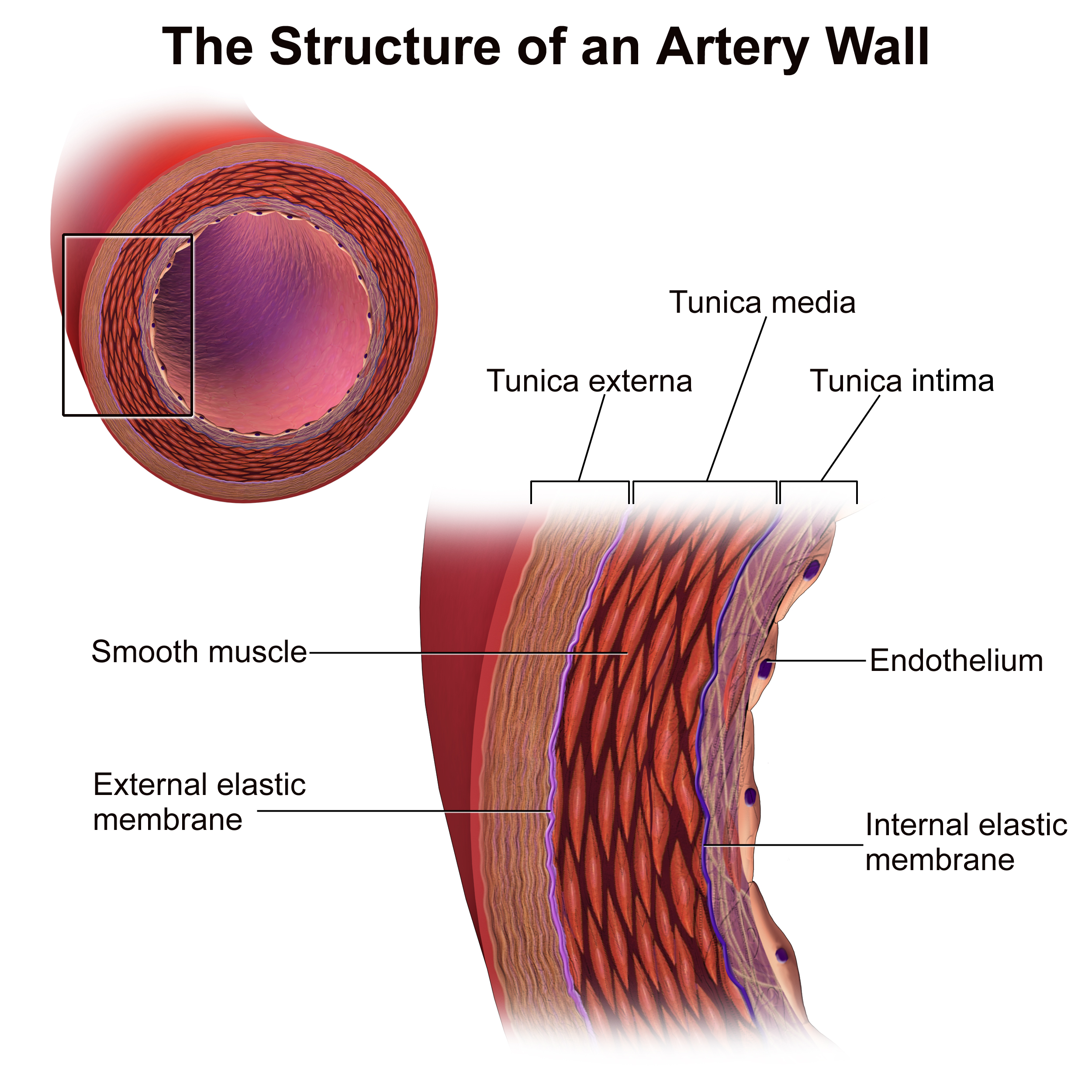
Tunica externa
outer layer; elastic and collagen fibers
Tunica media
middle layer; smooth muscle and elastic tissue
Tunica intima
inner layer; endothelium
atrium (atria)
the two upper chambers of the heart
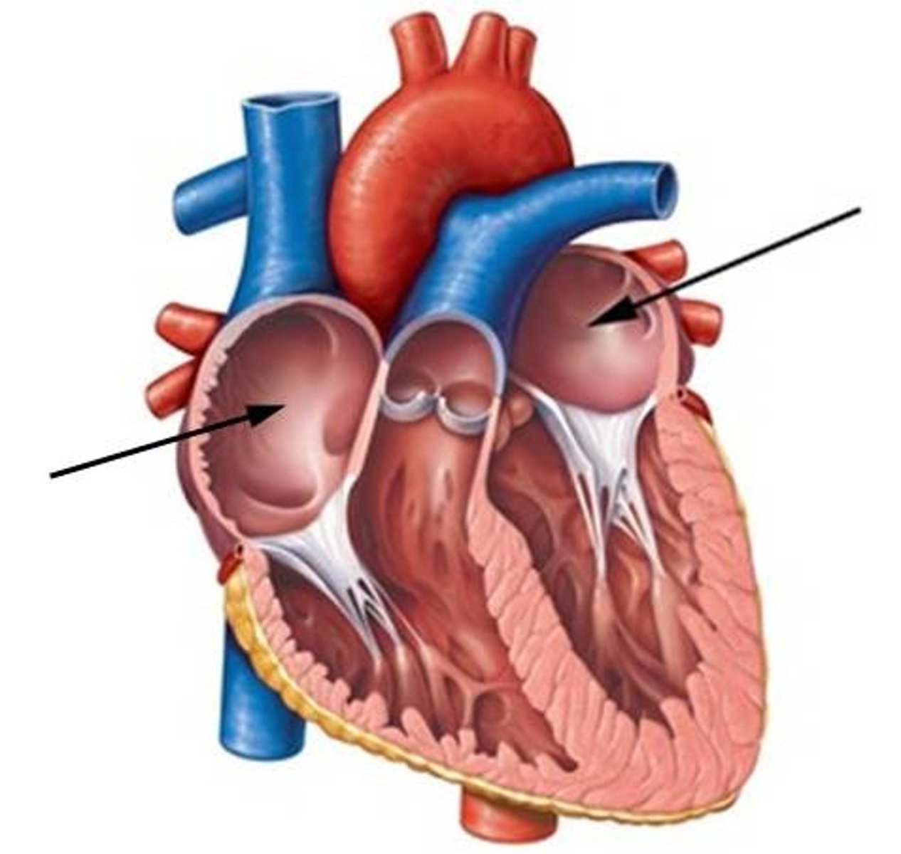
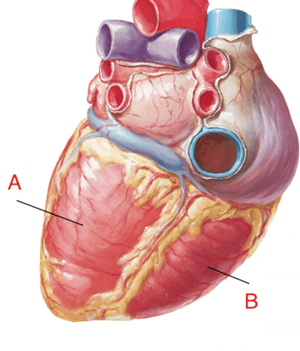
ventricles
the two lower chambers of the heart
right atrium
Receives deoxygenated blood from superior and inferior vena cava, and coronary sinus.
tricuspid valve
Blood passes from the right atrium to the right ventricle through the __ __
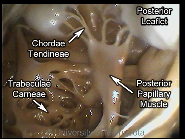
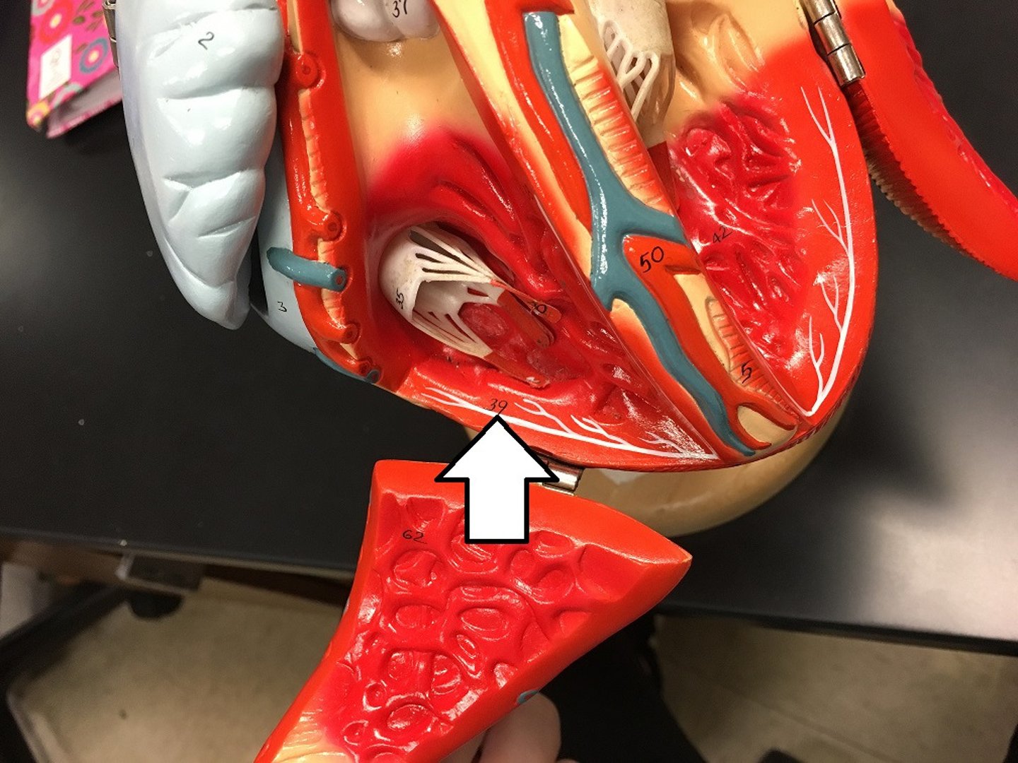
trabeculae carneae
raised bundles of cardiac muscle fibers; in the right ventricle
interventricular septum
partition between the right and left ventricles
right ventricle
blood passes from the __ __ through the pulmonary semi-lunar valve into the pulmonary trunk
pulmonary trunk
large artery which divides into right and left pulmonary arteries that lead to the lungs
left atrium
receives oxygenated blood from the lungs through the pulmonary veins
bicuspid valve
blood passes from left atrium to left ventricle through the __ __
left ventricle
oxygenated blood passes from the left ventricle through the aortic semi-lunar valve into the ascending aorta, then out to the body tissues/cells
papillary muscles
pulls on "heart strings" or chordae tendineae
chordae tendineae
fibrous cords that connect AV valves to papillary muscles
semilunar valves
aortic and pulmonary; blood flows into aorta and pulmonary trunk; as ventricles contract, SL valves open; blood exits the ventricles
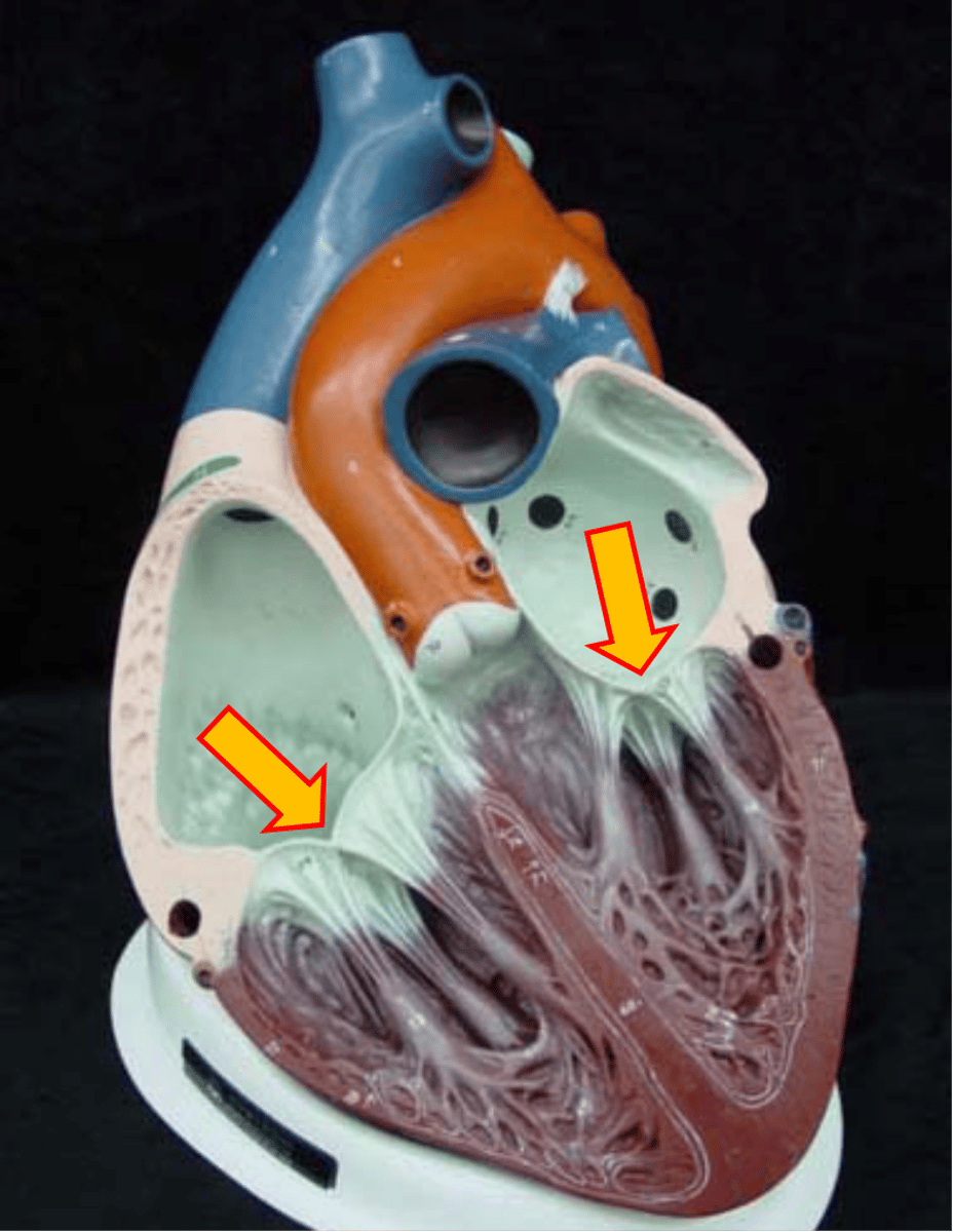
atrioventricular valves
bicuspid (mitral) and tricuspid; blood flows from atria into ventricles through AV valves; as ventricles contract, cusps of valves closes; closing prevents backflow into atria
SA Node
Pacemaker; generates impulses
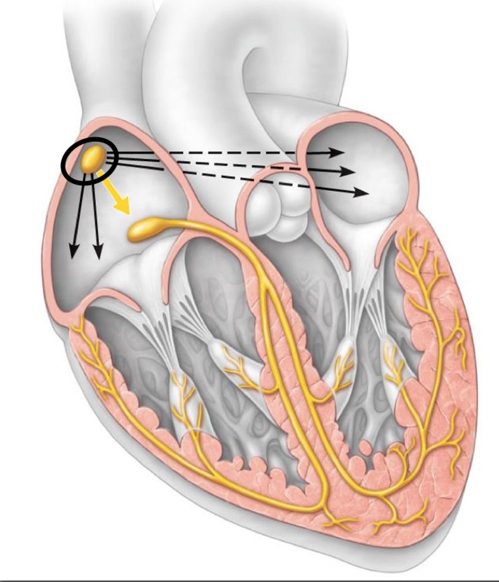
Purkinje fibers
stimulate the contractile cells of both ventricles; branches turn into "webs" called
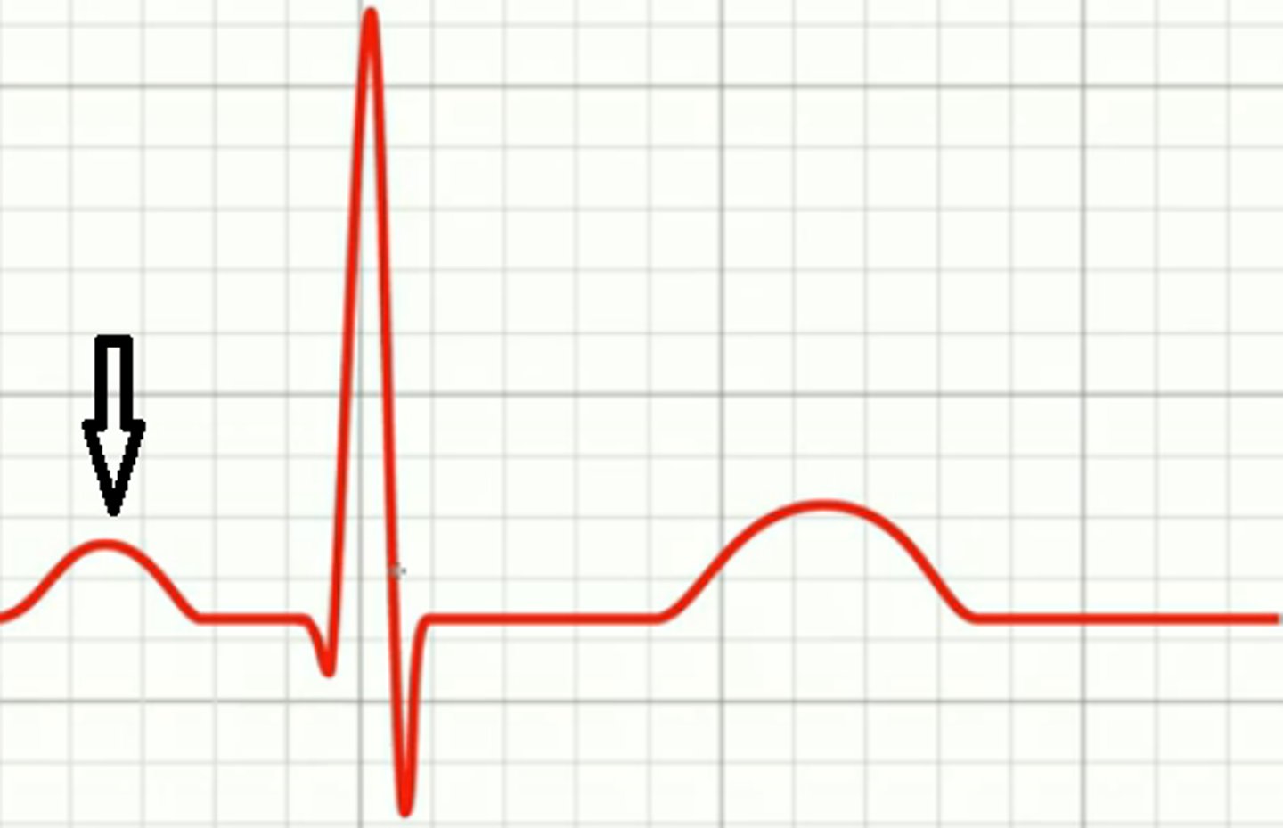
P wave
atrial depolarization (contraction); tells atria to contract
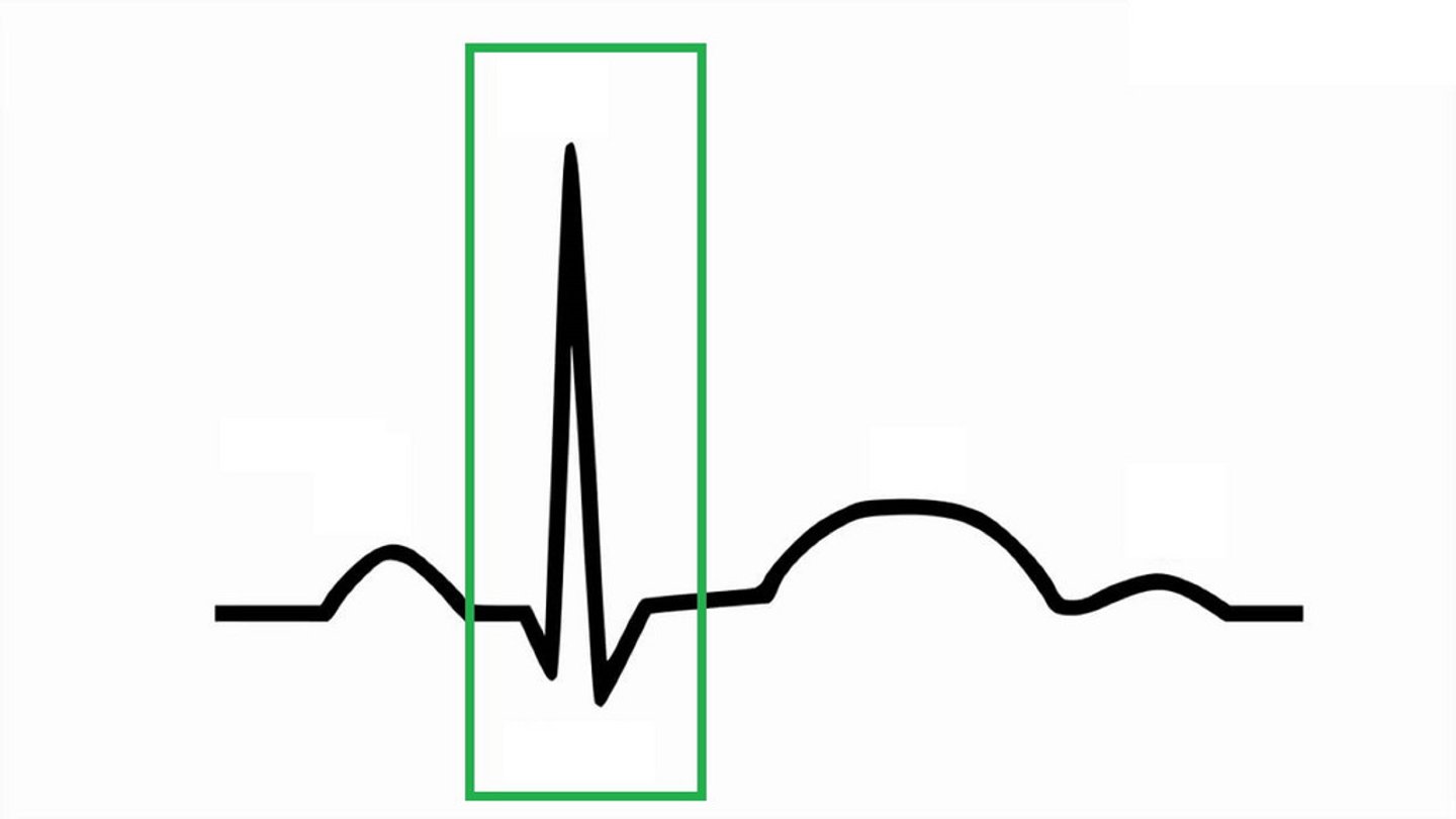
QRS complex
ventricular depolarization (contraction); tells ventricles to contract; atria relaxes here
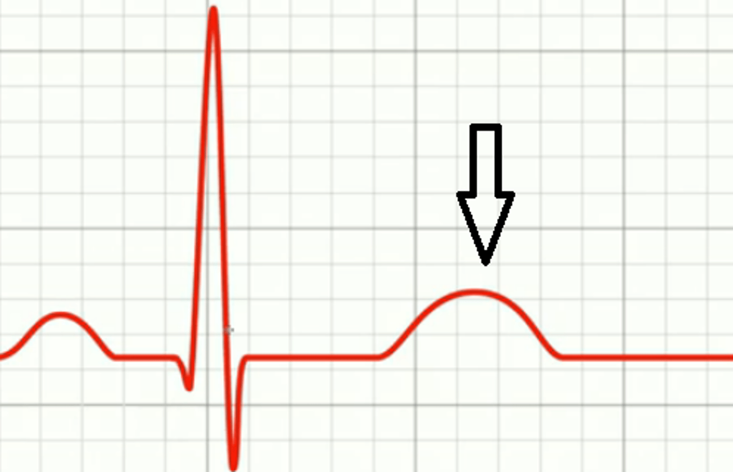
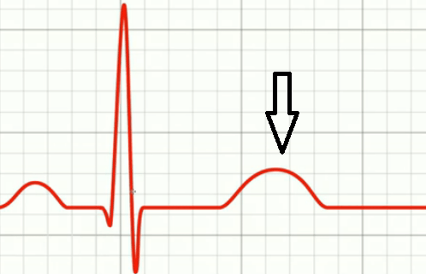
T wave
ventricular repolarization (relaxation); tells ventricles to relax; pulls blood in
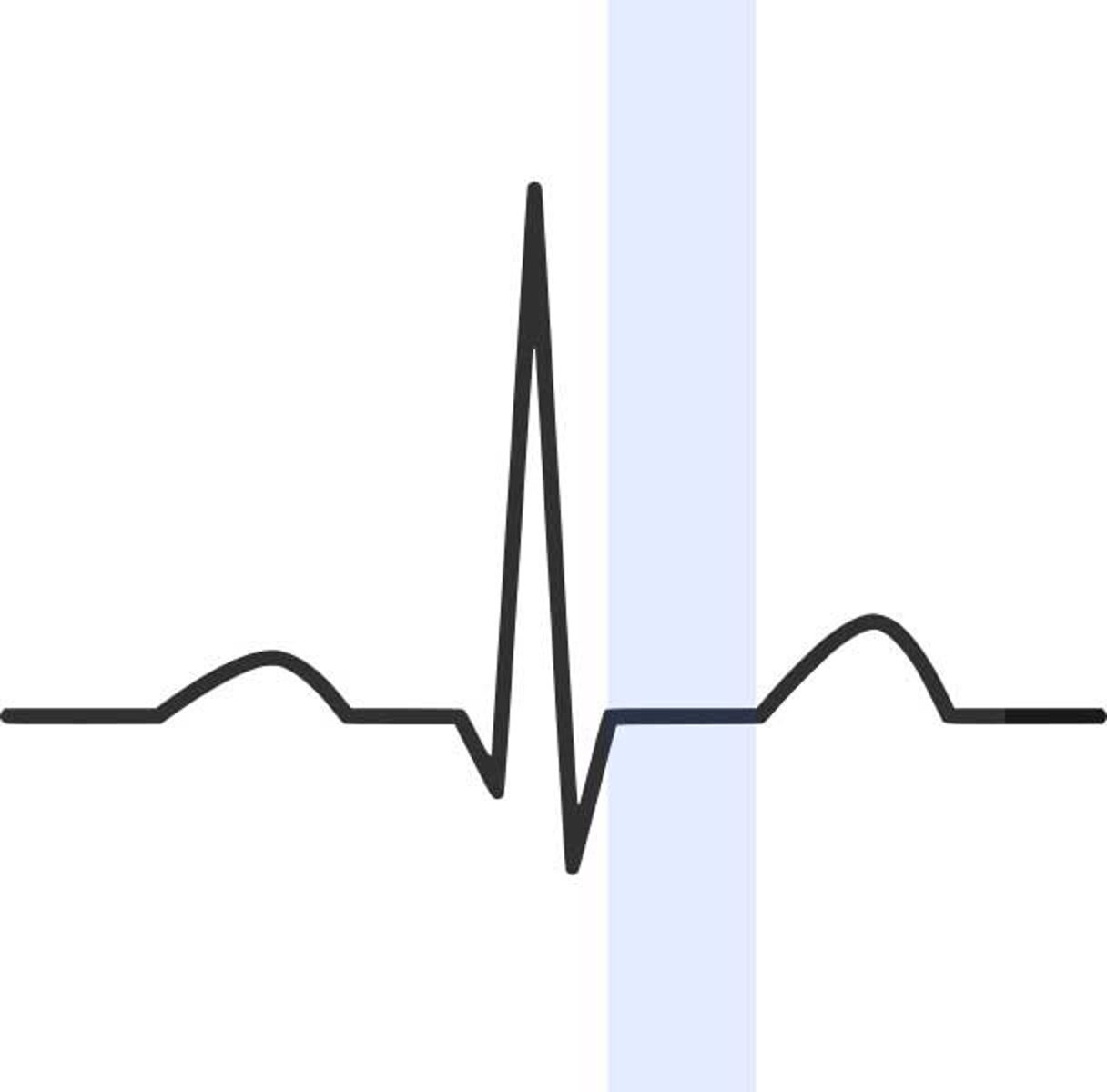
S-T segment
elevated during MI (myocardial infarction, heart attack)
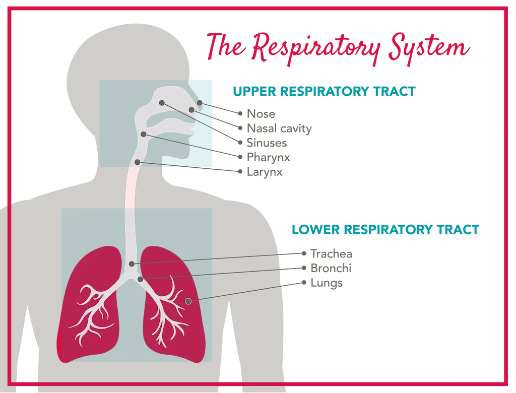
upper respiratory system
nose, nasal cavity, sinuses, pharynx, larynx
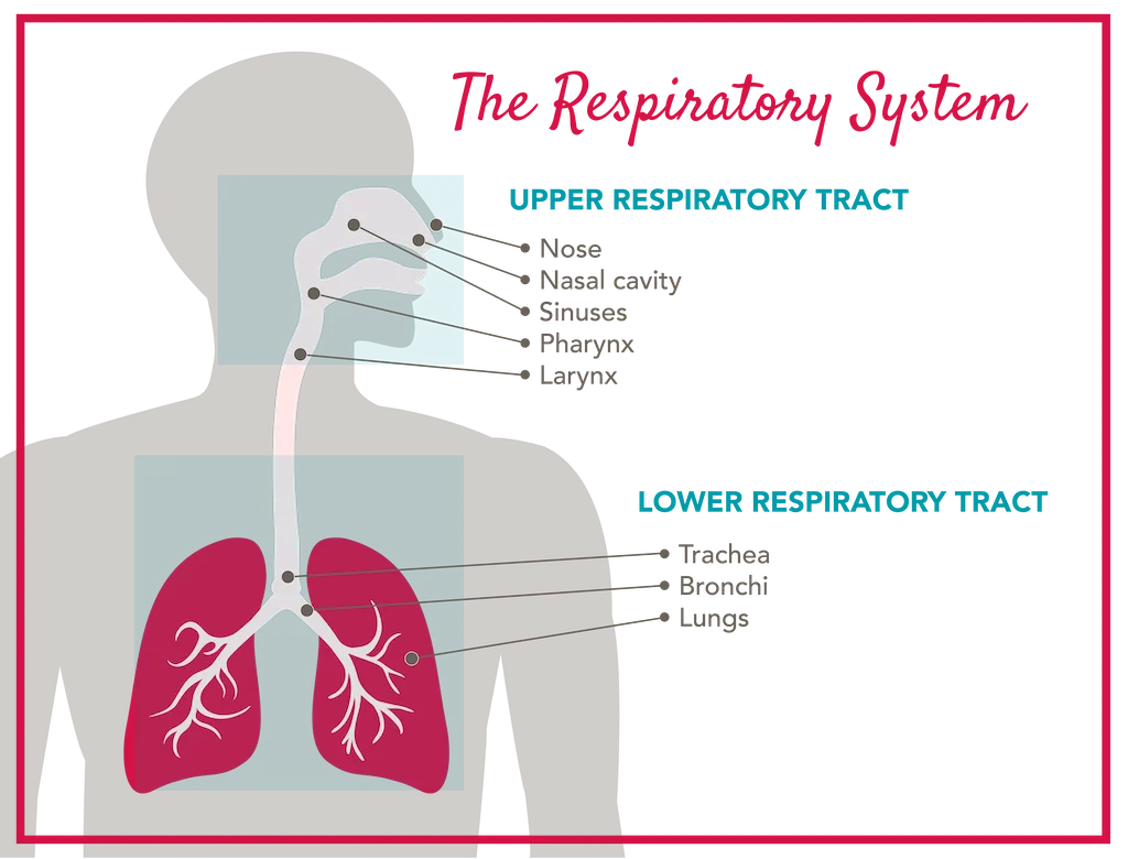
lower respiratory system
trachea. bronchi, lungs, alveoli
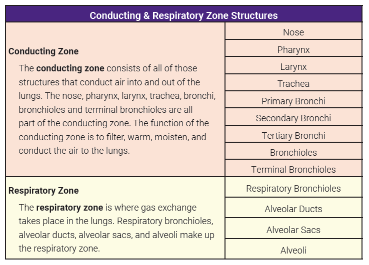
Conducting zone
air travels via tubes into and out of body; nose and nasal cavity to bronchioles
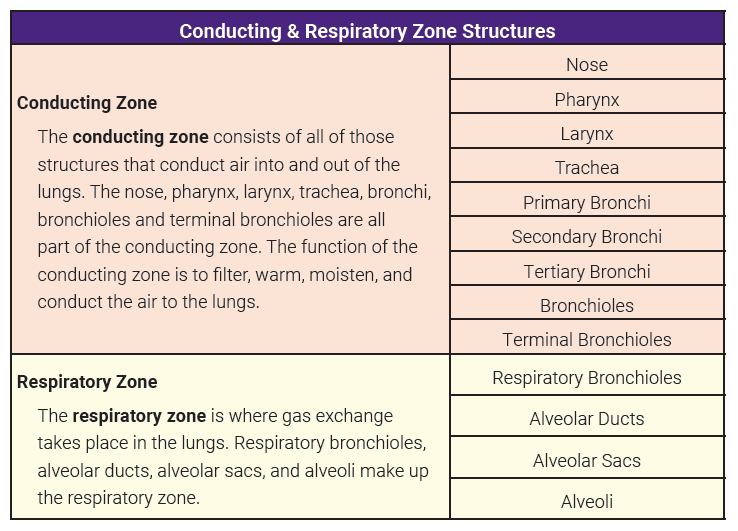
Respiratory zone
where gas exchange occurs; includes only structures that contain alveoli
Respiration
full breath; aka pulmonary ventilation
pulmonary gas exchange
movement of gases between lungs and blood; aka external respiration
tissue gas exchange
exchange of gases between blood and tissues; aka internal respiration
nares
nostrils
nasal conchae
scroll-shaped bony inside the nasal cavities; increases surface area
pharynx
passageway for food and air; throat; functions in speech resonance and air flow for proper sounds
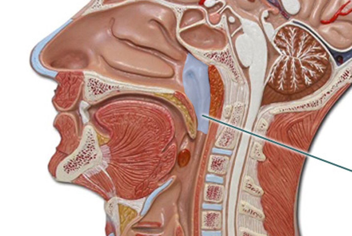
nasopharynx
air only
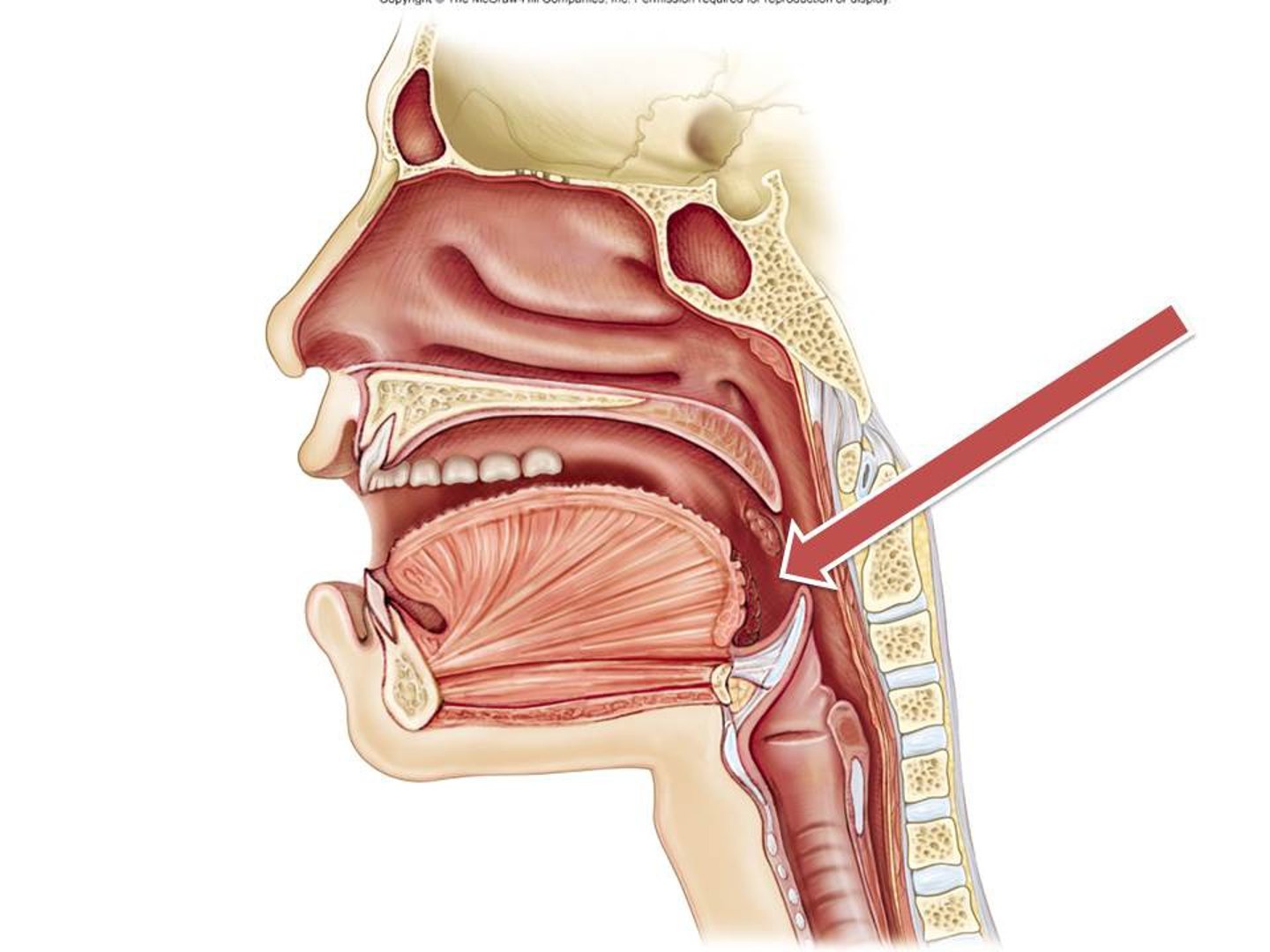
oropharynx
food and air
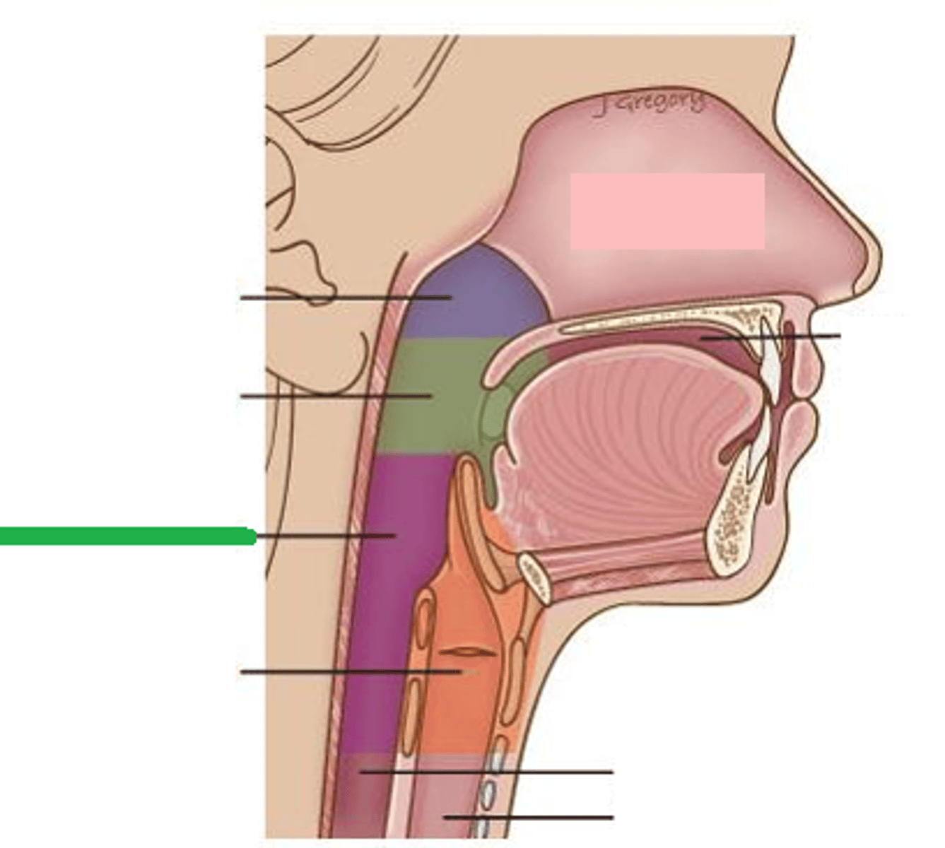
hypopharynx
food and air
larynx
voice box; keeps food and liquids out of respiratory tract
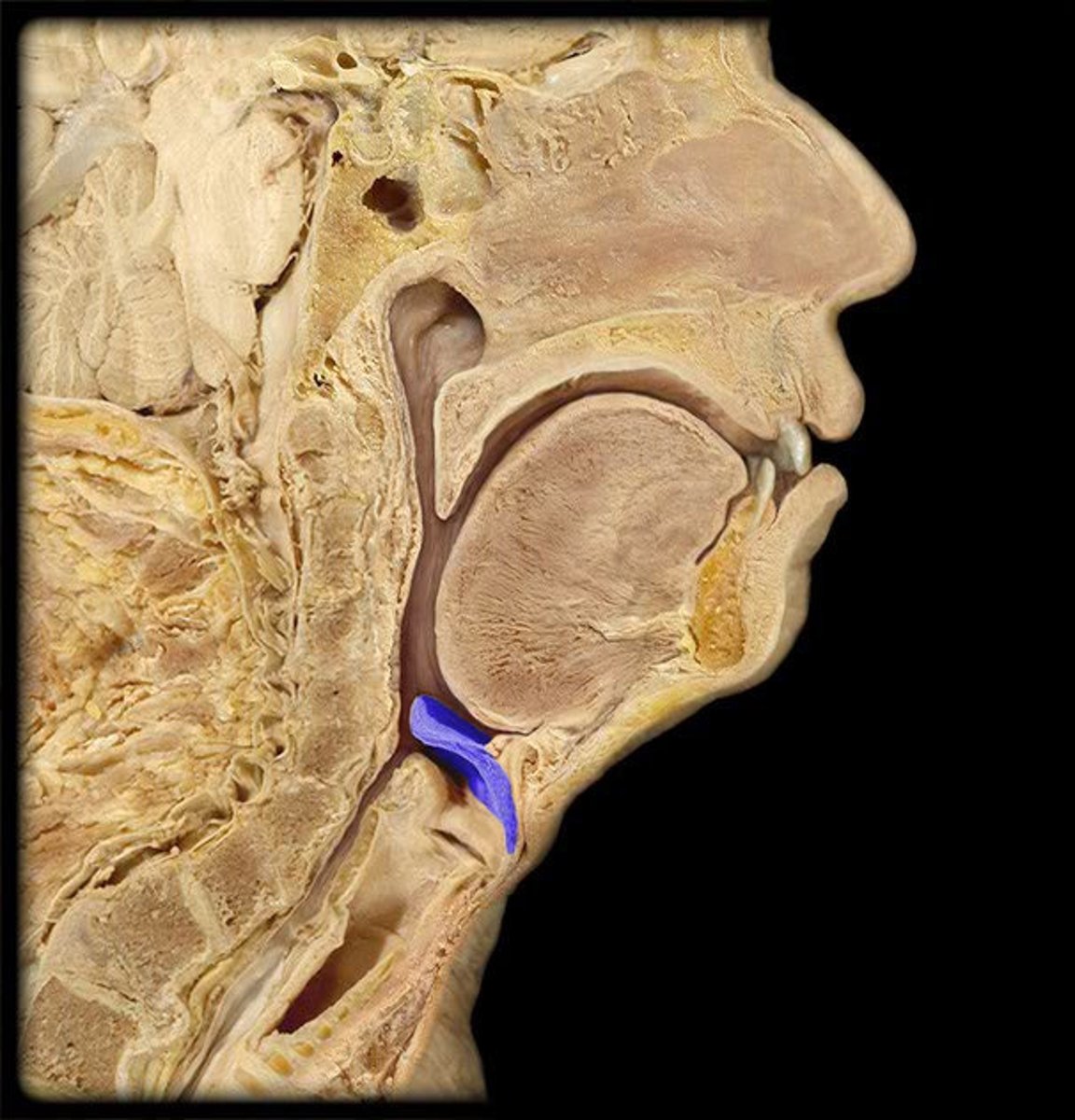
epiglottis
trap door that covers larynx while swallowing; keeps you from choking
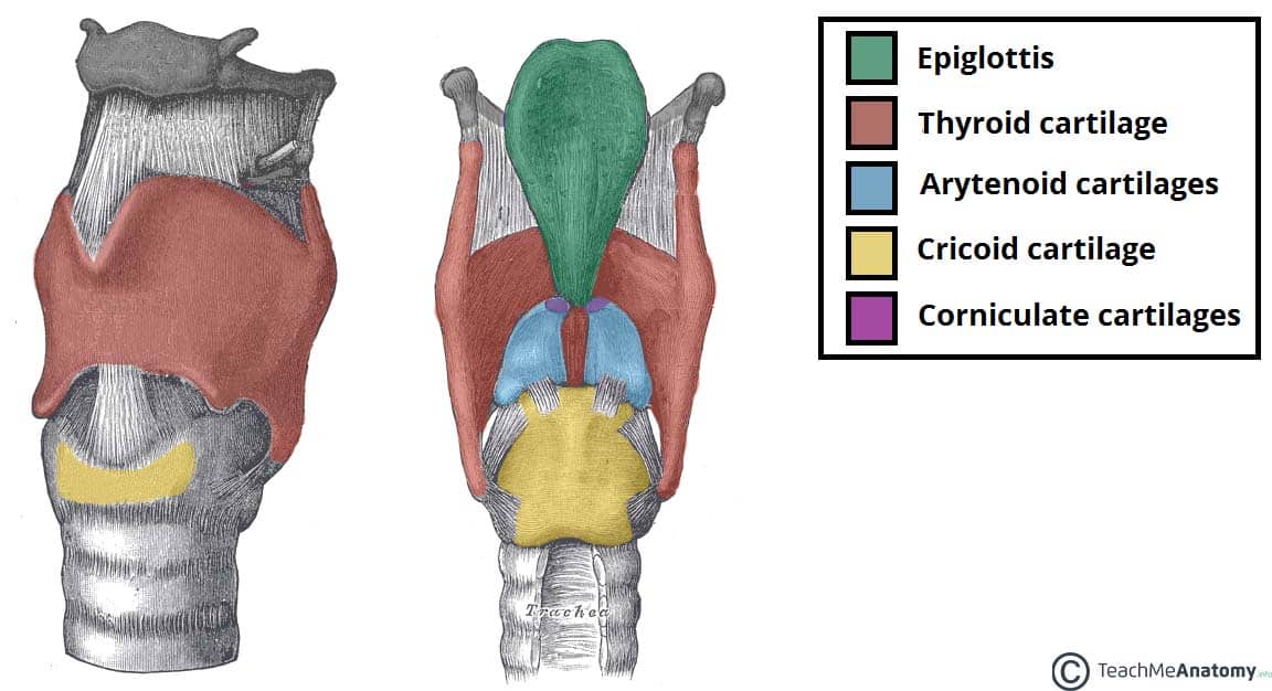
cricoid cartilage
landmark for tracheotomy (emergency airway; tube between thyroid cartilage and cricoid cartilage)
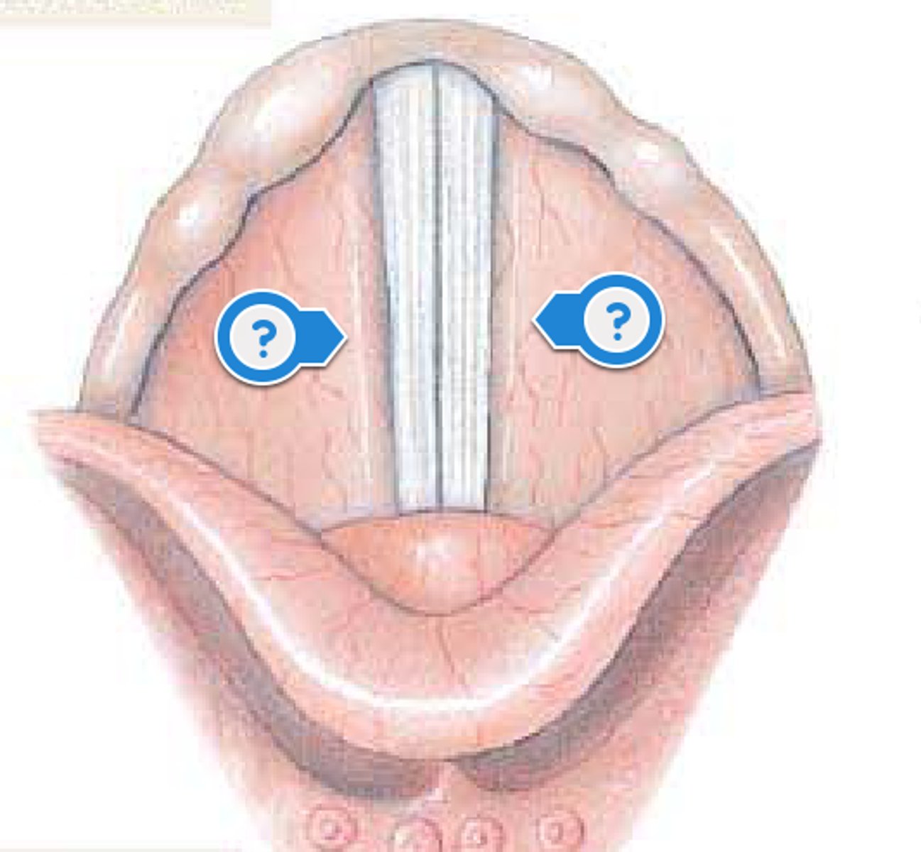
vestibular folds
false vocal cords; don't produce sound; close off glottis
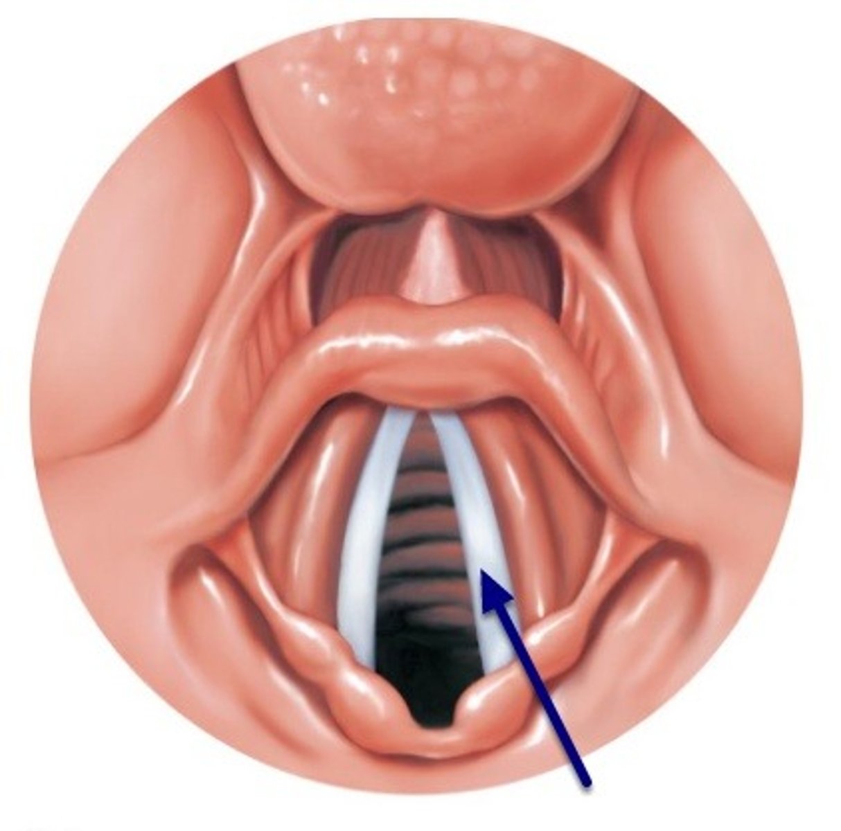
vocal folds
true vocal cords; speaking/singing; vocal ligaments vibrate to produce sound; testosterone increases size of larynx and deepens the voice of males
trachea
windpipe; extends from larynx to T5; has C rings to hold it open
carina
where trachea divides; inhalation of food, water, powders irritate carnia and trigger cough reflex; prevents damage to remaining respiratory tract
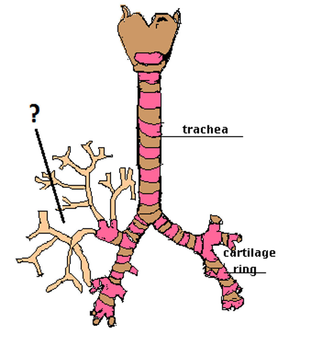
bronchial tree
trachea divides into left and right primary bronchi to the lungs; primary bronchi divide into secondary bronchi then to each lung lobe; secondary bronchi branch into tertiary bronchi and then to the bronchioles
bronchioles
smallest airways
alveolar ducts
respiratory bronchiole become __ __
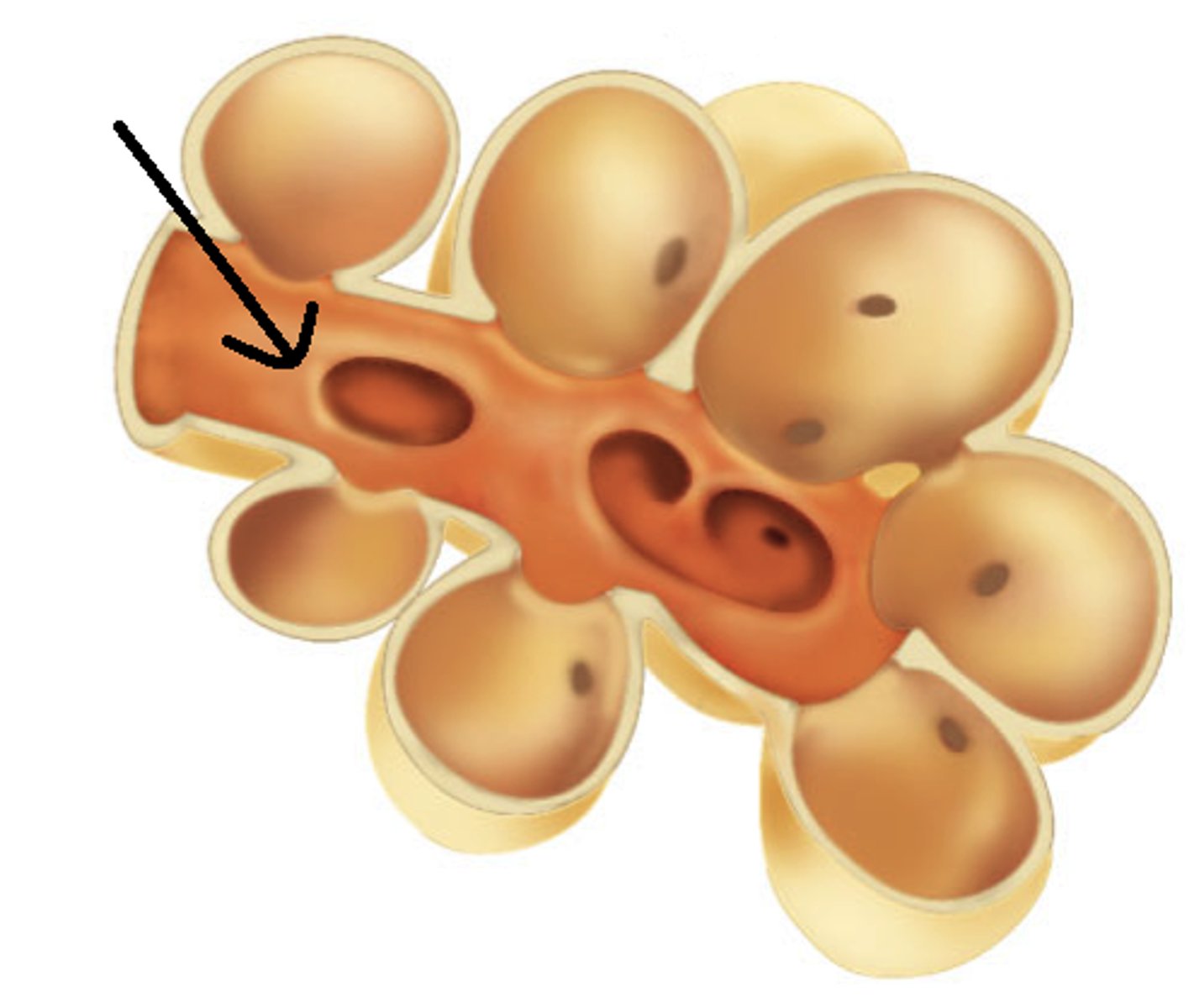
alveolar sacs
alveolar ducts end in __ __, grapelike clusters of alveoli; inspired air causes as exchange to occur
alveoli
final destination for inspired air
type I alveolar cells
squamous cells; 90% of cells in alveolar wall; very thin; allows for rapid diffusion of gases across cell membranes; huge amount of surface area; increases gas exchange efficiency
type II alveolar cells
small cuboidal cells; 10% of cells in alveolar wall; synthesize surfactant; chemical that reduces surface tension on alveoli; prevents alveoli from collapsing
alveolar macrophages
mobile phagocytes; clean up debris in alveoli
surfactant
sticky fluid made of phospholipids and lipoproteins; prevents alveoli from collapsing; decreases surface tension
visceral pleura, parietal pleura, pleural space
inner layer; adheres lungs, outer layer; attaches to thoracic wall and diaphragm, space between the two layers which is very small as the two layers are together and it technically contains pleural fluid
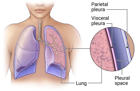
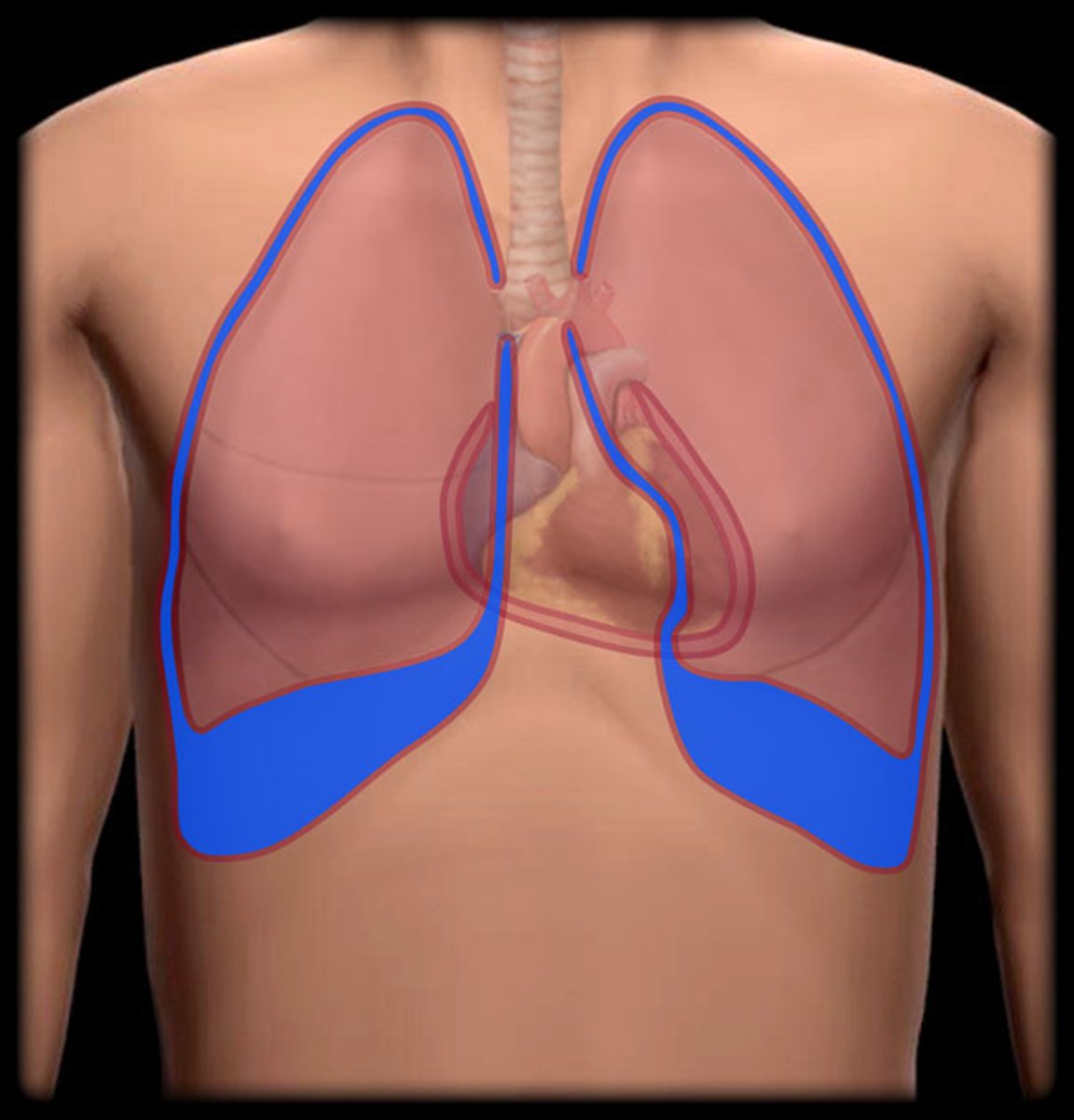
pleural fluid
between layers lubricates pleural membrane; prevents friction
lung lobes, left vs right
left lung has 2, right lung has 3
pulmonary ventilation
flow of air between atmosphere and lungs; when pressure in lungs is less than atmospheric pressure we breath in; when pressure inside the lungs is higher than atmospheric pressure, we breath out
average respiration
12-20 breaths/minute at rest
500mL air
one inhalation + one exhalation
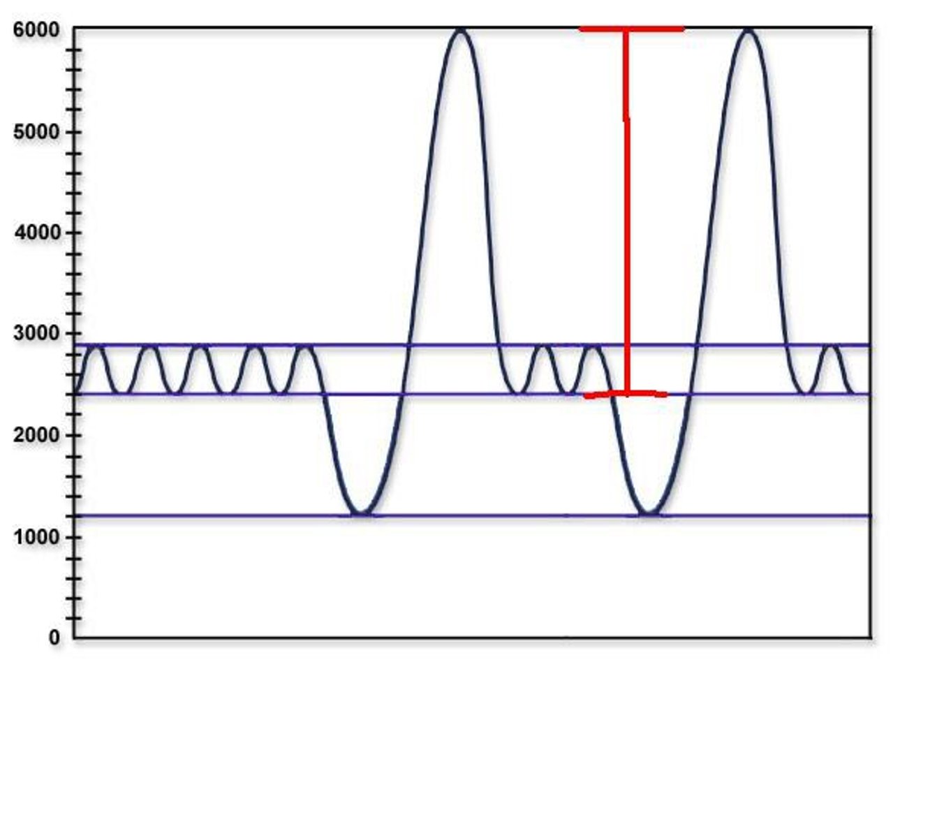
Tidal Volume (TV)
one volume of breath; amount of air entering/exiting the lungs in a single resting breath
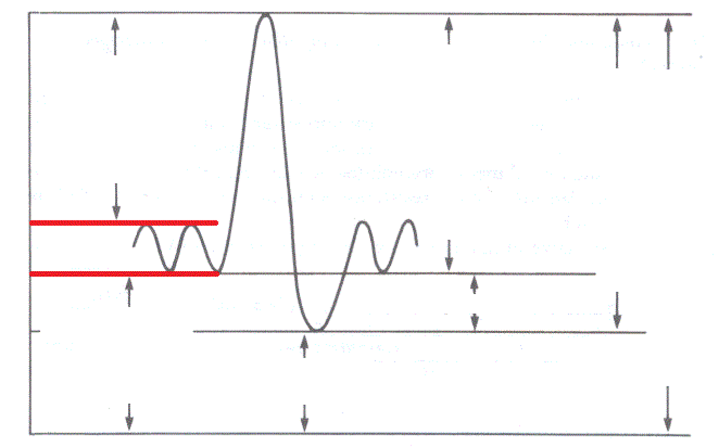
minute ventilation (MV)
total amount of air in and out of lungs in one minute
MV
12 breaths/min x 500mL = 6L/min
anatomical dead space
the volume of air inhaled that does not take part in gas exchange
Inspiratory reserve volume (IRV)
volume of air inspired beyond normal resting inhalation
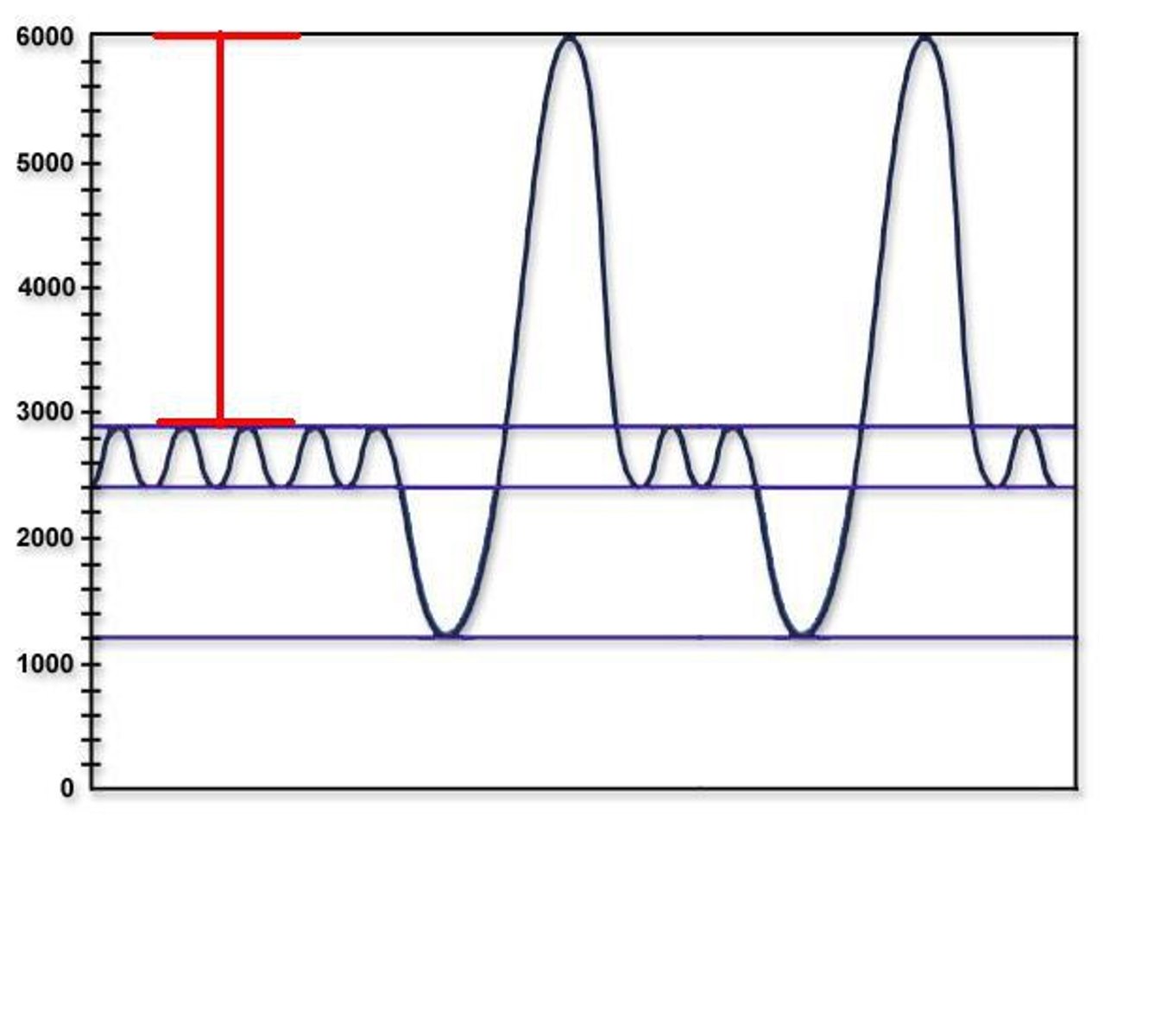
Expiratory reserve volume (ERV)
volume of air expelled beyond normal exhalation
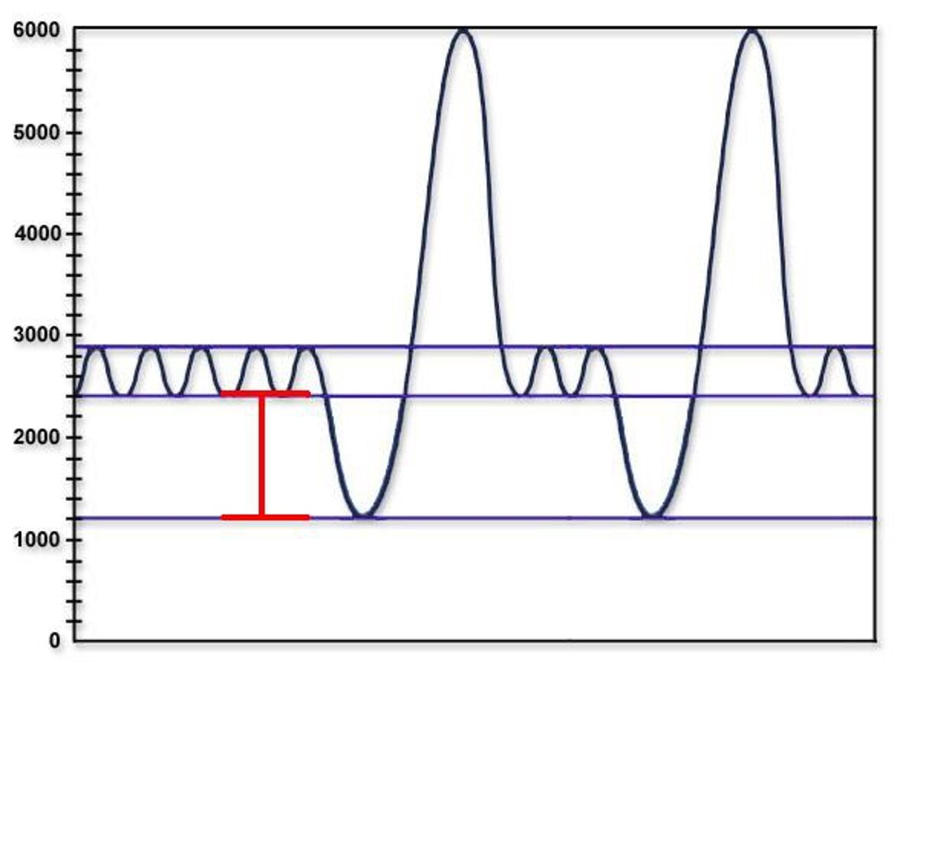
vital capacity (VC)
max vol of air expired in single breath following max inhalation; TV + ERV + IRV =
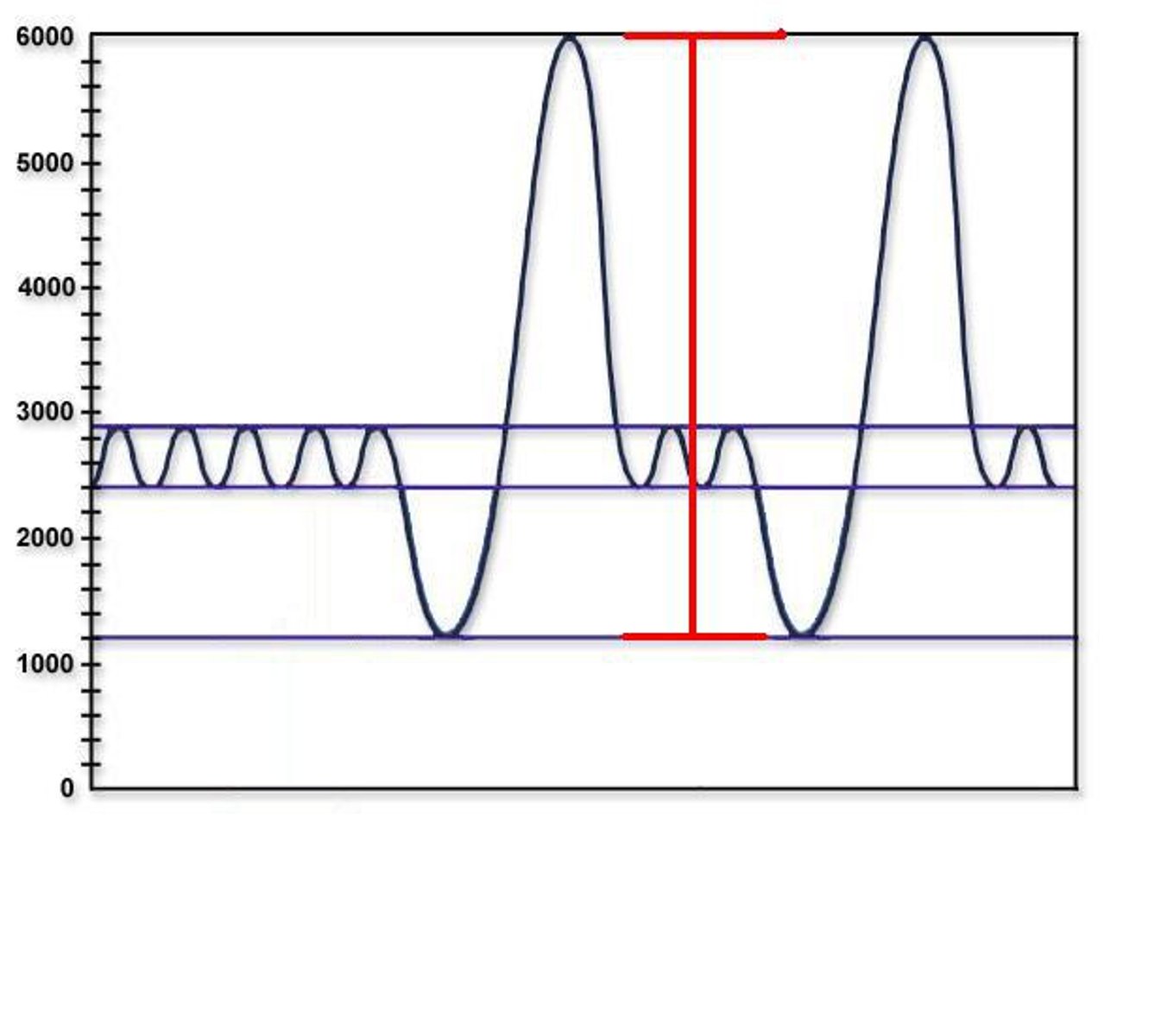
Residual volume (RV)
amount of air remaining in airways after ERV expelled
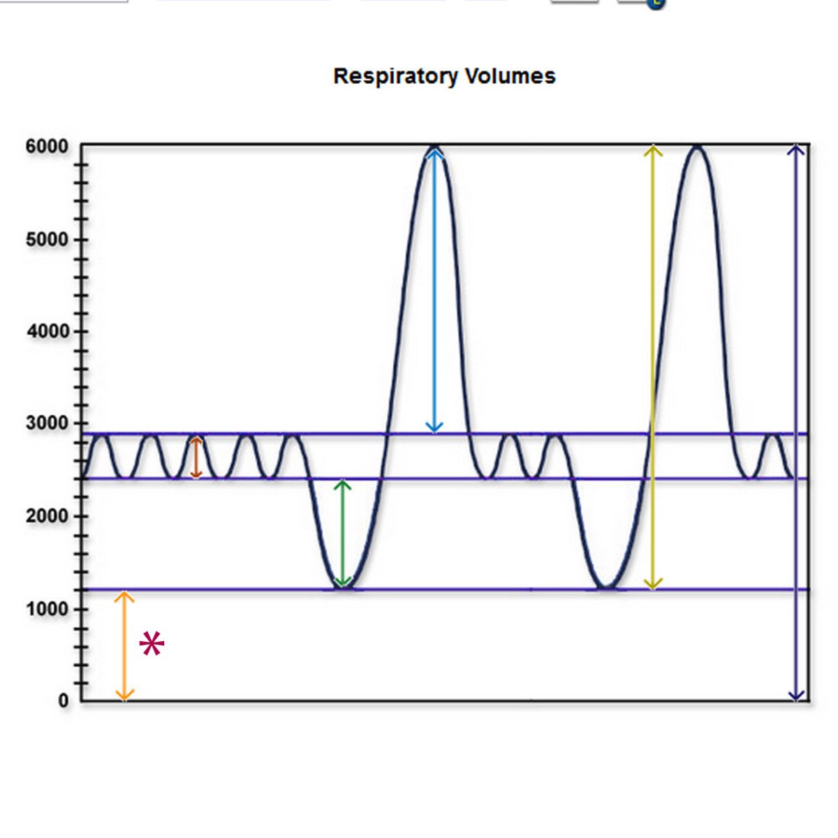
Inspiratory capacity (IC)
max amount of air that can be inspired; TV + IRV =
eupnea
normal quiet breathing
dyspnea
painful or labored
hyperventilating
negative feedback loop
COPD
often due to smoking, thereby preventable; defined as persistent airway obstruction that is not fully reversible
emphysema
destruction of structures of respiratory zone and loss of alveolar surface area
Normal lung (extended response Q)
malleable texture that felt like bubble gum; movement in each lobe during inhalation
emphysema lung (extended response Q)
thicker tissue that felt like an uncooked steak; prevents lung expiration; movement only in one lobe during inhalation
lung cancer
refers to tumors arising from epithelium that lines bronchi, bronchioles, and alveoli
pulse
expansion and recoil of an artery after blood has been pushed through it by the heart
blood pressure
force exerted on the blood vessel walls by the blood; higher in arteries; lower in veins
internal factors that affect blood pressure
viscosity of blood; more blood; faster heart rate; contraction strength (stronger); radius of blood vessels (smaller vessels)
outside factors that affect blood pressure
stress; smoking; being overweight; obstruction; cardiovascular conditions; exercise
systolic pressure
120mm Hg
diastolic pressure
80mm Hg
Mean Arterial Pressure (MAP)
1/3 (PP) =
Partial Pressure (PP)
systolic- diastolic
SA Node
action potentials are generated by authorhythmic cells
repolarization
the cells return back to original condition; relaxed
depolarization
contraction
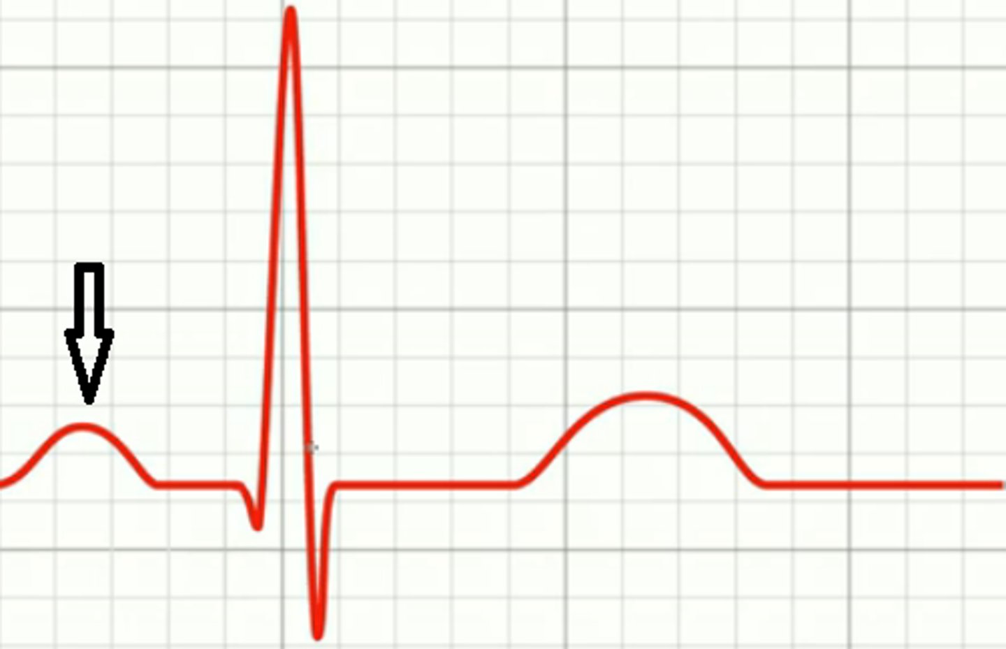
P wave
atrial depolarization; time measured in seconds; height of the wave is measured in mV
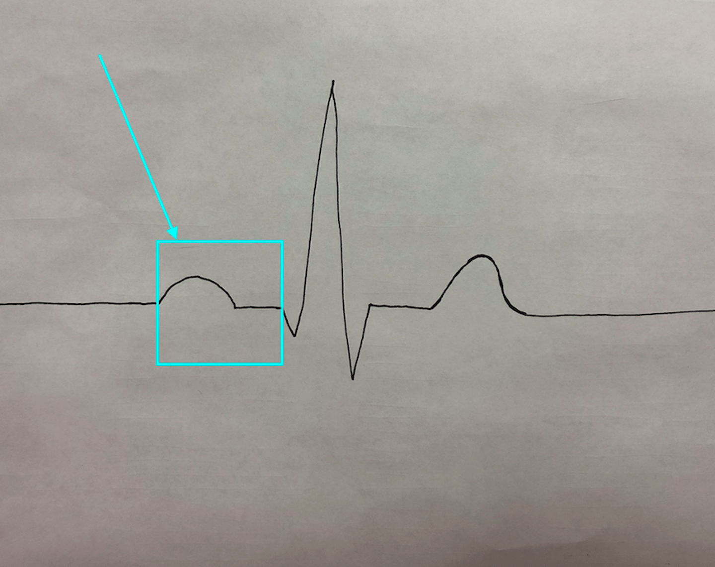
P-Q interval
time it takes to travel from SV node to ventricle muscle tissue; time measured in seconds
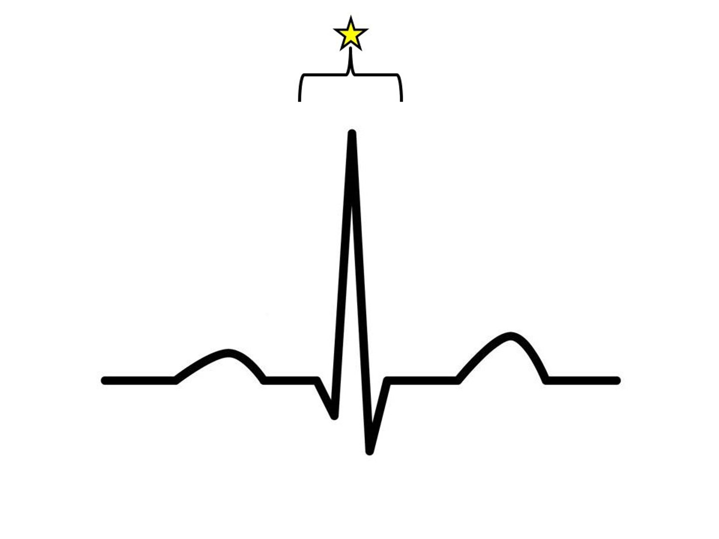
QRS complex
ventricular depolarization; time measured in seconds; height measured in mV
Q-T interval
time pushing; entire duration of ventricular depolarization + ventricular repolarization; aka ventricular systole; time measured in seconds
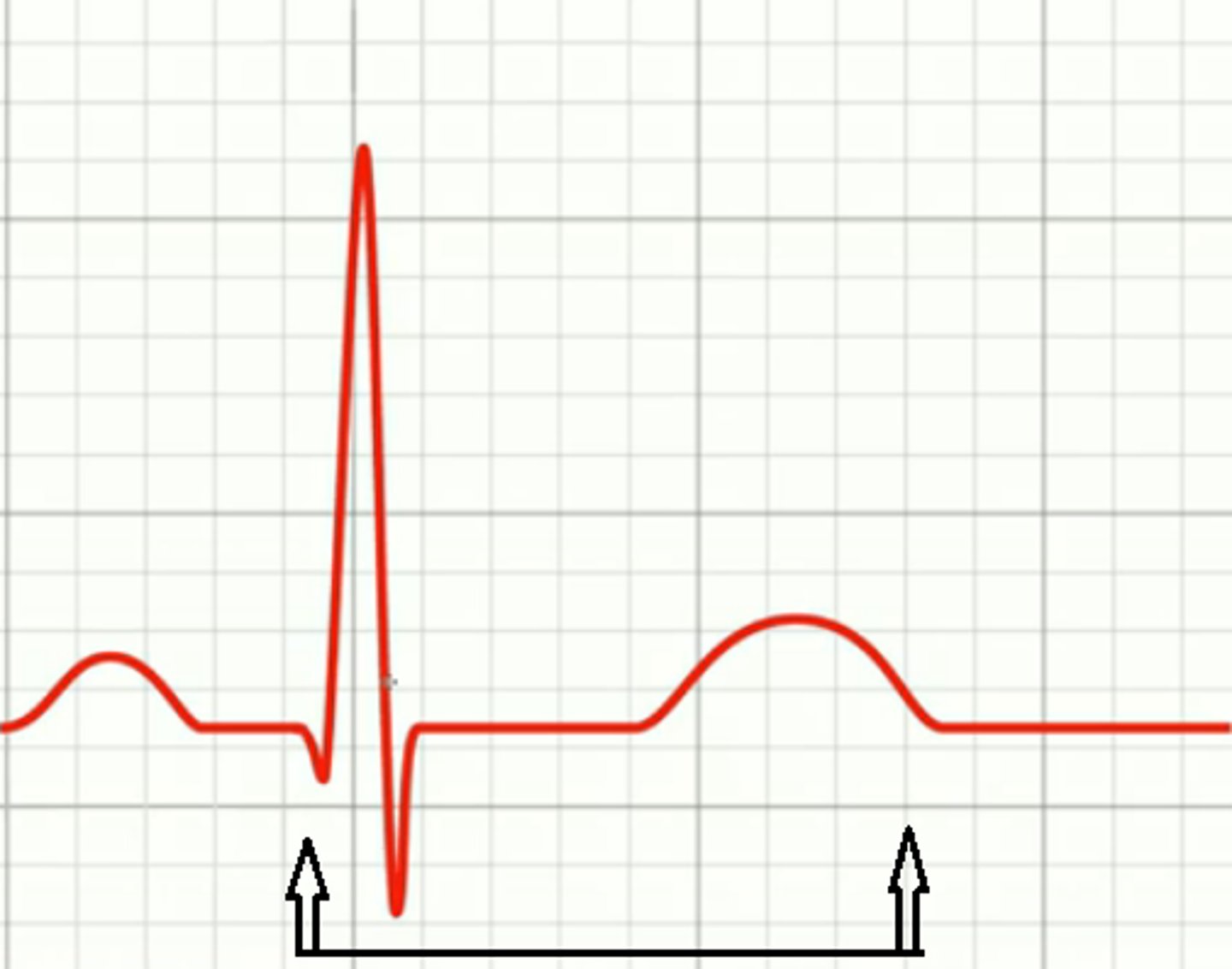
P-Q segment and S-T segment
time between different electrical signals; P-Q = time between atrial depolarization and ventricular depolarization; S-T = time between ventricular depolarization and ventricular repolarization
T-wave
ventricular repolarization (recovery); time measured in seconds; height measured in mV