Cranial OMM
 Call Kai
 Learn
Learn Practice Test
 Spaced Repetition
 Match
Match Flashcards
 Knowt Play
1/21

There's no tags or description
Looks like no tags are added yet.
Name | Mastery | Learn | Test | Matching | Spaced | Call with Kai | Chat |
|---|
No analytics yet
Send a link to your students to track their progress
22 Terms
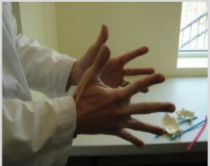
Physiologic. 2 transverse axes. Sphenoid and occiput (the midline bones) rotate opposite of each other. Fingers will point downward, palms move laterally, and fingers spread apart
Extra Info for Comp:
Occurs during inhalation. During flexion, sphenoid will rotate forward/anterior and occiput will rotate backward/posterior. Paired bones will externally rotate. Spheno-basilar junction will move superiorly.
AP diameter of cranium narrows. Lateral diameter of cranium widens.
As sphenoid flexes forward, it will pull on dural attachments (C2, C3, and S2) and cause counternutation of the sacrum (sacrum will rotate backwards and slightly superiorly)
Flexion (physiologic or not physiologic, axes, movement of sphenoid and occiput, and how to show that with air hands)
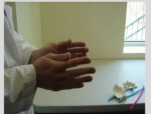
Physiologic. 2 transverse axes. Sphenoid and occiput (the midline bones) will rotate opposite of each other. Fingers will point upwards, palms move medially, and fingers approximate/come together
Extra Info for Comp:
Occurs during exhalation. During extension, sphenoid will rotate backward and occiput will rotate forward. Paired bones will internally rotate. Spheno-basilar junction will move inferiorly.
AP diameter of cranium lengthens. Lateral diameter of cranium shortens.
As occiput moves forward, it will release dural strain on the sacrum and cause nutation (sacrum will rotate forwards and slightly inferiorly)
Extension (physiologic or not physiologic, axes, movement of sphenoid and occiput, and how to show that with air hands)
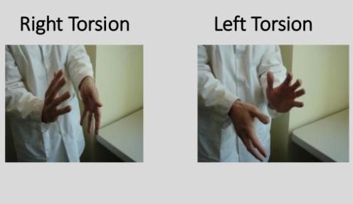
Physiologic, 1 AP axis, sphenoid and occiput will rotate in opposite directions, hands will rotate in opposite directions with one hand rotating up based on the naming of the diagnosis (i.e. right torsion will have right hand rotate superiorly) and the other will rotate down.
Extra Info for Comp:
Named after the direction in which the Greater Wing of Sphenoid will move cephalad/superiorly.
In a right torsion, sphenoid will rotate left (counterclockwise) around AP axis, bringing its right Greater Wing superiorly.
In a left torsion, sphenoid will rotate right (clockwise) around AP axis, bringing its left Greater Wing superiorly.
Torsion ( (physiologic or not physiologic, axes, movement of sphenoid and occiput, and how to show that with air hands)
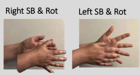
Physiologic. 2 parallel vertical axises and 1 AP axis. Sphenoid and occiput will sidebend in opposite directions around the parallel vertical axises but rotate in the same direction around the AP axis.
For a right sidebending and rotation: Right hand will move away from you (towards pt) with fingers spreading further apart. Left hand will move closer to you (away from pt) with fingers approximating/coming together
Extra Info for Comp:
Named after the side of convexity or where the hand “feels fuller.” This “fullness'“ comes from the widening of the sphenoid and occiput (midline bones).
Ex: in a right sidebending and rotation, the sphenoid and occiput will widen on the right side
Sidebending and Rotation (physiologic or not physiologic, axes, movement of sphenoid and occiput, and how to show that with air hands)

Non-physiologic. 2 parallel vertical axes. Sphenoid and occiput will rotate in the same direction. Named after side of pinkies. In a right lateral strain, both hands will lean right (i.e. pinkies point right). In a left lateral strain, both hands will lean left (i.e. pinkies point left)
Extra Info for Comp:
Index fingers will move in direction of greater wing of sphenoid while pinkies/5th fingers will move to the opposite side.
These strains are named for the position of the base of the sphenoid at the SBS, either left or right. In a right lateral strain, the base of the sphenoid moves to the right, while both index fingers move to the left.
As with all pathologic strains, the AP distance in lateral strains is unchanged. Shearing occurs on the SBS, but the base of the sphenoid moves in a left or right direction. A force on the side of the head in front of or behind the position of the SBS can cause this shearing strain.
For a right lateral strain, the sphenoid and occipital rotate left around their respective vertical axes. For a left lateral strain, they rotate right around their respective vertical axes. In a right lateral strain, the right greater wing of the sphenoid moves to the left and anteriorly, and the right posterior occipital moves to the right and anteriorly. At the SBS, the base of the sphenoid moves to the right
Lateral Strain (physiologic or not physiologic, axes, movement of sphenoid and occiput, and how to show that with air hands)
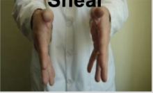
Non-physiologic. 2 transverse axises. Sphenoid and occiput will rotate in the same direction. Fingers will point down while hands rotate anteriorly.
Extra Info for Comp:
Vertical strain patterns do not change the AP diameter and result from a shearing force, either superiorly or inferiorly, acting on the SBS. Such a force can be generated by an uppercut force to the chin, for example, or a downward impact on the top of the head, whether from the front or back.
Named based on the position of the base of the sphenoid at the SBS relative to the occipital base. When the base of the sphenoid moves superiorly, it’s a superior strain.
Both sphenoid and occiput bones rotate anteriorly in a superior strain.
So, in a superior vertical strain, the sphenoid rotates forward or anteriorly, which moves the base of the sphenoid superiorly. Simultaneously, the occipital also rotates forward, shifting the base of the occipital inferiorly at the SBS.
Superior Vertical Shear/Strain (physiologic or not physiologic, axes, movement of sphenoid and occiput, and how to show that with air hands)

Non-physiologic. 2 transverse axises. Sphenoid and occiput will rotate in the same direction. Fingers will point up while hands rotate posteriorly.
Extra Info For Comp:
Vertical strain patterns do not change the AP diameter and result from a shearing force, either superiorly or inferiorly, acting on the SBS. Such a force can be generated by an uppercut force to the chin, for example, or a downward impact on the top of the head, whether from the front or back.
Named based on the position of the base of the sphenoid at the SBS relative to the occipital base. When the base of the sphenoid moves inferiorly, it’s an inferior strain.
In an inferior strain, both sphenoid and occiput bones rotate posteriorly
So, in an inferior vertical strain, the sphenoid rotates backward or posteriorly, which moves the base of the sphenoid inferiorly (base of the sphenoid or posterior aspect of the body is located behind the axis of motion). Simultaneously, the occipital also rotates backward, shifting the base of the occipital superiorly at the SBS.
Inferior Vertical Shear/Strain (physiologic or not physiologic, axes, movement of sphenoid and occiput, and how to show that with air hands)
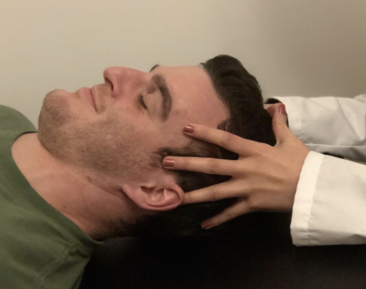
Your forearms should be on table.
Index fingers of each hand resting on the greater wings of the sphenoid bone (over the temples)
Middle fingers placed on the zygomatic processes of the temporal bones
Ring fingers resting on the mastoid processes of the temporal bones (directly behind the ear)
Fifth (little) fingers placed on the squama of the occipital bone
Palms, if possible, resting on the parietal bones
Thumbs resting against each other without contacting the patient’s skull
Vault Hold

Place your caudad/bottom hand under the patient’s occipital squama with the forearm resting on the table establishing a fulcrum.
Your cephalad/top hand bridges across the patient’s frontal bone, with the elbow resting on the table establishing a fulcrum.
The thumb and middle finger of your cephalad/top hand rests on the greater wings of the patient’s sphenoid (if the hand spread is too short, approximate the greater wings).
Extra Info:
Useful for:
Palpating the dura
Feeling dural attachments
Assessing what is occurring at the SBS.
Fronto-Occipital Hold

Hands gently cradle the posterior cranial fossa.
Let thumb tips slide antero-inferior to the mastoid tips so as not to compress them
In plain English: Basically you place your thumbs behind their ears
Extra Info:
Useful for sensing:
Posterior cranial fossa
Cranial base
Some vault motion
Cervicocranial/OA region
Becker Hold (modified version)

Basically: this one is where the pt is a little more active by slightly nodding their head
Full Technique:
You place the pads of both middle fingers on the posterior aspect of the cranium and slide these fingers down the occiput until they are against the posterior arches of the atlas.
You apply caudad/inferior pressure with both middle fingers to separate the facets from the condylar parts.
While you maintain this caudad/inferior pressure, the patient tucks the chin into the chest, making sure not to flex the neck. This is the nodding movement that occurs at the occipitoatlantal joint.
This motion carries the occipital condyles posteriorly, tenses the ligaments in the region, and stretches the contracted muscles in the occipital triangle.
You maintain this position while the patient holds one or more deep inspirations to their limit. This enhances articular release.
You then retest the rate and amplitude of the CRI as it manifests in the basioccipital region to assess the effectiveness of the technique. You can also assess occipitoatlantal motion for normalization.
OA Decompression

this is the one where you lean back and push your elbows (CONDYLES) together
Condylar Decompression

Hold the position until you feel the lateral angles of the frontal bones move more freely and symmetrically into external rotation, with expansion under your fingers and fuller amplitude over a few cycles.
You then gently release the frontal bones and reassess.
Frontal Lift (alternative)

Nasion Spread

Parietal Lift
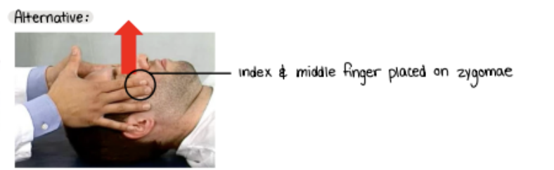
With a “plastic contact” on the bone, you apply a gentle anterior distraction (lift).
You pick up the slack until matching resistance is present at the sutures. Maintain this until an adequate release is felt bilaterally, indicated by less restriction and increased inherent motion felt at the zygomae.
You then gently release and reassess zygomatic motion.
Zygomatic Lift (Alternative)
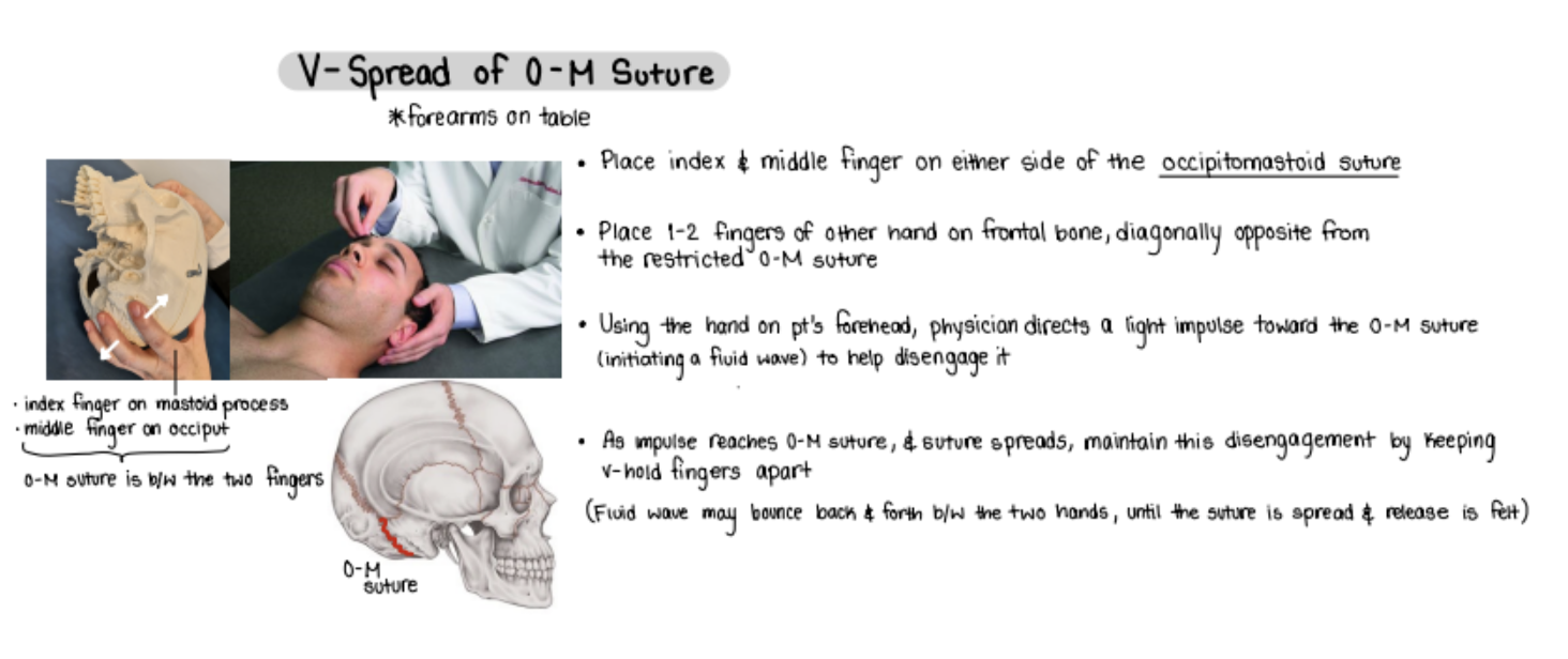
V-Spread

You may place your hands on the patient using a vault, fronto-occipital, or temporal contact.
After diagnosing the patient’s cranial strain pattern (somatic dysfunction named for the freedom), guide the cranium in the direction of the strain pattern to the point of greatest ease or freedom; this is an indirect treatment.
Monitor the CRI. The inherent forces eventually cause a slight increase toward the ease, followed by movement back to the original balance position, which is a sign of release.
You may use respiratory assist and/or direct the tide by dorsiflexing one or both feet to enhance the treatment.
If more than one pattern is present, you may treat each pattern individually or treat all patterns simultaneously, a process called stacking.
Indirect BMT

You may place your hands on the patient using a vault, fronto-occipital, or temporal contact.
After making a cranial strain pattern diagnosis, guide the cranium in the direction of the restrictive barrier of the cranial strain pattern.
Gently approach the barrier and maintain a light force until a release or enhanced CRI occurs. Then reassess.
You may use respiratory assist and/or direct the tide to enhance the treatment.
If more than one pattern is present, you may treat each pattern individually or treat all patterns simultaneously, a process called stacking.
Direct BMT

“volleyball hands”
encouraging extension and resisting flexion
CV4 (Compression of the 4th Ventricle)
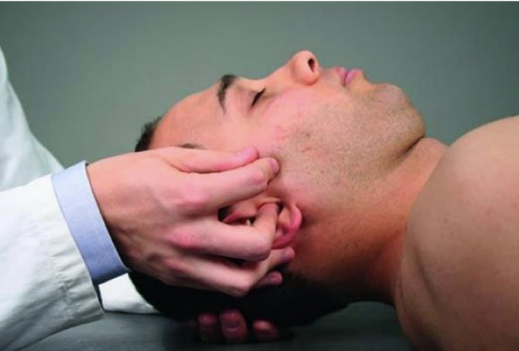
Your left hand cradles the patient’s occiput.
Your right thumb and index finger grasp the zygomatic portion of the patient’s right temporal bone, with the thumb cephalad (on top) and the index finger caudad (on bottom)
Your right middle finger rests on the external acoustic meatus of the ear.
Your right ring and little fingers rest on the inferior portion of the patient’s mastoid process.
Basically: This is the one where you put your middle finger in their ear
Unilateral Temporal Hold

Put patient into the vault hold
Index fingers of each hand resting on the greater wings of the sphenoid bone (over the temples)
Middle fingers placed on the zygomatic processes of the temporal bones
Ring fingers resting on the mastoid processes of the temporal bones (directly behind the ear)
Fifth (little) fingers placed on the squama of the occipital bone
Palms, if possible, resting on the parietal bones
Thumbs resting against each other without contacting the patient’s skull
What does reassessment look like for all the cranial treatments (i.e. what do you do after performing the treatment)?