Session 1- SLO✅
 Call Kai
 Learn
Learn Practice Test
 Spaced Repetition
 Match
Match Flashcards
 Knowt Play
1/56

There's no tags or description
Looks like no tags are added yet.
Name | Mastery | Learn | Test | Matching | Spaced | Call with Kai | Chat |
|---|
No analytics yet
Send a link to your students to track their progress
57 Terms
Describe the 8 diagnostic areas that aid in identifying pathological conditions.
Clinical diagnosis
Radiographic diagnosis
Historical diagnosis
Laboratory diagnosis
Microscopic diagnosis
Surgical diagnosis
Therapeutic diagnosis
Differential diagnosis
what is clinical diagnosis
How the lesion presents, including:
- Color
- Shape
- Location
- History of the lesion
what is radiographic diagnosis
The radiograph provides enough information to establish the diagnosis.
what is historical diagnosis
Includes-
- Personal history
- Family history
- Past and present medical and dental histories
- History of drug ingestion
- History of the presenting disease or lesion
what is Laboratory diagnosis
May include blood chemistries, urinalysis, and cultures.
what is Microscopic diagnosis
Often the main component of a definitive diagnosis. An adequate tissue sample is necessary.
what is Surgical diagnosis
The diagnosis is made using information gained during the surgical procedure.
what is Therapeutic diagnosis
Diagnosis is made by observing how a condition responds to treatment. Nutritional deficiencies are common conditions diagnosed this way.
Differential diagnosis
The point in the diagnostic process when the practitioner determines what test or procedure is needed to rule out suspected conditions and establish the definitive diagnosis.
OR
rule out the conditions originally suspected and establish the definitive or final diagnosis.
The 8 diagnostic areas provide information that helps determine what condition may be present and what testing is needed.
The dental hygienist's role includes what
- Being observant
- Collecting data
- Reviewing medical and dental health histories
- Reviewing the history of the lesion
- Performing clinical description and evaluation
- Reviewing biopsy and microscopy reports
Macule
An area distinguished by a color different from surrounding tissue. It is flat and does not protrude above the surface. A freckle is an example

Papule
A small, circumscribed lesion, usually less than 1 cm, that is elevated or protrudes above the surrounding tissue.

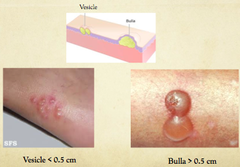
Vesicle
A small, elevated lesion less than 1 cm that contains serous fluid.
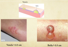
Bulla
A circumscribed, elevated lesion more than 5 mm in diameter. Usually contains serous fluid and looks like a blister.
Pustule
A circumscribed elevation containing pus.

Sessile
A lesion base that is flat or broad instead of stemlike.

Pedunculated
Attached by a stemlike or stalklike base, similar to a mushroom.
Lobulated
Made up of lobes or segments. The lobes may appear fused together.

Exophytic
An outgrowth; describes torus palatinus as an exophytic growth of normal compact bone. IN MIDLINE OF PALATE

Plaque
describing leukoplakia
Nodular
this term describes mandibular tori, which can be lobulated or nodular.

Cyanosis
cyanotic- color description of lesion: blue-black.
Erythema
An abnormal redness of the mucosa or gingiva.
Pallor
Paleness of the skin or mucosal tissues.
Fissured
A cleft or groove, normal or otherwise, showing prominent depth.

Corrugated
Wrinkled
Papillary
Resembling small, nipple-shaped projections or elevations found in clusters
Radiopaque
A light or white area on a radiograph caused by the inability of radiant energy to pass through the structure. The denser the structure, the lighter or whiter it appears.

Radiolucent
Black or dark areas on a radiograph. Radiant energy can pass through these structures.

Unilocular
Having one compartment or unit that is well defined or outlined.
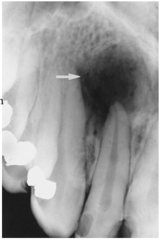
Multilocular
A lesion that extends beyond one distinct area and has many lobes or parts that are somewhat fused together.
Well-circumscribed
A lesion with specifically defined borders where the exact margins and extent can clearly be seen.
Diffuse
A lesion with borders that are not well defined, making the exact parameters difficult to detect.
Coalescence
The process by which parts of a whole join or fuse together to make one.
Root resorption
The apex of the tooth appears shortened or blunted and irregularly shaped. It can occur in response to a cyst, tumor, or trauma.

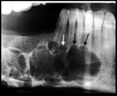
Scalloping around roots
A radiolucent lesion that appears to extend up the periodontal ligament and between the roots.
Crenated
a condition where the edges of the tongue appear notched, scalloped, or wavy
caused by irrigation from adjacent teeth
Indurated
soft tissue that has become abnormally hard, firm, or thick to the touch
Fluctuant
an abnormal oral mass or swelling that moves or compresses in a wave-like motion when pressed
How is visual observation used when describing a pathological lesion
used to evaluate how the lesion presents clinically, including:
- Color
- Shape/size
- Location
- History of the lesion
- Surface texture
- Radiographic appearance
how is palpation used when describing a pathological lesion
evaluating a lesion by feeling it with the fingers to determine the texture of the area.
Descriptive terms include:
- Soft
- Firm
- Semifirm
- Fluid filled
how is measurement used when describing a pathological lesion
uses the metric system to describe lesion size:
- Centimeter (cm) = one hundredth of a meter
- Millimeter (mm) = one thousandth of a meter
- A periodontal probe can help document the size or diameter of a lesion in millimeters.
Clinical Appearance and Etiology:
Fordyce Granules
Clinical appearance:
- Clusters of ectopic sebaceous glands
- Appear as yellow lobules in clusters
- Commonly found on the vermilion border of the lips and buccal mucosa
Treatment: None.
Clinical Appearance and Etiology:
Lingual Thyroid Nodule
Etiology/cause:
- Undescended, trapped remnants of thyroid tissue
Clinical appearance:
- A mass in the midline of the dorsal surface of the tongue
- Located posterior to the circumvallate papillae
- In the area of the foramen cecum
- Usually has a sessile base
- Approximately 2-3 cm wide
Predilection:
- Females
- Linked to hormonal changes
Important: The patient should be evaluated to determine whether the thyroid gland is present in its normal location.
Clinical Appearance and Etiology:
Mandibular Torus/Tori
Clinical appearance:
- Outgrowths of dense bone
- Found on the lingual aspect of the mandible
- Located in the area of the premolars above the mylohyoid ridge
- Usually bilateral
- Often lobulated or nodular
- Can appear fused together
- No predilection for either sex
Treatment: None unless they interfere with fabrication or placement of a prosthodontic appliance.
Clinical Appearance and Etiology:
Fissured Tongue
Clinical appearance:
- Dorsal surface of the tongue has deep fissures or grooves
Cause:
- Unknown
- Probably involves genetic factors
Occurrence: About 5% of the population.
Home care: Gently brush the tongue with a toothbrush to remove debris.
Treatment: None necessary.
Clinical Appearance and Etiology:
Palatal Torus/Torus Palatinus
Clinical appearance:
- An exophytic growth of normal compact bone
- Located in the midline of the hard palate
- May have different shapes and sizes
- May be lobulated
- Covered by normal soft tissue
Etiology:
- Inherited
- Gradual formation
- More common in women
Treatment: None unless it interferes with speech, swallowing, or a prosthetic appliance
Clinical Appearance and Etiology:
Median Rhomboid Glossitis
Clinical appearance:
- Flat or slightly raised
- Oval or rectangular
- Erythematous area in the center of the tongue
Possible association:
- May be associated with a chronic infection with Candida albicans
Treatment: Usually none necessary, but antifungal treatment may be used.
Clinical Appearance and Etiology:
Geographic Tongue / Migratory Glossitis
Also called erythema migrans.
Clinical appearance:
- Erythematous patches surrounded by a white or yellow border
- Diffuse areas without filiform papillae
- Fungiform papillae are distinctly present
- Depapillated areas show remission and changes
Possible causes/contributing factors:
- Genetic factors may play a role
- May be exacerbated by stress
Symptoms: Occasionally causes burning discomfort.
Treatment: Usually none.
Clinical Appearance and Etiology:
Gingival Melanin Pigmentation
Clinical appearance/etiology:
- Melanin is the pigment that gives color to the skin, eyes, hair, mucosa, and gingiva
Most commonly observed in dark-skinned individuals.
Clinical Appearance and Etiology:
Black Hairy Tongue
Clinical appearance:
- Papillae are brown-to-black
- Caused by chromogenic bacteria
Contributing factors:
- Tobacco
- Foods
- Hydrogen peroxide
- Alcohol
- Chemical rinses
Home care: Gently brush the tongue with a toothbrush to remove debris
Clinical Appearance and Etiology:
Retrocuspid Papilla
Clinical appearance:
- A sessile nodule
- Located on the gingival margin on the lingual aspect of the mandibular cuspids
- Only a few millimeters in size
- Seen more often in young individuals
- Resolves with age.
brown hairy tongue
a harmless, temporary condition where tiny bumps on the tongue grow long and trap food, bacteria, or stains
Clinical Appearance and Etiology:
Lingual Varicosities / Varices
Clinical appearance:
- Red-to-purple enlarged vessels or clusters
- Usually found on the ventral and lateral surfaces of the tongue
- Most commonly observed in individuals older than 60 years.
Clinical Appearance and Etiology:
White Hairy Tongue
Clinical appearance:
- Elongated filiform papillae are white
Cause:
- Increase in keratin production or
- Decrease in normal desquamation
Home care: Gently brush the tongue with a toothbrush to remove debris.
Clinical Appearance and Etiology:
Linea Alba
Clinical appearance:
- A white line
- Extends anteroposteriorly on the buccal mucosa
- Located along the occlusal plane
- May be bilateral
Associated factor:
- May be more prominent in patients with a clenching or bruxing habit.
Clinical Appearance and Etiology:
Leukoedema
Clinical appearance:
- A generalized opalescence on the buccal mucosa
- Most commonly observed in Black adults
- When the mucosa is stretched, the opalescence becomes less prominent
Treatment: None.