anatomy exam 4
 Call Kai
 Learn
Learn Practice Test
 Spaced Repetition
 Match
Match Flashcards
 Knowt Play
1/22

There's no tags or description
Looks like no tags are added yet.
Name | Mastery | Learn | Test | Matching | Spaced | Call with Kai | Chat |
|---|
No analytics yet
Send a link to your students to track their progress
23 Terms
GI embryology
GI tract is 1 tube that folds in on itself
day 30: embryo stops AP growth and starts folding laterally → yolk sac thins out to become vitelline duct into umbilical cord (gut tube formed by week 4 but all cells are pluripotent and organ differentiation is caused by a blast of retinoic acid)
dorsal mesentery: provides nutrents and attaches to gut wall (will become peritoneum)
foregut embryology
esophagus and trachea: respiratory diverticulum forms at 4 weeks and then the respiratory and esophageal separates (if they don’t separate properly it can lead to atresia)
superior 2/3 of esophagus = striated muscle (vagus nerve), inferior 1/3 smooth muscle (sphlancnic plexus)
stomach: at week 4 is just dilation of gut tube then undergoes 90 degrees clockwise rotation (left wall becomes anterior) → greater and lesser curvature, vagus nerve get dragged along
spleen: around week 5 it begins to form in dorsal mesogastrium (not connected to gut tube bc it’s mesodermal)
liver: begins at week 4 in ventral mesogastrium to intertwine with vitelline veins to anastamose → hepatic portal vein, bile duct created by liver’s connection to gut tube thinning, bud from bile duct will form gallbladder
pancreas: dorsal pancreatic bud in dorsal mesogastrium and ventral pancreatic bud and ventral mesogastrium, eventually they are moved next to each other and fuse to form pancreas (failure to fuse → pancreas divisum)
duodenum: in line with gut tube initially but gets pushed right and posterior by week 6, 1/3 is intraperitoneal
midgut embryology
begins as rapid elongation of gut tube following vitelline duct and superior mesenteric artery → primary loop
physiological herniation: necessary for development at 6 weeks midgut grows out of abdominal wall and into umbilicus following vitelline duct, cephalic limb grows the most, then 90 degree rotation around superior mesenteric artery, at 10 weeks abdominal cavity is bigger, so SI and LI stop growing and retreat into abdominal cavity, rotating total 270 degrees (including the first rotation)
meckel’s diverticulum- small part of vitelline duct persists → constricts intestine or extraperitoneal tissue attached to it (these are rare, most cases are asymptomatic)
intestinal malrotation (largely asymptomatic)
hindgut embryology
does not herniate or rotate
blood supply: inferior mesenteric artery
cloaca: opening for urogenital and anorectal system (fetal butthole), urorectal septum separates them to make poop hole and pee hole
watershed areas: areas that receive dual blood supply (duodenum, splenic flexure, anorectal canal) blood supply overlap at terminal vessels, high stress events → reduce blood flow
peritoneum
2 layers: parietal = lines abdominopelvic cavity, visceral peritoneum = surrounds viscera
in embryo = peritoneal cavity (in adults it’s a potential space)
peritoneal ligaments: peritoneum folds in on itself → omenta (connect organs) and mesenteries (connect organ to body wall)
greater omentum- between greater curve of stomach and transverse colon → gastrophrenic, gastrosplenic, gastrocolic ligaments
lesser omentum- between lesser curve of stomach and liver → gastrohepatic, hepatoduodenal ligament
compartments- omental bursa, supracolic (anterior to stomach, superior to transverse colon), infracolic (inferior to transverse colon)
omental foramen- how omental bursa communicates with greater sac
press on omental foramen to stop bleeding from cystic artery
blood supply to abdomen
3 arteries branch from abdominal aorta
foregut: esophagus → middle of duodenum, accessory digestive organs (supplied by celiac trunk)
midgut: middle of duodenum → 2/3 of transverse colon (supplied by branches of superior mesenteric artery)
hindgut: distal 1/3 trnasverse colon → anus (supplied by brances of inferior mesenteric artery)
foregut
esophagus: passes through diaphragm at T10, and joins stomach at gastroesophageal junction
stomach: fundus, body, pyloric canal (empties into duodenum)
pyloric sphincter controls stomach emptying
duodenum: c shaped loop of small intestine that curves around pancreas (L1-L3), superior (connects to pyloric sphincter), descending, horizontal, ascending, joins with jejunum at duodenal-jejunal junction
only proximal part is part of foregut
distal is part of midgut
pancreas: accessory digestive organ, in omental bursa, head, uncinate process, neck, body, tail, main pancreatic duct starts in tail and forms ampulla
spleen: not part of digestive system, derived from embryonic foregut, very delicate
midgut
jejunum- intraperitoneal, red, thick, highly vascular (more vasa recta)
ileum- intraperitoneal, pale pink, thin, more fat
in both jejunum and ileum mesenteries are organized into straight vessels (vasa recta) and arcades (loops)
hindgut
large intestine- tenia coli = thick bands of smooth muscle, haustra (pouches), omental appendices (fat hanging off)
poop: ileocecal valve, cecum, ascending colon, right colic flexure, transverse colon, left colic flexure, descending colon, sigmoid colon, rectum, anus
appendix- near ileocecal junction, appendicitis causes visceral referred pain around T10, then towards Mcburney point (somatic pain), psoas sign (flexion of thigh against resistance)
arteries of the abdomen
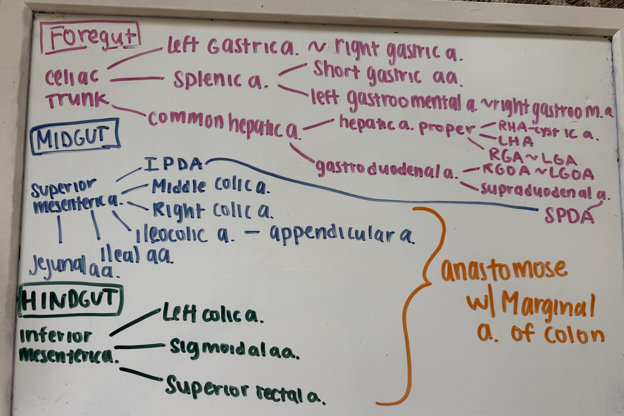
hepatic portal system
inferior and superior mesenteric veins and splenic vein → hepatic portal vein (to liver)
liver drained by hepatic veins which empty into IVC
anastomoses are important for collateral circulation but high BP can cause a lot of backup
portal hypertension: too high BP in portal system → blood backs up in portal veins and where there are anastomoses, there is also back up into systemic veins (esophageal varicose, caput medusae, anorectal varices)
liver
attached to diaphragm by coronary ligament
bare area of liver: has no peritoneum
anatomical lobes: left and right (caudate and quadrate) divided by falciform ligament
dual blood supply: 30% from proper hepatic artery, 70% from hepatic portal vein for filtration and nutrient processing, common bile duct travel in hepatoduodenal ligament
biliary tree: bile from liver → right and left hepatic duct → common hepatic duct → cystic duct → gallbladder (stored and concentrated) → during digestion bile leaves gb through cystic duct → common bile duct → hepatopancreatic ampulla
retroperitoneal organs
outside of parietal peritoneum, organs only have peritoneum on anterior aspects
left suprarenal vein and left gonadal vein empty into left renal vein but on right they empty directly into IVC
kidneys
retroperitoneal (right kidney is lower bc of liver)
suprarenal glands arterial supply: superior, middle, inferior suprarenal arteries
waste in kidneys emptied through ureters and filtered blood returns through renal veins
renal medulla: renal columns and pyramids (pee produced then exits through papilla into collecting system)
minor calyces, major calyces, renal pelvis, ureter, bladder
gonadal veins always anterior to ureters
diaphrgam and posterior abdominal wall
diaphragm openings- caval foramen for IVC (T8), esophageal hiatus (T10), aortic hiatus (T12)
medial arcuate ligament - sympathetic trunk, psoas major
lateral arcuate ligament - quadratus lumborum
quadratus lumborum- innervated by ventral rami of spinal nerves
psoas major- lumbar plexus
iliacus- femoral nerve
intermediate mesoderm
between paraxial and lateral plate mesoderm and connected to lateral plate mesoderm
differentiates to make glomeruli and tubing, kidney is diverticulum of celom but eventually separates
mesonephric duct: excretes urine, forms from above the heart down to the bladder
pronephros: primitive and never develops into functional excretory structures
mesonephros: ducts and tubules remain in adult but glomeruli disappear in adult (forms collecting system of metanephric kidney = calyces, CD, pelvis, ureter)
mesonephric ducts will form adult epididymis and vas deferens in men, paramesonephric ducts will form oviduct in females (diverticulum of uterus)
2 distillations: one in looping artery of glomerulus and one in tubules (no veins)
fluid accumulates outside artery and trapped outside bowman’s capsule
when urine enters distal convoluted tubule twists to make loop of henle and proximal convoluted tubule
metanephros: adult kidney with new glomeruli
blastema- forms excretory portion (bowman’s capsule, PCT, loop of henle, DCT)
uteric bud is a diverticulum of mesonephric duct (will eventually become excretory portion)
cloaca
cup at the bottom of embryo
3 tubes: anus, vagina, bladder
forms urorectal septum to separate anus and vagina
urogenital sinus is front sinus
bladder is composite organ and mesonephric trigone of bladder is formed by exstrophy (when mesonephric ducts turn inside out)
vascularization embryology
metanephric kidneys form low near the bladder and slowly rise to adult position and changes arterial supply based on where it is (stops when it hits adrenal gland), as kidney rises, ureter grows
urogenital system: male
male: bladder above prostate and inside prostate is orifice of prostatic utricle (urine passes on either side of tube)
female: in urethra when it empties inside vagina
embryological urogenital pathologies
VACTERL association: embryological pathologies (vertebral, anal atresia, CV abnormalities, tracheoesophageal fistula, esophageal atresia, renal anomalies, limb defects)
imperforate anus- dysfunction of urorectal septum (failure to pass meconium)
hydronephrosis- glomeruli don’t work → fluid accumulates in kidney → ureter expands
renal agenesis- kidney not formed
oligohydramnios- not enough amniotic fluid → deformed limbs, dry skin, face abnormal, pulmonary hypoplasia
polyhydrammnios- excess amniotic fluid caused by lack of swallowing from maternal diabetes
horseshoe kidney- asymptomatic but unified kidney prevented from reaching abdominal location by inferior mesenteric vein (high risk of kidney stones and UTIs)
sympathetic innervation of abdominal organs
foregut:
preganglionic cell bodies from T5-T9 (celiac ganglia)
postganglionic axons distribute with celiac trunk
midgut:
preganglionic cell bodies from T9-T12 (superior mesenteric ganglia)
postganglionic axons distribute with superior mesenteric artery
hindgut:
preganglionic cell bodies from T12-L2 (inferior mesenteric ganglia)
postganglionic axons distribute with inferior mesenteric artery
preganglionic pathway: start at lateral horn of T1-L2, through ventral horn, spinal nerve, ventral ramus, white ramus communicants, pass through paravertebral ganglia (NO SYNAPSE) through splanchnic nerve to subdiaphragmatic ganglia (some will descend to lower levels and leave at lower paravertebral ganglia)
greater splanchnic nerves- T5-T9, lesser splanchnic nerves- T10-T11, least splanchnic nerve- T12, lumbar splanchnic nerves- L1-L2
parasympathetics to abdominal organs
most of parasympathetic innervation still comes from vagus nerve → posterior and anterior vagal trunks → pass with arteries/sympathetics to organs they innervate
some (hindgut) will be innervated by S3-S4:
cell bodies in lateral horn, ventral root, spinal nerve, ventral ramus, then just leave ventral ramus as pelvic splanchnic nerves → inferior hypogastric plexus
enteric nervous system
intrinsic part of autonomic nervous system (both plexuses controlled by extrinsic and enteric nervous system)
submucosal plexus: regulates secretion and degree of blood flow
myenteric plexus: controls motility
hirchsprung disease: birth defect where neural crest cells fail to migrate → lack of ganglia in distal bowel → failure to pass meconium, abdominal distention
visceral sensory from abdominal organs
usually follows path of sympathetic outflow backwards (pelvic organs are exception)